Cardiac fibromas are the second most common benign primary tumour of the heart following rhabdomyoma in the children. Reference Cordinhã1 However, a number of patients with cardiac fibroma are asymptomatic Reference Cordinhã1 ; the clinical features depend on the size and location and include chest pain, arrhythmia, low cardiac output due to outflow tract obstruction, and sudden cardiac death. Reference Cordinhã1,Reference Shenthar2 Furthermore, it is known that sports are associated with an increased risk for sudden death in athletes who are affected by cardiovascular conditions predisposing to life-threatening arrhythmias. Reference Steriotis3 We report an asymptomatic 10-year-old boy diagnosed with left ventricular fibroma, who was admitted to our clinic for cardiac examination for competitive sports.
Case report
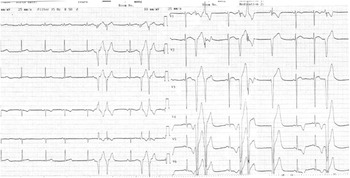
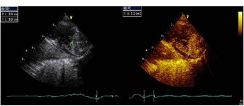
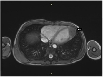
A 10-year-old boy was referred to the paediatric cardiology department by his general practitioner for cardiac examination before participation in competitive sports. He was asymptomatic. There was no family history of syncope, cardiac arrest, sudden death, or cardiomyopathy. He was found to have extrasystoles with auscultation. There was no murmur and no thrill. No other abnormal physical examination findings were detected. His systemic blood pressure was 100/70 mmHg. X-ray radiography showed no cardiomegaly; pulmonary vascularity was normal. The electrocardiogram showed premature ventricular contractions originated from inferior of ventricle with deep S waves in the inferior leads DII, DIII, aVF (Fig 1). Complete blood count, blood urea, electrolytes, and thyroid functions were normal. Detailed two-dimensional transthoracic echocardiography examination demonstrated a 20 mm × 30 mm sized mass at the left ventricle apex myocardium (Fig 2a). The mass was immobile, invaded into myocardial tissue. It was observed that it protrudes slightly into the left ventricle. There was no valve involvement and wall motion defect. Systolic functions of left ventricle were normal. After that, three-dimensional transthoracic echocardiography was performed and confirmed that egg-shaped mass measuring 2.3 by 2.8 cm along the apical portion of lateral wall (Fig 2b). It was thought that premature ventricular contraction might be related to this apical located mass. To determine its features, cardiac MRI was performed. MRI showed a 31 × 29 × 28 mm sized tumour compatible with fibroma, located in the myocardium of left ventricle apex extending to lateral wall (Fig 3). Tumour was demonstrated low signal intensity on T1 and T2 sequences, late contrast enhancement was detected. On rhythm holter, the monitored premature ventricular contractions were 30% of total heart beat and 112 couplet premature ventricular contractions were seen at 166 bpm. There was no ventricular tachycardia detected on rhythm holter. In the exercise test, premature ventricular contractions were observed at above 160 beats/min. Because of frequent premature ventricular contractions seen in rhythm holter and detecting premature ventricular contraction at high heart rates, antiarrhythmic therapy was initiated. We were monitoring the patient by medical treatment with flecainide and propranolol. Flecainide was initiated 100 mg/m2/day, propranolol was initiated 1 mg/kg/day, gradually increased to 2 mg/kg/day. Rare PVCs, not exceeding 5% of total beat, were detected in the rhythm holter performed after the medical treatment was started; couplet PVCs and ventricular tachycardia were not observed. The patient was recommended effort restriction and his arrhythmia is under control with medical treatment for following 2 years.
The electrocardiogram showed couplet premature ventricular contractions originated from inferior of ventricle.

( a ) 2D transthoracic echocardiographic examination demonstrated a 20 mm × 30 mm sized mass at the left ventricle apex myocardium. ( b ) Three-dimensional transthoracic echocardiography demonstrated that egg-shaped mass measuring 2.3 by 2.8 cm along the apical portion of lateral wall.

MRI demonstrated a 31 × 29 × 28 mm sized tumour located in the myocardium of left ventricle apex extending to lateral wall.

Discussion
Sports are associated with an increased risk for sudden death in athletes who are affected by cardiovascular conditions predisposing to life-threatening ventricular arrhythmia during exercise. Cardiac screening with non-invasive examinations is a very important tool for identification of possible pathology and for the characterisation of electrical instability. Rhythm and conduction abnormalities are the first cardiovascular causes of sports disqualification. Reference Steriotis3 Cardiac tumours are a rare cause of arrhythmias in clinical practice. Reference Shenthar2
The most common origins of PVC are the ventricular outflow tracts. Reference Oomen, Dekker and Meijer4 However, on cardiac screening for competitive sports, inferior originated PVC was detected in our patient. Like these atypical originated PVCs when detected, it should be kept in mind that there may be a structural anomaly like mass. In such cases, examination with echocardiography will be helpful in recognising structural anomalies and if necessary, advanced investigation methods such as CT or MRI can be used. Our patient’s, who was detected mass with echocardiography, MRI results gave findings in favour to fibroma.
Primary cardiac tumours are rare. 75% of cardiac tumours are benign and have a reported incidence of 0.03–0.32% Reference Rajput, Bishop and Limaiem5 Cardiac fibromas found in one quarter of cases, amongst tumours identified at postmortem in hearts of children between 1 and 15 years Reference Bédard, Becker and Gatzoulis6 Cardiac fibromas are the second most common benign cardiac tumours in the children Reference Rajput, Bishop and Limaiem5 which arise from heart fibroblasts. Reference Zheng7 These tumours are mostly located in the left ventricular free wall or interventricular septum and less frequently in the right ventricle or atrial free wall. Reference Maleszewski and Burke8 In our patient, it appeared as a mass extending to the lateral wall of the left ventricle, which is the most common localisation for fibromas. Clinical symptoms and signs of the tumour are chest pain, cardiomegaly, arrhythmias, and even sudden mortality. In some cases, cardiac tumours are asymptomatic as in our patient. Due to the non-specific nature of symptoms, many are diagnosed following an echocardiographic finding, Reference Cordinhã1 as in the case presented.
Cardiac tumours are associated with wide variety of arrhythmias depending on the tumour type and the location of involvement. The arrhythmias resulting from cardiac tumours include atrial ectopics, atrial flutter, atrial fibrillation, Wolff–Parkinson–White syndrome, ventricular ectopics, ventricular tachycardia, torsades de pointes, atrioventricular blocks, and sudden death. The mechanism can be re-entry or triggered automaticity. In fibroma, clinically significant arrhythmias are more common than in any other cardiac tumours. Some form of cardiac arrhythmia is present in 64% of cases. Very frequent monomorphic PVCs and couplet PVCs were observed in our patient as well. Ventricular tachycardia and sudden death may occur in up to 30% of patients with cardiac fibroma. Reference Shentar9
Two-dimensional transthoracic echocardiography provides information in terms of mass localisation and size. Two-dimensional transthoracic echocardiography may underestimate the size of cardiac masses; the accurate size is possible only if a plane chances to encompass the complete long axis of the mass, but that may not occur especially the mass is mobile. With 3D echocardiography, the whole extent of mass is contained in the data and that would provide more reliable measurements. Reference Asch, Biegansky, Panza and Weissman10 However, in our patient, there is no significant difference between two-dimensional transthoracic echocardiography and three-dimensional transthoracic echocardiography measurements. This may be due to immobility of the mass. In literature, 3D transoesophageal echocardiography was utilised to define the morphologic and spatial characteristics of cardiac and paracardial tumours in vivo, establish their relationships with adjacent structures, and assess the haemodynamic effects. Reference Zavaleta, Morales, Vargas-Barro, Keirns and Fraustro11
Using advanced imaging techniques such as CT and MRI helps to determine the characteristics of the cardiac mass. Cardiac CT often demonstrates partial calcifications in fibromas. Reference Yılmaz, Demir, Önur, Yılbazbayhan and Dursun12 We preferred MRI for further examination in our patient because of superior tissue contrast, versatility in image planes Reference Zavaleta, Morales, Vargas-Barro, Keirns and Fraustro11 , and radiation free. MRI is an important imaging technique in the evaluation of cardiac tumours. T1-weighted, T2-weighted, and gadolinium-enhanced sequences are used for anatomic definition and tissue characterisation. Because of their fibrous nature, the tumours are usually homogeneous and hypointense on T2-weighted MR and generally isointense or hypointense on T1-weighted images. Appearances after administration of gadolinium contrast material can demonstrate heterogeneous enhancement. Reference Sparrow, Kurian, Jones and Sivananthan13 MRI findings of our patient were compatible with fibroma.
Different management strategies are suggested in the literature including surgical resection, single ventricle palliation, cardiac transplant, and conservative treatment with antiarrhythmic medications. Reference Rajput, Bishop and Limaiem5 Cases showing the efficacy of amiodarone and/or beta-blockers in cardiac fibroma-associated arrhythmias have been reported in the literature. Reference Rajput, Bishop and Limaiem5,Reference Miyake, Del Nido and Alexander14,Reference Jones, Ramcharan and Chaudhari15 Our presentation is original because it is a publication showing the efficacy of flecainide and beta-blocker in cardiac fibroma-associated PVCs. In cardiac tumours, the mechanism can be re-entry or triggered automaticity. Abnormal automaticity is thought to play a role in cases of elevated extracellular potassium, low intracellular pH, and catecholamine excess. Reference Gaztanaga, Marchlinski and Betensky16 Flecainide blocks fast inwards sodium channels and slowly unbinds during diastole, prolonging the refractory period of the heart. Flecainide also prevents delayed rectifier potassium channels from opening, lengthening the action potential through ventricular and atrial muscle fibres.Reference Andrikopoulos, Pastromas and Tzeis17 Therefore, we started to our patient flecainide and beta-blocker combination treatment to minimise the risk of arrhythmia and symptoms. Our patient’s tumour was not amenable to resection due to its extensive invasion of myocardial tissue. To avoid cardiac surgery complications, it was decided to follow up in our patient because the fibroma that invaded the left ventricular apicolateral myocardial tissue was asymptomatic, life-threatening ventricular tachycardia was not observed, and PVCs were controlled with antiarrhythmic therapy. And his arrhythmia is under control with medical treatment for 2 years.
Conclusion
In conclusion, applications before participation in sports should be carefully evaluated. Electrocardiogram assessment of all individuals should be done. Electrocardiogram can provide important clues for serious diseases. The patients with arrhythmia on electrocardiogram require further examination with echocardiography. In our patient, the diagnosis of cardiac mass was made by echocardiography. Evaluations by CT or MRI provide the identification, location, and surrounding structures of the tumour. Step-by-step cardiac examination and if necessary imaging are vital for diagnosing patient before participation in sports. Sports are essential for health, but sudden deaths are known to occur during sports. Detailed examinations are necessary for professional sports. In this case, the importance of detailed cardiac examination with electrocardiogram and imaging techniques in asymptomatic individuals before participating in sports was emphasised.
Acknowledgements
We thank the parents of the boy described for allowing us to share his details, and than Karabekir Ercan MD, Department of Radiology for performing MRI.
Financial support
This research received no specific grant from any funding agency, commercial, or not-for-profit sectors.
Conflicts of interest
None.
Ethical standards
Informed consent was obtained from the patient’s family for publication of this case report and any accompanying images.