INTRODUCTION
Cryptosporidium is a protozoan parasite in humans and animals and has a worldwide distribution. Cryptosporidium infection has been implicated as a cause of multiple waterborne and foodborne outbreaks of diarrhoea (MacKenzie et al. Reference MacKenzie, Hoxie, Proctor, Gradus, Blair, Peterson, Kazmierczak, Addiss, Fox and Rose1994; Fayer et al. Reference Fayer, Morgan and Upton2000; Slifko et al. Reference Slifko, Smith and Rose2000). Humans can also acquire the infection through direct contact with humans (anthroponotic transmission) or animals (zoonotic transmission). Although cryptosporidiosis causes self-limiting diarrhoea in immunocompetent persons, it may lead to life-threatening diarrhoea in immunocompromised patients (DuPont et al. Reference DuPont, Chappell, Sterling, Okhuysen, Rose and Jakubowski1995; Kjos et al. Reference Kjos, Jenkins, Okhuysen and Chappell2005). The disease has also been considered as a predictor of malnutrition in early childhood (Huang and White, Reference Huang and White2006).
The genus Cryptosporidium is a multispecies complex. To date, approximately 27 species and more than 60 genotypes have been identified (Fayer and Santin, Reference Fayer and Santin2009; Fayer, Reference Fayer2010; Traversa, Reference Traversa2010; Elwin et al. Reference Elwin, Hadfield, Robinson, Crouch and Chalmers2012). Most of the species or genotypes infect a particular host with a few notable exceptions. Cryptosporidium parvum infects humans and several species of animals, and is the major species responsible for zoonotic cryptosporidiosis in many developed countries. Another species, C. hominis, commonly infects humans and is responsible for anthroponotic transmission, with high prevalence in developing countries compared with industrial nations (Raccurt, Reference Raccurt2007; Xiao, Reference Xiao2009). Subtyping of C. parvum based on 60 kDa glycoprotein (gp60) gene identified at least 12 subtype families (IIa – IIl). The zoonotic IIa subtype is predominant in developed nations and rarely isolated from humans in developing nations where the anthroponotic IIc subtype family is the most common cause of human C. parvum infections. The C. parvum IIa subtype family has a high genetic diversity with IIaA15G2R1 being predominant in calves and the major subtype infecting human and cattle in some countries (Xiao, Reference Xiao2009, Reference Xiao2010).
The epidemiology of Cryptosporidium in Middle Eastern countries is different from other developing countries as earlier studies reported that C. parvum had higher prevalence than C. hominis in Saudi Arabia (Al-Brikan et al. Reference Al-Brikan, Salem, Beeching and Hilal2008), Kuwait (Sulaiman et al. Reference Sulaiman, Hira, Zhou, Al-Ali, Al-Shelahi, Shweiki, Iqbal, Khalid and Xiao2005), Jordan (Hijjawi et al. Reference Hijjawi, Ng, Yang, Atoum and Ryan2010), Turkey (Tamer et al. Reference Tamer, Turk, Dagci, Pektas, Guy, Guruz and Uner2007), and Iran (Nazemalhosseini-Mojarad et al. Reference Nazemalhosseini-Mojarad, Haghighi, Taghipour, Keshavarz, Mohebi, Zali and Xiao2011). However, in nearby Egypt, C. hominis was more prevalent than C. parvum (Eida et al. Reference Eida, Eida and El-Desoky2009; Abd El Kader et al. Reference Abd El Kader, Blanco, Ali-Tammam, Abd El Ghaffar, Osman, El Sheikh, Rubio and de Fuentes2011). Subtype analysis based on gp60 has shown that the IId subtype family is the predominant subtype in Middle Eastern countries whereas the IIc subtype family is the dominant C. parvum in developing countries (Xiao, Reference Xiao2010).
In Yemen, cryptosporidiosis is an endemic disease with prevalence ranging from 1% to 50% (Al-Shibani et al. Reference Al-Shibani, Azazy and El-Taweel2009; Al-Shamiri et al. Reference Al-Shamiri, Al-Zubairy and Al-Mamari2010; Alyousefi et al. Reference Alyousefi, Mahdy, Mahmud and Lim2011). Currently, there are no data on Cryptosporidium species and subtype families in Yemen. This study aims at identifying Cryptosporidium species and subtype families prevalent in Yemen, a poorer Middle Eastern country. Findings generated from this study will improve our understanding of molecular epidemiology of cryptosporidiosis not only in a developing country like Yemen but also in the larger Middle Eastern region.
MATERIALS AND METHODS
Sample collection
Fecal samples were collected during December 2008 to March 2009 from 3 main hospitals in Sana'a City, the capital of Yemen, including the Al-Jomhury Hospital, Al-Kuwait Hospital and Alzahrawy Clinical Center. Participation in the study was voluntary and each participant was asked to provide informed consent after a brief explanation of the research objectives by an attending physician or nurse. If the patient was a child, written informed consent was obtained from his/her parents. Permission was also obtained from the authorities of each hospital. The study protocol was approved by the Research and Ethical Committee of the Faculty of Medicine, University of Malaya, Malaysia (MEC RF. No: 782.9). Fecal samples were collected from all those who attended the outpatients clinics in hospitals, referred to the laboratory for fecal examination, and consent to participation in the study. Fecal specimens were collected in wide-mouthed screw-capped containers, mixed with 2·5% potassium dichromate, and stored at 4 °C until DNA extraction. The minimum sample size required for this study was 246 individuals. The expected sample size was calculated according to the following parameters – expected prevalence of Cryptosporidium at 20% (Al-Shibani et al. Reference Al-Shibani, Azazy and El-Taweel2009), confidence level at 95%, and absolute precision at 0·05 (Lwanga and Lemeshow, Reference Lwanga and Lemeshow1991). However, 335 individuals were eventually included.
Questionnaire
Collection of samples was done simultaneously with data gathering via a pre-tested questionnaire, which included socio-demographical information such as age, gender and general area of residence, health practices, behavioural habits (i.e. washing hands before eating, washing hands after defecation, and washing fruits and vegetables), and health conditions including the occurrence and history of symptoms (e.g. diarrhoea, nausea, vomiting and abdominal pain). Environmental factors such as water supply and direct contact with animals were also included. The age of participants was categorized into 2 groups: ⩽12 years and >12 years. Detection of Cryptosporidium in this study was by PCR since this approach was shown to have higher sensitivity than modified Ziehl-Neelsens stain (Starke et al. Reference Stark, Al-qassab, Barratt, Stanley, Roberts, Marriott, Harkness and Ellis2011).
DNA extraction
Fecal samples were washed by centrifugation with distilled water to remove potassium dichromate used in fecal storage. DNA was extracted directly from approximately 0·25 g of fecal samples using the PowerSoil® DNA Isolation Kit (MO BIO Laboratories, USA) and the manufacturer's recommended procedures. DNA was eluted in 50 μl of elution buffer and kept in the freezer at −20 °C until use.
Cryptosporidium genotyping
A partial polymorphic region of 18 s rRNA gene was amplified according to Nichols et al. (2003). In the primary reaction, a 655 to 667 bp fragment was amplified depending on the species of Cryptosporidium or C. parvum genotype, using a 27-mer forward primer (N-DIAGF; 5′-CAA TTG GAG GGC AAG TCT GGT GCC AGC-3′) and a 26-mer reverse primer (N-DIAGR2; 5′-CCT TCC TAT GTC TGG ACC TGG TGA GT-3′). The secondary PCR amplified a 435 bp fragment using the forward primer (CPB-DIAGF; 5′-AAG CTC GTA GTT GGA TTT CTG-3′) and the reverse primer (CPB-DIAGR; 5′-TAA GGT GCT GAA GGA GTA AGG-3′) that were previously developed by Johnson et al. (Reference Johnson, Pieniazek, Griffin, Misener and Rose1995). The primary and the secondary PCRs were run in a total volume of 50 μL containing 200 μM of each of the 4 deoxynucleoside triphosphates (dNTP) (Fermentas, Ontario, Canada), 0·2 μM of each primer, 400 μg mL−1 of non-acetylated bovine serum albumin (New England Biolabs, Ipswich, USA), 3·5 mM of MgCl2 (Fermentas, Ontario, Canada), 2·5 U of Taq polymerase (New England Biolabs, Ipswich, USA) and 1× ThermoPol PCR buffer (New England Biolabs, Ipswich, USA). Two μL of DNA template were used in the primary PCR whereas 5 μL of the primary PCR product was used as template in the secondary PCR. The cycling conditions were as follows: hot start at 95 °C for 5 min followed by 35 cycles of denaturing at 94 °C for 30 s, annealing at 60 °C for 1 min and extension at 72 °C for 30 s, and a final extension was at 72 °C for 10 min. The secondary PCR had similar cycling conditions except that the annealing temperature in the secondary PCR was 68 °C instead of 60 °C. To minimize cross-contamination, DNA extraction, PCR reaction and post-PCR manipulation were performed in 3 separate laboratory areas using 3 separate sets of micropipettes and tips. Genomic DNA positive for Cryptosporidium was used as the positive control in each run of PCR. Distilled water was used as the negative control.
Cryptosporidium subtyping
A nested-PCR was used to amplify the partial gp60 gene (Strong et al. Reference Strong, Gut and Nelson2000; Mallon et al. Reference Mallon, MacLeod, Wastling, Smith, Reilly and Tait2003). In the primary PCR, a fragment of ∼980–1000 bp was amplified using primers gp15-ATG and gp15-STOP. Subsequently, a ∼450 bp fragment was amplified in the secondary PCR using the primers gp15-15A and gp15-15E. The primary and the secondary PCRs were done in a total volume of 50 μL containing 200 μM of each of the 4 dNTP (Fermantas), 0·4 μM of each primer, 3 mM of MgCl2 (Fermentas), 2·5 U of Taq polymerase (New England Biolabs), and 1× ThermoPol PCR buffer (New England Biolabs). Two μL of DNA template were used in the primary PCR whereas 5 μL of the primary PCR product was used as the template in the secondary PCR. The cycling conditions were as follows: hot start at 94 °C for 5 min, followed by 40 cycles of denaturing for 30 s at 94 °C, annealing for 45 s at 55 °C and extension for 1 min at 72 °C, and a final extension at 72 °C for 10 min. The secondary PCR had 35 cycle conditions with annealing and extension times for 30 s for each 1 instead of 45 s and 1 min, respectively. The PCR products were resolved on 2% agarose gel and stained with Sybr Safe (Invitrogen, USA) for both 18S rRNA and gp60 PCR products.
DNA sequencing and subtype determination
PCR products were purified using the QIAquick Gel Extraction Kit (QIAgen, Germany), according to the manufacturer's instructions, and sequenced in both directions using the secondary PCR primers. The sequencing was carried out in an automated DNA sequencer (3130x1 Genetic Analyzer, Applied Biosystems, USA) using the BigDye Terminator v3.1 Cycle sequencing Kit. Sequences were edited and the consensus sequences were created using BioEdit software (http://www.mbio.ncsu.edu/bioedit/bioedit.html). The consensus sequences were aligned with reference sequences downloaded from the GenBank database using ClustalX (http://www.clustal.org/). Cryptosporidium parvum and C. hominis gp60 subtypes were named by counting the number of the trinucleotide repeats of TCA (A), TCG (G) and TCT (T) and the ACATCA repeat (R) after the trinucleotide repeats as described previously (Sulaiman et al. Reference Sulaiman, Hira, Zhou, Al-Ali, Al-Shelahi, Shweiki, Iqbal, Khalid and Xiao2005). DNA sequences from this study were deposited in the GenBank database under Accession numbers JX032678–JX032693.
Statistical analysis
Data were analysed using the SPSS program for Window version 11.5 (SPSS Inc. Chicago, IL, USA). Chi-square test was used to assess the association between dependent and independent variables and 95% confidence intervals were calculated. Fisher exact test was used where applicable. Significance of the difference was defined as P < 0·05.
RESULTS
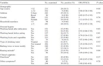
Among the 335 study participants, 42% were males and 58% were females. The age of patients ranged from 1 to 80 years with a median of 22 years. The 18 s rRNA-based nested PCR detected Cryptosporidium in 33 (9·9%) of the 335 patients. Females had a slightly higher infection rate of Cryptosporidium than males (10·4% vs 9·3%). A higher prevalence of cryptosporidiosis was seen in patients who had a bath at least twice a week (14%) compared with those who had a bath less than twice a week (8·5%). Patients who drank untreated water had a higher infection rate (10·7%) than patients who use treated drinking water (8·7%). However, these differences were not significant. The result of the statistical analysis is shown in Table 1.
Prevalence and distribution of Cryptosporidium in patients seeking healthcare in Sana'a City
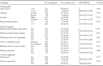
a Rearing animals was defined as the traditional rearing which involves sending animals to mountains and valleys for grazing and caring of animals in shelters within the house vicinity.
# Diarrhoea was defined as loose stools for 3 consecutive times/day.
* Other symptoms included vomiting, abdominal pain and/or nausea, Fisher exact test was used for calculating the significance in this variable.
The 18 s rRNA PCR products of all 33 positive samples were successfully sequenced. All except 1 (97%) were identified as C. parvum; the remaining 1 (3%) was identified as C. hominis. All samples positive for Cryptosporidium parvum/hominis (33 samples) were subjected to PCR amplification targeting the partial sequence of the gp60 gene. Of these, 21 samples produced PCR amplicons, which were sequenced in both directions. The partial sequence of gp60 gene was successfully sequenced for 7 isolates of C. parvum and the isolate of C. hominis. Sequence analysis of the gp60 gene identified all 7 C. parvum isolates as the IIaA15G2R1 subtype of the IIa subtype family. Compared with sequences deposited in the GenBank database, the sequence of the IIa subtype family from this study matched the IIaA15G2R1 subtype from a human in Australia (GenBank Accession no. JF727795) (Waldron et al. Reference Waldron, Dimeski, Beggs, Ferrari and Power2011) the UK (GenBank accession no. HQ149037) (Hadfield et al. 2011) and Iran (GenBank Accession no. AB560747) (Nazemalhosseini-Mojarad et al. Reference Nazemalhosseini-Mojarad, Haghighi, Taghipour, Keshavarz, Mohebi, Zali and Xiao2011). The C. hominis isolate was identified as the IeA11G3T3 subtype of the Ie subtype family which matched gp60 gene sequence of the IeA11G3T3 subtype of the Ie subtype family isolated from humans in the UK (GenBank Accession no. GU214354) (Pangasa et al. Reference Pangasa, Jex, Nolan, Campbell, Haydon, Stevens and Gasser2010) and Canada (GeneBank Accession no. DQ192509) (Trotz-Williams et al. Reference Trotz-Williams, Martin, Gatei, Cama, Peregrine, Martin, Nydam, Jamieson and Xiao2006).
DISCUSSION
In the current study the prevalence of Cryptosporidium was 9·9% by PCR, which is 10 times higher than our previous report based on the microscopical examination of fecal smears stained with Ziehl–Neelsen stain (Alyousefi et al. Reference Alyousefi, Mahdy, Mahmud and Lim2011). This difference could be attributed to the higher sensitivity of PCR. The superiority of PCR compared with microscopy was previously reported (Johnson et al. Reference Johnson, Pieniazek, Griffin, Misener and Rose1995; Stark et al. Reference Stark, Al-qassab, Barratt, Stanley, Roberts, Marriott, Harkness and Ellis2011). In addition, it was demonstrated that the threshold of the 100% sensitivity of Ziehl–Neelsen staining technique is 500 000 oocysts per gram of feces (Weber et al. Reference Weber, Bryan, Bishop, Wahlquist, Sullivan and Juranek1991).
This study is the first report of Cryptosporidium species and subtypes in Yemen. The current results showed that all isolates were C. parvum except 1 isolate, which was identified as C. hominis. These findings are consistent with previous reports in other Middle Eastern countries (Sulaiman et al. Reference Sulaiman, Hira, Zhou, Al-Ali, Al-Shelahi, Shweiki, Iqbal, Khalid and Xiao2005; Tamer et al. Reference Tamer, Turk, Dagci, Pektas, Guy, Guruz and Uner2007; Al-Brikan et al. Reference Al-Brikan, Salem, Beeching and Hilal2008; Hijjawi et al. Reference Hijjawi, Ng, Yang, Atoum and Ryan2010; Xiao, Reference Xiao2010; Nazemalhosseini-Mojarad et al. Reference Nazemalhosseini-Mojarad, Haghighi, Taghipour, Keshavarz, Mohebi, Zali and Xiao2011). In Saudi Arabia, a neighbouring country of Yemen, of the 31 human fecal samples examined, Al-Brikan and his colleagues detected C. parvum and C. hominis in 15 and 13 samples, respectively (Al-Brikan et al. Reference Al-Brikan, Salem, Beeching and Hilal2008). Subsequently, in the same country, another study found C. parvum in 43 of 53 samples examined (Nazemalhosseini-Mojarad et al. Reference Nazemalhosseini-Mojarad, Feng and Xiao2012). Similar results have also been reported in Kuwait where C. parvum was the predominant causative agent for cryptosporidiosis in children (Sulaiman et al. Reference Sulaiman, Hira, Zhou, Al-Ali, Al-Shelahi, Shweiki, Iqbal, Khalid and Xiao2005; Iqbal et al. Reference Iqbal, Khalid and Hira2011). A molecular epidemiological study conducted in Jordan identified C. parvum and C. hominis in 22 and 20 specimens, respectively (Hijjawi et al. Reference Hijjawi, Ng, Yang, Atoum and Ryan2010). In Iran, collective data generated from 4 molecular epidemiological studies showed a predominance of C. parvum (Nazemalhosseini-Mojarad et al. Reference Nazemalhosseini-Mojarad, Feng and Xiao2012). In contrast, 2 molecular epidemiological studies reported the presence of C. parvum and C. hominis in Egypt with the predominance of C. hominis (Abd El Kader et al. Reference Abd El Kader, Blanco, Ali-Tammam, Abd El Ghaffar, Osman, El Sheikh, Rubio and de Fuentes2012). It has been reported that C. hominis is the most common species in developing countries with the common occurrence of Ia, Ib, Id and Ie subtype families. In Europe and Australia, and North America, the IIa subtype family is the dominant C. parvum, suggesting zoonotic transmission of Cryptosporidium may be common in these countries. Although C. parvum is prevalent in some developing countries, the anthroponotic IIc subtype family is the most common subtype family of C. parvum in humans in these countries (Xiao, Reference Xiao2010).
It is our understanding that geographical distribution of Cryptosporidium species is not a conclusive indication of differences in the transmission of cryptosporidiosis. Although C. parvum has been known as a zoonotic species, most subtype families of C. parvum identified (IIb, IIc, IIe – IIl) in humans are probably not zoonotic in origin (Xiao, Reference Xiao2010). In this study, all C. parvum subtyped belonged to IIaA15G2R1 subtype of the IIa subtype family. The subtype IIaA15G2R1 is the major subtype of C. parvum in cattle in developed countries (Alves et al. Reference Alves, Xiao, Sulaiman, Lal, Matos and Antunes2003; Wu et al. Reference Wu, Nagano, Boonmars, Nakada and Takahashi2003; Sulaiman et al. Reference Sulaiman, Hira, Zhou, Al-Ali, Al-Shelahi, Shweiki, Iqbal, Khalid and Xiao2005; Abe et al. Reference Abe, Matsubayashi, Kimata and Iseki2006; Cohen et al. Reference Cohen, Dalle, Gallay, Di Palma, Bonnin and Ward2006; Trotz-Williams et al. Reference Trotz-Williams, Martin, Gatei, Cama, Peregrine, Martin, Nydam, Jamieson and Xiao2006; Feng et al. Reference Feng, Ortega, He, Das, Xu, Zhang, Fayer, Gatei, Cama and Xiao2007; Thompson et al. Reference Thompson, Dooley, Kenny, McCoy, Lowery, Moore and Xiao2007; Broglia et al. Reference Broglia, Reckinger, Cacciò and Nöckler2008; Wielinga et al. Reference Wielinga, de Vries, van der Goot, Mank, Mars, Kortbeek and van der Giessen2008). This subtype has also been found in humans in the USA (Santin et al. Reference Santin, Trout and Fayer2008), Canada (Budu-Amoako et al. Reference Budu-Amoako, Greenwood, Dixon, Sweet, Ang, Barkema and McClure2012), the UK (Chalmers et al. Reference Chalmers, Elwin, Thomas, Guy and Mason2009), the Netherlands (Wielinga et al. Reference Wielinga, de Vries, van der Goot, Mank, Mars, Kortbeek and van der Giessen2008), Slovenia (Soba and Logar, Reference Soba and Logar2008), Portugal (Alves et al. Reference Alves, Xiao, Antunes and Matos2006), Ireland (Thompson et al. Reference Thompson, Dooley, Kenny, McCoy, Lowery, Moore and Xiao2007), Japan (Abe et al. Reference Abe, Matsubayashi, Kimata and Iseki2006), Australia (Ng et al. 2008), and Malaysia (Iqbal et al. Reference Iqbal, Lim, Surin and Sim2012). However, it has been rarely reported in humans in developing countries, where most C. parvum infections are caused by the anthroponotic IIc subtype family (Xiao, Reference Xiao2010).
In Middle Eastern countries, the subtype family IIa is responsible for some C. parvum infections in humans. It was reported in 7 of 22 C. parvum-infected patients from Iran (Nazemalhosseini Mojarad et al. Reference Nazemalhosseini-Mojarad, Haghighi, Taghipour, Keshavarz, Mohebi, Zali and Xiao2010), 3 of 13 patients from Jordan (Hijjawi et al. Reference Hijjawi, Ng, Yang, Atoum and Ryan2010), and 1 of 37 patients from Saudi Arabia (Nazemalhosseini-Mojarad et al. Reference Nazemalhosseini-Mojarad, Feng and Xiao2012). Another zoonotic C. parvum subtype family, IId, is also common in humans in this region (Nazemalhosseini-Mojarad et al. Reference Nazemalhosseini-Mojarad, Feng and Xiao2012). The predominance of the zoonotic subtype families of C. parvum IIa and IId in the Middle Eastern regions suggests that animal-to-human transmission may be a common transmission route of Cryptosporidium. However, this postulation may be premature in view of findings in 2 studies conducted in Kuwait (Sulaiman et al. Reference Sulaiman, Hira, Zhou, Al-Ali, Al-Shelahi, Shweiki, Iqbal, Khalid and Xiao2005; Iqbal et al. Reference Iqbal, Khalid and Hira2011) where there was no contact with farm animals by most of the study participants and desalinated seawater was the source of drinking water in the study area. As contact with animals in the context of illness course is a key requirement to document the occurrence of the zoonotic transmission, the authors suggested that the source of C. parvum infection in Kuwait was from humans.
In Yemen, the situation is different from other Middle Eastern countries whereby people in Yemen have very close contact with animals, especially in the rural and suburban areas. It is a very common practice to keep animals inside or next to houses rather than on farms. In addition, local veterinarian services are also limited, thus local or imported animals are not routinely screened for parasitic diseases. These circumstances provide a suitable environment for the transmission of zoonotic diseases. Thus, the fact that all C. parvum isolates successfully subtyped belong to the IIa subtype family may represent potential evidence for the possible zoonotic transmission of Cryptosporidium in Yemen. However, the small number of the C. parvum isolates subtyped has limited the strength of this conclusion, and more conclusive evidence should come from case-controlled studies and genotyping/subtyping of Cryptosporidium spp. from humans and animals in the same household or locality.
ACKNOWLEDGEMENTS
We thank the staff of the participating hospitals for the collection of samples and data. This work was supported by University of Malaya funded projects (RG302/11HTM and HIR J-00000-73587).