Introduction
The dwarf tapeworm Rodentolepis ( = Hymenolepis) nana (von Siebold, 1852) (Cestoda: Hymenolepididae) is a common parasite of rodents, humans and other primates (Baer & Tenora, Reference Baer and Tenora1970; Ghandour et al., Reference Ghandour, Zahid, Banaja, Kamal and Bouq1995; Melo et al., Reference Melo, Neri, Ferreira, Souza and Menezes1997). How horizontal parasite transmission among vertebrate hosts might contribute to survival in the wild is still not fully elucidated. There is still no consensus regarding host specificity of R. nana despite many attempts to clarify the question (Ferretti et al., Reference Ferretti, Gabriele and Palmas1981; Duclos & Richardson, Reference Duclos and Richardson2000; Macnish et al., Reference Macnish, Morgan, Behnke and Thompson2002a, Reference Macnish, Morgan, Monis and Behnkeb).
In contrast to other cestodes, R. nana is able to complete a direct life cycle when a suitable vertebrate host is accidentally infected through ingestion of its eggs. The embryos initially develop in the intestinal villi and later cysticercoids emerge into the intestinal lumen, attach to the epithelium, and complete their development to gravid adults in 15–20 days. The biology and pathogenicity of R. nana, as well as aspects of the parasite interaction with vertebrate host immune systems, mainly mouse and rat strains, have been investigated extensively (Andreassen, Reference Andreassen1997; Ito, Reference Ito1997; Siles-Lucas & Hemphill, 2002).
The Mongolian jird Meriones unguiculatus Milne-Edwards, 1867 (Rodentia: Muridae) has quite recently been introduced to pet stores and laboratory animal facilities in Brazil without an accurate evaluation of its helminth parasite fauna, as Pinto et al. (Reference Pinto, Gomes, Menezes, Muniz-Pereira and Noronha2003) pointed out. Although natural parasitism of M. unguiculatus by R. nana has been reported previously in specimens of a pet shop colony in the USA (Lussier & Loew, Reference Lussier and Loew1970), as well as parasitism of other rodent breeding colonies in Brazil (Pinto et al., Reference Pinto, Vicente, Noronha, Gonçalves and Gomes1994, Reference Pinto, Gonçalves, Gomes and Noronha2001; Gonçalves et al., Reference Gonçalves, Pinto, Vicente, Noronha and Gomes1998), there are still no studies on the host–parasite relationship of R. nana in experimental infections of M. unguiculatus.
The present paper reports an interspecies transmission study to determine if a mouse-derived R. nana strain can be transmitted experimentally to M. unguiculatus and if the parasite can complete a normal life cycle and adapt to this unnatural host after a series of passages through immunosuppressed jirds.
Materials and methods
Parasite
The R. nana strain used was first obtained in 2003 from faeces of a naturally infected colony of outbred Swiss mice. The parasite has been kept ever since in our laboratory through direct infection with eggs from faeces of another mouse group infected 20–30 days earlier.
Animal
Meriones unguiculatus of both sexes aged 6–8 weeks old at the time of infection and obtained from an outbred stock colony maintained at the Department of Parasitology, Universidade Federal de Minas Gerais, were used as hosts. All animals were housed in plastic cages containing wood shavings. Commercial rodent chow and water were supplied ad libitum throughout the experimental period.
Infection methods
Faecal material from infected donor Swiss mice and/or immunosuppressed jirds was processed initially as described by Hoffman et al. (Reference Hoffman, Pons and Janer1934). Three infection methods were employed in the present study:
(1) Approximately 10 ml suspensions containing up to 1500 eggs/ml were evenly distributed onto the bottom of plastic cages. The jird groups were then housed in these contaminated cages for 48 h and transferred to and maintained in clean cages throughout the experimental period thereafter.
(2) Approximately 5 ml suspensions containing up to 1500 eggs/ml were homogeneously distributed on common milk biscuits. The jird groups were fed infected food exclusively for 48 h or less, until the complete ingestion of the biscuits. After that, they were fed a routine food supply until the end of the experiment.
(3) Three 100 μl aliquots of suspension were examined by microscope. The mean number of eggs per suspension volume was calculated and adjusted with 0.85% physiological saline solution to a concentration of approximately 200 eggs/ml saline. Each jird host was orally inoculated with 100 eggs in suspension with a 1 ml hypodermic syringe fitted with a blunt intragastric needle.
Experimental design of jird infections
In order to investigate the suitability of M. unguiculatus as a host for R. nana, 14 experiments were conducted. From experiments 1–3, groups of five jirds were infected orally through direct contact with faecal material of mouse origin (infection method 1). Control mouse groups were infected under identical conditions. Experiments 4–13 were conducted in parallel with the adaptation study (see below). In these experiments, groups of five jirds were infected with spiked milk biscuits (infection method 2). The faecal material used in these trials was of mouse (experiment 4) or immunosuppressed jird origin (experiments 5–13). Immunosuppressed jird groups were used as controls. In the final experiment (14), 18 jirds and 18 control immunosuppressed jirds were orally inoculated with 100 R. nana eggs (infection method 3). All transmission experiments were monitored daily by the technique described by Hoffman et al. (Reference Hoffman, Pons and Janer1934) until the 40th day post-infection (DPI). The presence of R. nana eggs in faecal samples at any time of the monitored period was taken as evidence of infection.
A group of ten jirds was used to evaluate the adaptation of R. nana to M. unguiculatus hosts. A daily 10 mg/kg subcutaneous injection of dexamethasone was given to half the group from the day of infection to the end of the experiment. The other half of the group was used as a control and did not receive any dexamethasone treatment. Regardless of their status (dexamethasone treated or not) jirds were orally infected through contact with milk biscuit contaminated with R. nana eggs derived from mouse donors (infection method 2). In a subsequent experiment, R. nana eggs from faeces of jird donors infected about 20–30 days earlier and treated with dexamethasone were used in a novel infection trial with new recipient jirds grouped and infected under identical conditions in nine repeats. The course of infection with R. nana was monitored through the survey of 10–30 faecal pellet samples from dexamethasone-treated and control groups collected daily from 10–40 DPI and analysed by the technique described by Hoffman et al. (Reference Hoffman, Pons and Janer1934).
Results
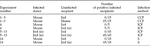
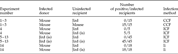
The results of the experimental infections are summarized in table 1. Jird groups not treated with dexamethasone did not pass R. nana eggs in faeces during the 40-day infection period. Control animals used in all transmission experiments were positive for R. nana eggs in all cases.
Summary of experimental trials (isolate origin, mode of infection and number of infected jirds) during Rodentolepis nana infection of Meriones unguiculatus. is, Immunosuppressed with a daily subcutaneous injection of dexamethasone at a dose of 10 mg/kg; CCF, contact with contaminated faeces; ICF, ingestion of contaminated food; Ii, intragastric inoculum.
As observed previously, no R. nana eggs were detected in faecal samples of M. unguiculatus groups not treated with dexamethasone in any passage. However, complete parasite development was observed in dexamethasone-treated jirds in all passages. Most often, jirds continuously passed eggs in faeces uninterruptedly, with the exception of scattered negative faecal samples recorded during some passages.
These series of ten passages in dexamethasone-treated jirds had a range of prepatent periods from 10 to 17 DPI with an average of 12 ± 2 days. The length of the patent period ranged from 18 to 30 DPI, with an average of 25 ± 4 days.
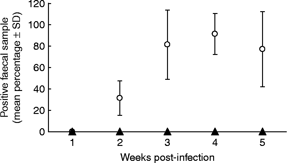
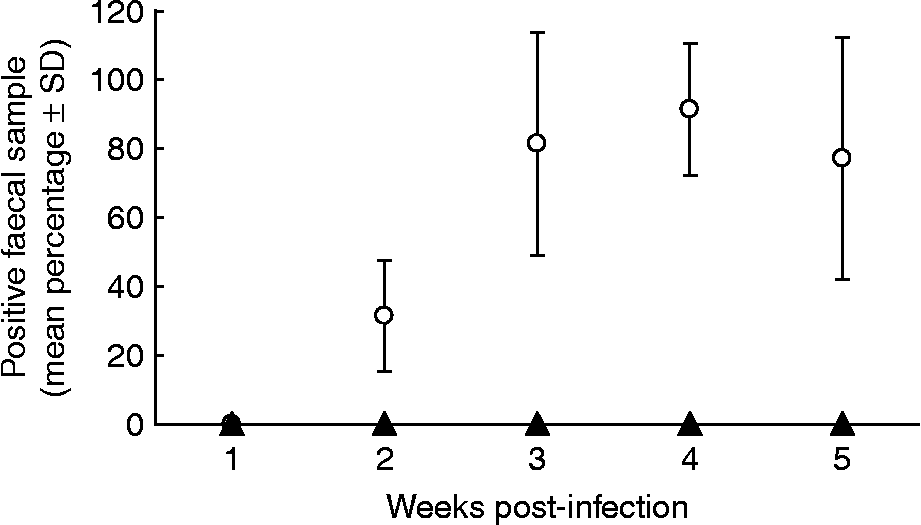
The population pattern of daily faecal egg output in both M. unguiculatus groups during the adaptation experiments is presented in fig. 1. In dexamethasone-treated jirds, faecal egg output began by the second week post-infection, with an average of 31.43% positive faecal samples. The mean percentage of positive faecal samples increased to 81.43% in the third week post-infection, peaking in the fourth week (91.43%). In the subsequent week the mean percentage of faecal samples positive for R. nana eggs dropped to 77.14%, but this was still a high percentage of positive samples.
Mean percentage of faecal samples positive for Rodentolepis nana eggs in Meriones unguiculatus groups, whether submitted to a daily subcutaneous injection of dexamethasone at a dose of 10 mg/kg (open circles; test) or not (closed triangles; control).
Discussion
During its adaptation to a potentially susceptible host, the tapeworm R. nana is challenged by a physiologically and biochemically complex environment. The environmental adversity might impose limitations on the parasite in any of its developmental stages, from hatching and activation of the hexacanth embryos to the development of the cysticercoids in intestinal villi, to the establishment and maturation of the adult parasite in the intestinal lumen (Ferretti et al., Reference Ferretti, Gabriele and Palmas1981; Ito, Reference Ito1984, Reference Ito1997).
Published data show that different vertebrate hosts have variable levels of susceptibility to R. nana and other Hymenolepididae species (Woodland, Reference Woodland1924; Larsh, Reference Larsh1944; Schiller, 1959a, b; Litchford, Reference Litchford1963; De Rycke, Reference De Rycke1966; Turton, Reference Turton1971; Hopkins et al., Reference Hopkins, Subramanian and Stallard1972; Isaak et al., Reference Isaak, Jacobson and Reed1977; Murray et al., Reference Murray, Foster and Passmore1984; van Haeren & de Rycke, 1984; Ishih et al., Reference Ishih, Nishimura and Sano1992; Stradowski, Reference Stradowski2004). It has been found that non-immunosuppressed M. unguiculatus have low susceptibility to the mouse isolates of R. nana used herein, even when different infection methods are employed. Likewise, no adaptation of R. nana to M. unguiculatus hosts, not treated with dexamethasone, was evident, at least during the ten-passage trials carried out in the present work. Conversely, Ferretti et al. (Reference Ferretti, Gabriele and Palmas1981) showed that an R. nana strain isolated from human faeces could adapt to CF1 mice after ten passages and suggested that it could be explained by an acclimatization process rather than genetic mutation, due to the few parasite generations involved.
Regardless of the low susceptibility of M. unguiculatus to R. nana, the parasite was able to establish, grow and develop into gravid adults in dexamethasone-treated M. unguiculatus. In fact, Johnson & Conder (Reference Johnson and Conder1996) studied the infectivity of Hymenolepis diminuta in M. unguiculatus, which is also an unnatural host for this parasite, and observed that hydrocortisone-treated jirds harboured relatively more worms and that they persisted longer than in non-treated jirds. Additionally, in experimental infections of different rat strains with R. nana isolates maintained exclusively in mice, Ito (Reference Ito1982, Reference Ito1983, Reference Ito1997) and Ito & Kamiyama (Reference Ito and Kamiyama1987) recorded low susceptibility to the parasite, which is able to establish and develop into cysticercoids in the rat intestinal tissue normally, but thereafter fails to mature into luminal adults. However, corticosteroid-treated rats allowed the complete maturation of worms to adults when treated in the expected luminal phase of the parasite.
In the present work, the R. nana life span recorded in dexamethasone-treated M. unguiculatus ranged within the expected values based on studies in other suitable hosts; however, the mean patent period was slightly longer and the mean prepatent period somewhat shorter than that observed in R. nana infections of mice and rats (Shorb, Reference Shorb1933; Hunninen, Reference Hunninen1935; Barbosa, Reference Barbosa1975) and in H. diminuta and H. pseudodiminuta infections of rats (Ishih et al., Reference Ishih, Sekijima, Asakawa, Tenora and Uchikawa2003). The results obtained herein suggest that dexamethasone might have influenced either the onset of the host immune response or parasite development directly, allowing earlier faecal egg output and longer worm persistence.
The faecal egg output pattern in M. unguiculatus under the daily treatment protocol suggests that the parasite is able to maintain egg production throughout the monitored life cycle period. The negative periods recorded in some passages, which were shorter than the prepatent period, might be explained by asynchronous parasite development; as some worms of an infection conclude their egg production there may be a lack of time during which the immature worms are completing their development. This replacement process might be continuous in some hosts, while in others only a few worms might mature at a time, producing the observed negative periods when no eggs are passed in faeces, as verified by Shorb (Reference Shorb1933) and Hunninen (Reference Hunninen1935) during R. nana experimental infections of mice and rats, and Pacheco et al. (Reference Pacheco, Neri, Frahia and Melo2003) studying natural parasitism in Callicebus nigrifons (Primates: Cebidae).
Thus, to elucidate whether these results indicate that R. nana adapts less well to M. unguiculatus than to other rodent hosts, further studies are needed. It will be particularly interesting to examine the link between the immunogenicity of R. nana and the capacity of various hosts, including M. unguiculatus, to respond immunologically to infection.