Introduction
Fourth branchial arch anomalies are extremely rare and constitute less than 3 per cent of all branchial arch anomalies.Reference Neff, Kirse and Pranikoff1 They usually present within the first two decades of life. An understanding of early embryological processes is essential in order to appreciate how these anomalies present, and also to distinguish between first, second, third and fourth arch disorders.
In rare cases, fourth branchial pouch anomalies present as an airway problem in neonates.Reference Sharma, Razif, Hamzah, Dharap, Mahbar and Kamal2–Reference Nicollas, Ducroz, Garabedian and Triglia5 Reported cases have also been associated with a neck mass.
The objective of this article is to present a case of fourth branchial cleft pouch which presented with stridor and no external neck swelling.
Case report
An 18-day-old neonate was referred to the paediatric otolaryngology department at Great Ormond Street Hospital, London, from a district hospital, with stridor and failure to thrive.
The child was born prematurely at 35 weeks (weight 2.460 kg). He was delivered by an emergency Caesarean section due to fetal bradycardia and meconium-stained liquor with prolonged rupture of membranes (three days). The Apgar score at birth was 5 at 1 minute and 10 at 10 minutes. At birth, the baby's respiratory effort was poor and his cry was noted to be weak. He was supported with intermittent positive pressure ventilation for an apnoeic episode lasting 30 seconds, following which he improved.
Postnatally, the baby was managed in the special care baby unit for 24 hours with intermittent ventilatory support, and was then transferred to a transitional care unit.
However, three days later the baby deteriorated and became cyanotic, and was returned to the special care baby unit to be supported with continuous positive airways pressure ventilation.
A week after birth, the baby's stridor became more obvious and frequent, and clinically he appeared more distressed. Feeding was also a problem, and associated with constant desaturations.
The child was transferred to our centre at 18 days of age. He had intermittent stridor but was in no acute distress and was spontaneously ventilating in air. Neck examination was unremarkable, with no externally palpable lump or inflammatory or infective process. He was being fed with a nasogastric tube.
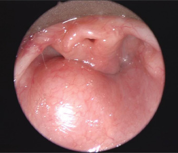
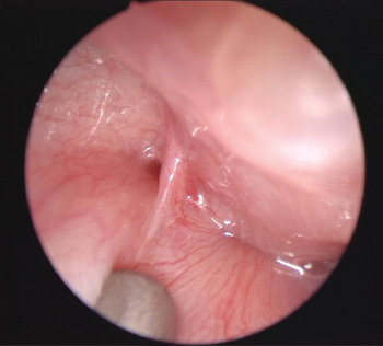
The child underwent an endoscopic examination using microlaryngoscopy and bronchoscopy. This revealed a ‘cystic looking’ swelling arising from the posterolateral pharyngeal wall on the left side (Figure 1). This swelling was pushing the whole laryngeal framework to the right. Structurally, the supraglottis, glottis and trachea were all normal. We also noted a sinus in the left pyriform fossa (Figure 2).
Microlaryngoscopic view showing swelling of the left posterolateral pharyngeal wall displacing the larynx.

Microlaryngoscopic view showing the sinus opening in the left pyriform fossa.

An urgent magnetic resonance imaging (MRI) scan of the neck revealed a cyst with an air–fluid level, which lay within the left posterior pharyngeal wall and extended down parallel to the airway, pushing the larynx medially. There were no identifiable lymphatic malformations or cystic lesions. The thyroid gland was normal. A diagnosis of a fourth branchial pouch sinus was made, and the decision was made to take the child back to the operating theatre the following day to treat the problem endoscopically.
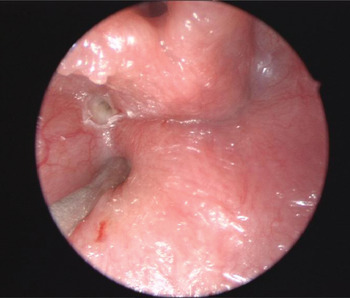
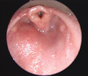
At surgery, the swelling was aspirated (withdrawing fluid and air) and the sinus opening in the left pyriform fossa was closed using monopolar diathermy (Figure 3). The laryngeal displacement resolved following aspiration (Figure 4).
Microlaryngoscopic view showing closure of the sinus opening after diathermy treatment.

Microlaryngoscopic view showing reduction of swelling and resolution of laryngeal displacement after aspiration.

Post-operatively, the child made a good recovery. His stridor completely disappeared and oral feeding was established. The child was observed for another 24 hours and then discharged home.
Six weeks later, repeated microlaryngoscopy and bronchoscopy showed complete resolution of the pharyngeal swelling. At this stage, the child had no airway symptoms and was feeding normally.
Discussion
Branchial anomalies arise as a result of abnormal development of the branchial apparatus leading to vestigial remnants which may develop into cysts, sinuses or fistulae. The six paired branchial arches develop during the fourth to sixth week of intra-uterine development. Each arch is covered externally by ectoderm, forming the cleft, and internally by mesoderm, forming the pouch. The fourth arch creates the following structures in the neck: the cricopharyngeal, inferior pharyngeal constrictor and cricothyroid muscles; the cuneiform and thyroid cartilages; the superior laryngeal nerve; the aortic arch and right subclavian artery; the superior parathyroid glands; and the thymus.
Fourth branchial pouch sinuses are extremely rare, and the current presentation of a neonate with stridor but no external neck mass is unique. Previous reports of neonatal respiratory compromise due to a fourth branchial cleft anomaly have included an additional clinical feature: a lateral cervical mass.Reference Sharma, Razif, Hamzah, Dharap, Mahbar and Kamal2, Reference Pereira, Losh, Dwight and Poole3, Reference Ford, Balakrishnan, Evans and Bailey6, Reference Evans, Marinello and Dodson7 Usually, children with a fourth branchial cleft anomaly present with a left-sided, recurrent neck abscess or swelling. The present case highlights an unusual presentation of this anomaly, with stridor but without any external swelling.
The clinical significance of the present case is illustrated further by two previously reported cases: an adult presenting with a fourth branchial pouch sinus cyst which ruptured and caused mediastinitis;Reference Neff, Kirse and Pranikoff1 and a neonate with a fourth branchial cleft fistula tracking into the mediastinum.Reference Evans, Marinello and Dodson7
A consistent feature of fourth branchial cleft anomalies is that they are predominantly (95–97 per cent) left-sided.Reference Godin, Kearns, Pransky, Seid and Wilson8 The reason for this is still not understood. It is thought to be due to the more complex and longer pathway of the fourth branchial tract on the left side compared with the right. Some authors have suggested it is a result of asymmetrical vascular agenesis during embryogenesis.Reference Shugar and Healy9
Correct diagnosis of fourth branchial anomalies by early investigation is fundamental to preventing recurrence and treatment complications. Computed tomography (CT) scanning combined with a barium swallow has been proven useful in identification of a tractReference Yang, Cohen, Everts, Smith, Caro and Andersen4, Reference Liberman, Kay, Emil, Flageole, Nguyen and Tewfik10, Reference Nicoucar, Giger, Harrison, Jaecklin and Dulgerov11 and its relationship to surrounding structures.Reference Pereira, Losh, Dwight and Poole3, Reference Nicollas, Ducroz, Garabedian and Triglia5 Direct laryngoscopy may be used to visualise the origin of the tract, to confirm the diagnosis and to treat by cauterisation to close the tract.Reference Nicoucar, Giger, Harrison, Jaecklin and Dulgerov11 Most studies have confirmed that CT and MRI scanning are both useful modalities in the initial evaluation of fourth branchial cleft anomalies.
In the present case, as the child had presented with airway symptoms it was not considered safe to request a barium study because of the risk of aspiration. Also, at that stage the child's swallowing had not been assessed. An MRI scan was performed as, at that stage, we did not know whether the posterior pharyngeal swelling was connected to the branchial pouch anomaly or was a concurrent, separate pathology. Also, the radiologists recommended an MRI scan in view of this modality's superior soft tissue delineation.
• Fourth branchial pouch anomalies are extremely rare
• Neonatal stridor without neck swelling is a novel presentation of fourth branchial cleft sinus
• In the presented case, magnetic resonance imaging plus direct laryngoscopy enabled successful diagnosis and treatment
• Endoscopic cauterisation of the sinus led to a good outcome with no complications
Historically, the treatment of congenital fourth arch sinuses has been surgical, in the form of formal excision of the whole sinus or fistula tract as well as thyroid lobectomy for thyroid involvement as curative treatment.Reference Pereira, Losh, Dwight and Poole3, Reference Nicollas, Ducroz, Garabedian and Triglia5, Reference Nicoucar, Giger, Harrison, Jaecklin and Dulgerov11, Reference Hamoir, Rombaux, Cornu and Clapuyt12 However, due to the anatomical course of the tract, complete resection remains challenging.Reference Sharma, Razif, Hamzah, Dharap, Mahbar and Kamal2, Reference Yang, Cohen, Everts, Smith, Caro and Andersen4
More recently, successful outcomes have been achieved using endoscopic techniques, which are reliable, quick, simple and have fewer post-operative complications.Reference Evans, Marinello and Dodson7, Reference Leboulanger, Ruellan, Nevoux, Pezzettigotta, Denoyelle and Roger13 A recent review of management of fourth branchial arch anomalies in children recommended treatment with cauterisation rather than open neck surgery, to reduce complications.Reference Nicoucar, Giger, Harrison, Jaecklin and Dulgerov11 Recurrence rates are similar with both procedures.
In the reported case, treatment with cautery had a good outcome.
Conclusion
Fourth branchial pouch anomalies are extremely rare and can often be diagnosed incorrectly or late. Neonatal stridor in the absence of neck swelling is truly a novel presentation of a fourth branchial cleft sinus. The presented case illustrates the fact that MRI scanning combined with direct laryngoscopy can lead to successful diagnosis as well as treatment. In this child, endoscopic cauterisation of the sinus led to a good outcome with no complications.