Introduction
Since ancient times, humans have relied on natural products for their basic life needs. Despite global development, the rural communities in various countries still rely on traditional systems of medicine for primary health care (Karunamoorthi et al. Reference Karunamoorthi, Jegajeevanram, Vijayalakshmi and Mengistie2013). Traditional medical practices include animal-, plant-, and mineral-based medicines, massage, spiritual therapies, and other practices specific to various regions, traditions, and cultures (Tesfahuneygn and Gebreegziabher Reference Tesfahuneygn and Gebreegziabher2019). Among traditional medical practices, medicinal plants are considered very useful, affordable, and economical due to the presence of constituents that help treat various infections (Sumathi and Parvathi Reference Sumathi and Parvathi2010). The use of plants for therapeutic purposes dates back at least 5000 years, with evidence from Indian, Chinese, Egyptian, Greek, and Roman civilisations (Bamola et al. Reference Bamola, Verma and Negi2018). Medicinal plants act as a source of both traditional and modern medicine. According to the 2001 World Health Organization (WHO) report, about 80% of the world’s rural population relies on traditional medicines as their primary remedies (Abd El-Ghani Reference Abd El-Ghani2016; WHO, 2001). Despite the 20th-century prominent development in synthetic organic medicinal products, more than 25% of the medicines in industrialised countries are still derived directly or indirectly from plant sources (Newman et al. Reference Newman, Cragg and Snader2000). Traditionally, crude plant extracts are widely used in ethnomedicine for the treatment of various ailments due to their perceived therapeutic properties. However, advanced chemical and pharmacological techniques could play pivotal roles in the discovery of potential therapeutic compounds from folk medicine; many have already been identified, but others still need to be isolated (Ahmed Reference Ahmed2015).
Plants produce many secondary metabolites that have no role in their primary biological functions, but are involved in other vital processes such as environmental adaptation and defence against various biotic and abiotic stimuli (Ozyigit et al. Reference Ozyigit, Dogan, Hocaoglu-Ozyigit, Yalcin, Erdogan, Yalcin, Cabi and Kaya2023). Plant secondary metabolites comprise a diverse group of bioactive compounds, including alkaloids, steroids, flavonoids, phenols, terpenoids, saponins, coumarins, chalcones, tannins, and quinones, which are synthesised through complex biosynthetic pathways and play important roles in physiological processes, ecological interactions, and biological activities (Pang et al. Reference Pang, Chen, Wang, Gao, Li, Guo, Xu and Cheng2021; Twaij et al. Reference Twaij and Hasan2022). Secondary metabolites, or phytoconstituents, can be extracted using various traditional and modern techniques, such as ultrasound-assisted and supercritical fluid extraction. Moreover, advanced techniques like high-performance liquid chromatography (HPLC) and liquid chromatography/mass spectrometry (LC/MS) have improved the qualitative and quantitative assessment of phytochemicals (Marrelli Reference Marrelli2021). The phytochemicals of over 300,000 plant species have been elucidated, showing diverse properties and structures (Patle et al. Reference Patle, Shrivas, Kurrey, Upadhyay, Jangde and Chauhan2020). These phytochemicals possess significant therapeutic potential benefits due to their diverse pharmacological effects in human health (Prakash et al. Reference Prakash, Gupta and Sharma2012).
The use of traditional medicinal plants for therapeutic purposes is deeply rooted in indigenous knowledge and cultural practices (Chukwuma et al. Reference Chukwuma, Soladoye and Feyisola2015). Moreover, the demand for drugs from plant sources is increasing daily. Therefore, medicinal plants need to be systematically investigated for the treatment of various diseases (Sumathi and Parvathi Reference Sumathi and Parvathi2010). This requires screening of medicinal plants for promising biological activities. In this regard, local communities in the Swat region of Pakistan were invited to participate in the study (Khan et al. Reference Khan, Swennen, Panda, Schoofs and Luyten2025b), and information on the plants’ traditional uses was compared with the available literature. Based on a thorough search of various databases, antibacterial, antifungal, and antiviral activities of extracts from the selected medicinal plants have been widely reported by many researchers; however, information on anthelmintic activity is lacking for most of these plant extracts.
Therefore, the present study investigated the anthelmintic potential of selected medicinal plant extracts along with their phytochemical screening and thin-layer chromatographic profiling. This work could support the traditional use of folk medicine and lead to the development of novel and effective anthelmintic drug candidates. Accordingly, 17 medicinal plants were selected to investigate their anthelmintic activity based on ethnobotanical relevance and frequent reports of traditional use in the Swat region of Pakistan for treating various microbial and parasitic infections, as well as gastrointestinal disorders. Multiple extraction solvents with varying polarities were used to obtain a broad range of phytoconstituents. Initially, the anthelmintic (AH) activity of extracts obtained using 5 different solvents (hexane, acetone, ethanol, methanol, or water) from the 17 selected plants was comparatively evaluated to identify variations in activity among solvent extracts. The solvent showing the highest overall activity was selected for subsequent studies. The AH activity of the selected plant extracts was further characterised by calculating IC50 values to determine their relative potency. Furthermore, the selected acetone extracts were subjected to qualitative phytochemical screening and thin-layer chromatographic (TLC) profiling for chemical characterisation. Among the tested extracts, acetone extracts demonstrated comparatively higher anthelmintic activity and were therefore selected for detailed phytochemical and chromatographic analyses. This study provides scientific evidence supporting the traditional use of these medicinal plants and may contribute to the identification of potential candidates for further pharmacological investigation.
Materials and methods
Chemicals and reagents
All solvents and chemicals were procured from Sigma-Aldrich Co. (USA), except for ethanol, obtained from Fischer Chemicals (UK), and Bacto™ peptone, sourced from Lab M Ltd. (Lancashire, UK). Sterile deionised water was prepared using a Milli-Q Reagent Water System (MA, USA).
Study design and plant materials collection
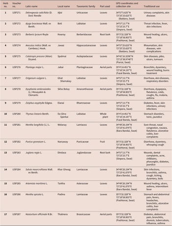
The plant materials were collected from the Swat region of Pakistan at different times of the day to ensure availability and representativeness, based on their traditional use by local communities for treating various infections as a primary health care resource. In this study, a total of 17 plants were collected after obtaining permission from the concerned authorities and traditional knowledge holder community members. The plants were identified at the Center for Plant Science and Biodiversity, University of Swat, Pakistan, by following standard authentication methods. The voucher specimens are deposited in the Herbarium of the University of Swat with their complete details as shown in Table 1, adapted from our previous study (Khan et al. Reference Khan, Panda, Hu, Schoofs and Luyten2025a).
List of the collected traditional medicinal plants of the Swat region of Pakistan
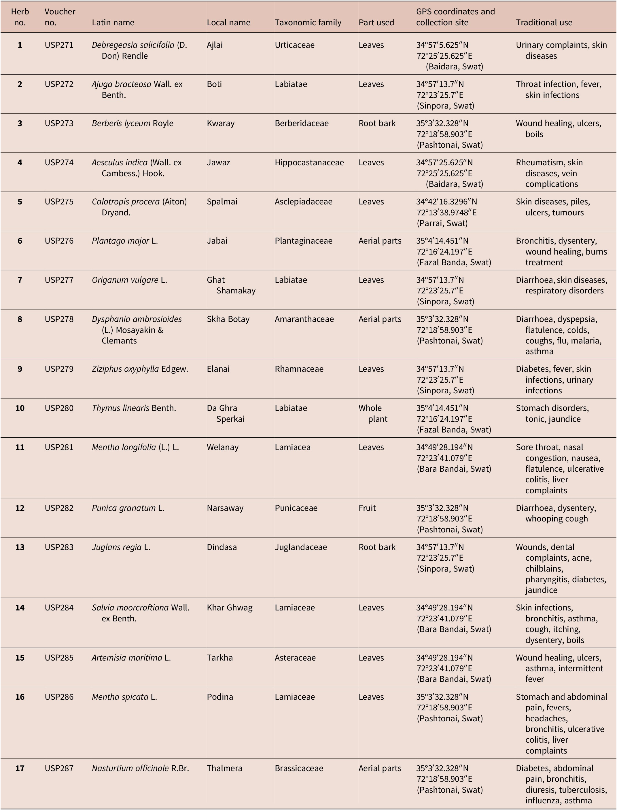
Table 1. Long description
The table contains 17 entries with the following data points:
* Herb 1: Voucher U S P 271, Debregeasia salicifolia, Local name Ajlai, Family Urticaceae. Part used: Leaves. Site: Baidara Swat (34 degrees 57 minutes 5.625 seconds North, 72 degrees 25 minutes 25.625 seconds East). Uses: Urinary complaints, skin diseases.
* Herb 2: Voucher U S P 272, Ajuga bracteosa, Local name Boti, Family Labiatae. Part used: Leaves. Site: Sinpora Swat (34 degrees 57 minutes 13.7 seconds North, 72 degrees 23 minutes 25.7 seconds East). Uses: Throat infection, fever, skin infections.
* Herb 3: Voucher U S P 273, Berberis lyceum, Local name Kwaray, Family Berberidaceae. Part used: Root bark. Site: Pashtonai Swat (35 degrees 3 minutes 32.328 seconds North, 72 degrees 18 minutes 58.903 seconds East). Uses: Wound healing, ulcers, boils.
* Herb 4: Voucher U S P 274, Aesculus indica, Local name Jawaz, Family Hippocastanaceae. Part used: Leaves. Site: Baidara Swat. Uses: Rheumatism, skin diseases, vein complications.
* Herb 5: Voucher U S P 275, Calotropis procera, Local name Spalmai, Family Asclepiadaceae. Part used: Leaves. Site: Parrai Swat (34 degrees 42 minutes 16.3296 seconds North, 72 degrees 13 minutes 38.9748 seconds East). Uses: Skin diseases, piles, ulcers, tumours.
* Herb 6: Voucher U S P 276, Plantago major, Local name Jabai, Family Plantaginaceae. Part used: Aerial parts. Site: Fazal Banda Swat (35 degrees 4 minutes 14.451 seconds North, 72 degrees 16 minutes 24.197 seconds East). Uses: Bronchitis, dysentery, wound healing, burns treatment.
* Herb 7: Voucher U S P 277, Origanum vulgare, Local name Ghat Shamakay, Family Labiatae. Part used: Leaves. Site: Sinpora Swat. Uses: Diarrhoea, skin diseases, respiratory disorders.
* Herb 8: Voucher U S P 278, Dysphania ambrosioides, Local name Skha Botay, Family Amaranthaceae. Part used: Aerial parts. Site: Pashtonai Swat. Uses: Diarrhoea, dyspepsia, respiratory issues, malaria, asthma.
* Herb 9: Voucher U S P 279, Ziziphus oxyphylla, Local name Elanai, Family Rhamnaceae. Part used: Leaves. Site: Sinpora Swat. Uses: Diabetes, fever, skin and urinary infections.
* Herb 10: Voucher U S P 280, Thymus linearis, Local name Da Ghra Sperkai, Family Labiatae. Part used: Whole plant. Site: Fazal Banda Swat. Uses: Stomach disorders, tonic, jaundice.
* Herb 11: Voucher U S P 281, Mentha longifolia, Local name Welanay, Family Lamiacea. Part used: Leaves. Site: Bara Bandai Swat (34 degrees 49 minutes 28.194 seconds North, 72 degrees 23 minutes 41.079 seconds East). Uses: Sore throat, nausea, ulcerative colitis, liver complaints.
* Herb 12: Voucher U S P 282, Punica granatum, Local name Narsaway, Family Punicaceae. Part used: Fruit. Site: Pashtonai Swat. Uses: Diarrhoea, dysentery, whooping cough.
* Herb 13: Voucher U S P 283, Juglans regia, Local name Dindasa, Family Juglandaceae. Part used: Root bark. Site: Sinpora Swat. Uses: Wounds, dental complaints, acne, diabetes, jaundice.
* Herb 14: Voucher U S P 284, Salvia moorcroftiana, Local name Khar Ghwag, Family Lamiaceae. Part used: Leaves. Site: Bara Bandai Swat. Uses: Skin infections, bronchitis, asthma, dysentery.
* Herb 15: Voucher U S P 285, Artemisia maritima, Local name Tarkha, Family Asteraceae. Part used: Leaves. Site: Bara Bandai Swat. Uses: Wound healing, ulcers, asthma, fever.
* Herb 16: Voucher U S P 286, Mentha spicata, Local name Podina, Family Lamiaceae. Part used: Leaves. Site: Pashtonai Swat. Uses: Stomach pain, fevers, headaches, liver complaints.
* Herb 17: Voucher U S P 287, Nasturtium officinale, Local name Thalmera, Family Brassicaceae. Part used: Aerial parts. Site: Pashtonai Swat. Uses: Diabetes, abdominal pain, bronchitis, tuberculosis, influenza.
Source: (Khan et al. Reference Khan, Swennen, Panda, Schoofs and Luyten2025b)
Extraction of plant materials
The plant materials were dried in the absence of sunlight for about 1 week at ambient temperature during the collection period (April–June) in Swat, Pakistan, following standard drying procedures to maintain their green colour and preserve volatile oils (Panda et al. Reference Panda, Padhi, Leyssen, Liu, Neyts and Luyten2017). The dried materials were ground into powder form using an electric grinder and were stored in plastic bags in a 4 °C cold room. For extraction, 1 gram of powder from each plant was transferred into separate sterile 15 mL Falcon tubes, each containing 10 mL of hexane, acetone, ethanol, methanol, or water. The tubes containing the plant materials and solvents were subjected to repeated sonication and vortexing 4 times, with a 6-hour interval between each. Afterward, the tubes were centrifuged for 10 minutes at 3500 rpm, and the supernatants were transferred to 1.5 mL Eppendorf tubes in 1 mL aliquots, then dried. The extracts were stored at 4 °C for future use (Liu et al. Reference Liu, Kipanga, Mai, Dhondt, Braeckman, De Borggraeve and Luyten2018).
Anthelmintic test
Culture, maintenance, and synchronisation of Caenorhabditis elegans strains
The methodology for the culture, maintenance, and synchronisation of Caenorhabditis elegans (strain N2, wild type) for anthelmintic testing was adapted primarily from our previous work (Liu et al. Reference Liu, Kipanga, Mai, Dhondt, Braeckman, De Borggraeve and Luyten2018; Yaghoobi et al. Reference Yaghoobi, Moridi Farimani, Khan, Asadollahi, Omrani, Luyten and Hu2025) with some modifications. The worms were grown at 20 °C on NGM (Nematode Growth Medium) in Petri dishes with a lawn of the bacterium E. coli OP50. The worms were synchronised using a modified alkaline bleaching method. Initially, S-basal buffer (4.68 g NaCl, 0.896 g K2HPO4, 4.744 g KH2PO4, 0.8 L demineralised water) was used to wash culture plates containing eggs and egg-laying adults, followed by incubation in fresh bleaching solution, and three subsequent washes with S-basal buffer. Afterward, 3.5 mL of worm suspension was added to 1.5 mL alkaline hypochlorite solution (1 mL of bleach and 0.5 mL of 5 M NaOH) and was shaken for 5 minutes. This was followed by the addition of 5 mL of 60% (w/v) sucrose solution and 1 mL of buffer. The mixture was then centrifuged at 800 rpm for 3 minutes, and the eggs were collected and washed 5 times with S-basal buffer. In a light-free environment, the eggs in S-basal buffer were kept on a rotator at 20 °C overnight to hatch into L1 larvae (L1s). After incubation for 24 hours, L1s were transferred onto a fresh NGM plate seeded with an E. coli lawn, and grown at 20 °C (L2s). After 48 hours, ‘L4’ larvae were isolated from the plates and washed 4 times with S-basal buffer to prepare them for use in the anthelmintic test.
Anthelmintic assay
The anthelmintic assay was carried out in a 96-well microplate with flat-bottom wells using a WMicroTracker apparatus, following the method described in our previous study (Panda et al. Reference Panda, Padhi, Leyssen, Liu, Neyts and Luyten2017). Plant extracts prepared in five solvents with varying polarities (hexane, acetone, ethanol, methanol, and water) were tested for anthelmintic potential using C. elegans as a model organism for the anthelmintic screening. Briefly, synchronised C. elegans L4 larval stages (15 μL, approx. 45 worms) were seeded into a 96-well microplate containing 184 μL of E. coli culture (OD = 0.5 at 620 nm). Following larval seeding, 1 μL of plant extract (40 mg/mL) was added to the wells containing a final assay volume of 200 μL, resulting in a final extract concentration of 200 μg/mL. In the negative controls, 1 μL of DMSO or distilled water was used, giving a final DMSO concentration of 0.5% (v/v). Organic solvent extracts were reconstituted in DMSO, whereas aqueous extracts were prepared in distilled water, and the corresponding solvent controls (DMSO for organic extracts and distilled water for aqueous extracts) were included to ensure that the solvents did not affect anthelmintic activity. In addition, 1 μL of levamisole was used as a positive control (final concentration 50 μM). The 96-well microplate was then incubated in a WMicroTracker apparatus at 20 °C for 20 hours, during which the apparatus recorded worm movements every 30 minutes. All anthelmintic assays were carried out in duplicate to confirm results, and the mean values from the two experiments were used. The half-maximal inhibitory concentration (IC50) values for anthelmintic assays were determined by preparing two-fold serial dilution series (up to 64-fold) of the test samples in DMSO or water in a V-shaped 96-well plate, followed by the respective tests. The IC50 values were calculated using GraphPad Prism 11.0.0 (San Diego, CA, USA) by fitting the anthelmintic dose-response data to a nonlinear model of log(inhibitor concentration) versus response. To estimate the relative anthelmintic activity, the 20-hour average movement percentage in wells containing test samples with the extract was compared with that of the solvent control. Based on this reduction in larval movement relative to the negative control, anthelmintic activity (%) was calculated using the given formula.

where:
T = the proportion of moving worms in the treatment group
C = the proportion of moving worms in the negative control
Phytochemical analysis
The phytochemical analysis of acetone extracts from selected traditional medicinal plants in the Swat region of Pakistan was conducted to identify their secondary metabolites. The acetone extracts were selected for phytochemical analysis due to their lower cytotoxicity (Khan et al. Reference Khan, Panda, Hu, Schoofs and Luyten2025a) and potent anthelmintic potential. The crude plant extracts were screened for the presence of commonly reported phytochemical constituents, including alkaloids, steroids, flavonoids, phenols, terpenoids, saponins, coumarins, chalcones, tannins, and quinones, using conventional qualitative methods (Khan et al. 2025c).
Detection of alkaloids
Alkaloids were detected by using Wagner’s reagent test. A few drops of Wagner’s reagent were added to 1 mL of the filtrate of the extract treated with 1% aqueous HCl on a steam bath. The formation of a creamy or brown-reddish precipitate confirms the presence of alkaloids.
Detection of saponins
The Foaming test was used to detect saponins in the plant extracts. In this test, 3 mL of the extracts were vigorously shaken with 3 mL of distilled water. The formation of foam or froth upon shaking indicates a positive test.
Detection of steroids
The Salkowski test was used for the detection of steroids. 2 mL of the extract was mixed with 2 mL of chloroform, followed by the addition of concentrated H2SO4. A red colour observed in the chloroform layer indicates the presence of steroids.
Detection of flavonoids
The Alkaline reagent test was used to detect flavonoids in the samples. A few drops of dilute HCL were added to a mixture of 1 mL of extract and 2 mL of 2% sodium hydroxide (NaOH). The colour change from yellow to colourless indicates the presence of flavonoids.
Detection of phenols
The ferric chloride test was used to detect phenols. One mL of the extract was treated with 2 mL of 5% ferric chloride (FeCl3). The development of a bluish-green colour signifies the presence of phenols.
Detection of terpenoids
The Salkowski test was used to detect terpenoids. 5 mL of the extract was mixed with 2 mL of chloroform, followed by the addition of 3 mL of concentrated sulfuric acid. A red coloration signifies the presence of terpenoids.
Detection of coumarins
The NaOH test was used to detect coumarins in the plant samples. 2 mL of the extracts was mixed with 3 mL of 10% sodium hydroxide (NaOH) solution. A yellow coloration signifies the presence of coumarins.
Detection of chalcones
The detection of chalcones was tested by introducing 1 mL of the extracts into 2 mL of a 5% ammonia (NH3) solution. The test was considered positive upon the formation of a red colour.
Detection of tannins
The detection of tannins in the plant extracts was assessed by stirring 2 mL of plant extract with 3 mL of distilled water, followed by the addition of 5 drops of 10% FeCl3 solution. The formation of a dark blue precipitate indicates the presence of tannins.
Detection of quinones
The concentrated HCl test was used to detect the presence of quinones in the plant extracts. One mL of the extract was added to 2 mL of concentrated hydrochloric acid (HCl). The appearance of a green colour confirms the presence of quinones.
Thin layer chromatographic (TLC) profiling
The TLC profiling of the selected plant extracts was carried out as per the protocol adopted from our previous study (Khan et al. 2025c). Thin-layer chromatography (TLC) profiling was performed to further assess the separation patterns and chemical diversity of compounds in the active acetone extracts following preliminary phytochemical screening. The crude extracts of the selected medicinal plants were redissolved in acetone to a concentration of 10 mg/mL using repeated vortexing and sonication. The particle-free solution (5 μL) of the selected extracts was applied to TLC silica gel glass plates (Sigma-Aldrich, Germany; 2.5 × 7.5 cm) using a fine-bore glass capillary tube over a marked pencil line on the lower side of the plate. The spotted plates were dried at ambient temperature. The sample-loaded plates were then placed in a glass chamber containing a mixture of solvents for the chosen mobile phases, viz., hexane and ethyl acetate (6:4) and (3:7), to optimise the separation of the mixture components. The developed plates were dried in a fume hood. The dried plates were visualised under ultraviolet (UV) light at 254 nm. The Rf value of each spot was calculated as:

Data processing and analyses
Data analysis was performed to evaluate and summarise the anthelmintic activity of the plant extracts based on their inhibitory effects on C. elegans movement. An anthelmintic activity threshold of ≥40% inhibition was used for classification, where inhibition was calculated as the reduction in worm movement relative to the corresponding solvent control (DMSO for organic solvent extracts and distilled water for aqueous extracts). The inhibition value (IV) represents the percentage reduction in worm movement relative to the corresponding solvent control. Solvent-wise activity was calculated as the proportion of active extracts obtained with each solvent relative to the total number of extracts prepared with that solvent, using the following formula: Solvent-wise activity (%) = (number of active extracts obtained with a specific solvent/total number of extracts prepared with that solvent) × 100. The overall anthelmintic potential of each plant was determined based on the proportion of active extracts relative to the total number of extracts tested per plant, using the following formula: Overall anthelmintic potential activity (%) = (number of active extracts obtained from a plant/total number of extracts tested for that plant) × 100.
The data from anthelmintic tests of the plant extracts were analysed using the web tool ClustVis (https://biit.cs.ut.ee/clustvis) to generate hierarchical clustering heatmaps. For heatmap production, the parameters were set as: data import, upload file, detect delimiter, detect column and row annotations, no quotes, NA; pre-processing options: no transformation, maximum percentage for rows and columns, row centring, no scaling, NIPALS PCA (Nonlinear Iterative Partial Least Squares Principal Component Analysis), and heatmap generation.
Statistical comparison of solvent-wise anthelmintic activity was performed using the chi-square test of independence based on the number of active (≥40% inhibition) and non-active extracts. Statistical significance was considered at p < 0.05.
The graphs, tables, and data from phytochemical screening and TLC profiling were prepared using Microsoft Office.
Results
Anthelmintic activity
The anthelmintic activity of 85 extracts obtained from 17 selected medicinal plants using 5 different solvents was evaluated against C. elegans. Several extracts were found to be significantly active (inhibition ≥ 40%) against C. elegans. The most prominent average inhibition of C. elegans motility over a 20-hour interval was shown by acetone extracts of M. longifolia (65%), B. lyceum (64%), N. officinale (63%), P. granatum (62%), S. moorcroftiana (61%), A. bracteosa (60%), C. procera (60%), A. indica (58%), Z. oxyphylla (56%), M. spicata (52%), A. maritima (50%), D. salicifolia (47%), P. major (47%), and O. vulgare (47%), ethanol extracts of M. longifolia (74%), S. moorcroftiana (57%), D. salicifolia (53%), C. procera (51%), A. maritima (51%), P. granatum (47%), P. major (47%), O. vulgare (46%), A. bracteosa (45%), D. ambrosioides (44%), and Z. oxyphylla (41%), hexane extracts of M. spicata (69%), A. indica (61%), P. granatum (60%), D. salicifolia (59%), B. lyceum (57%), A. maritima (54%), O. vulgare (53%), M. longifolia (48%), J. regia (44%), and A. bracteosa (41%), methanol extracts of A. maritima (76%), P. granatum (63%), B. lyceum (60%), C. procera (60%), M. longifolia (53%), A. bracteosa (52%), T. linearis (49%), J. regia (49%), and Z. oxyphylla (42%), and water extracts of P. major (64%), N. officinale (62%), A. bracteosa (58%), J. regia (56%), M. spicata (51%), S. moorcroftiana (47%), P. granatum (44%), and A. indica (42%) as shown in the form of a heatmap in Figure 1 as average inhibition values and in Supplementary Table S1 as inhibition values over a 20-hour interval as well as average inhibition values.
Heatmap and hierarchical clustering of the average anthelmintic activity of selected medicinal plant extracts against C. elegans.
Note: Hierarchical clustering was performed based on similarities in anthelmintic activity among extraction solvents and plant extracts. Plant names are shown on the right side of the heatmap, while the X-axis represents the extraction solvents used. The color scale represents percentage anthelmintic activity against C. elegans, ranging from −40 to 40. Higher positive values (red colors) indicate a greater reduction in C. elegans motility compared with the solvent control, reflecting stronger anthelmintic activity, whereas negative values (blue colors) indicate extracts showing lower or no reduction in motility compared with the solvent control and do not indicate enhanced worm locomotor activity.
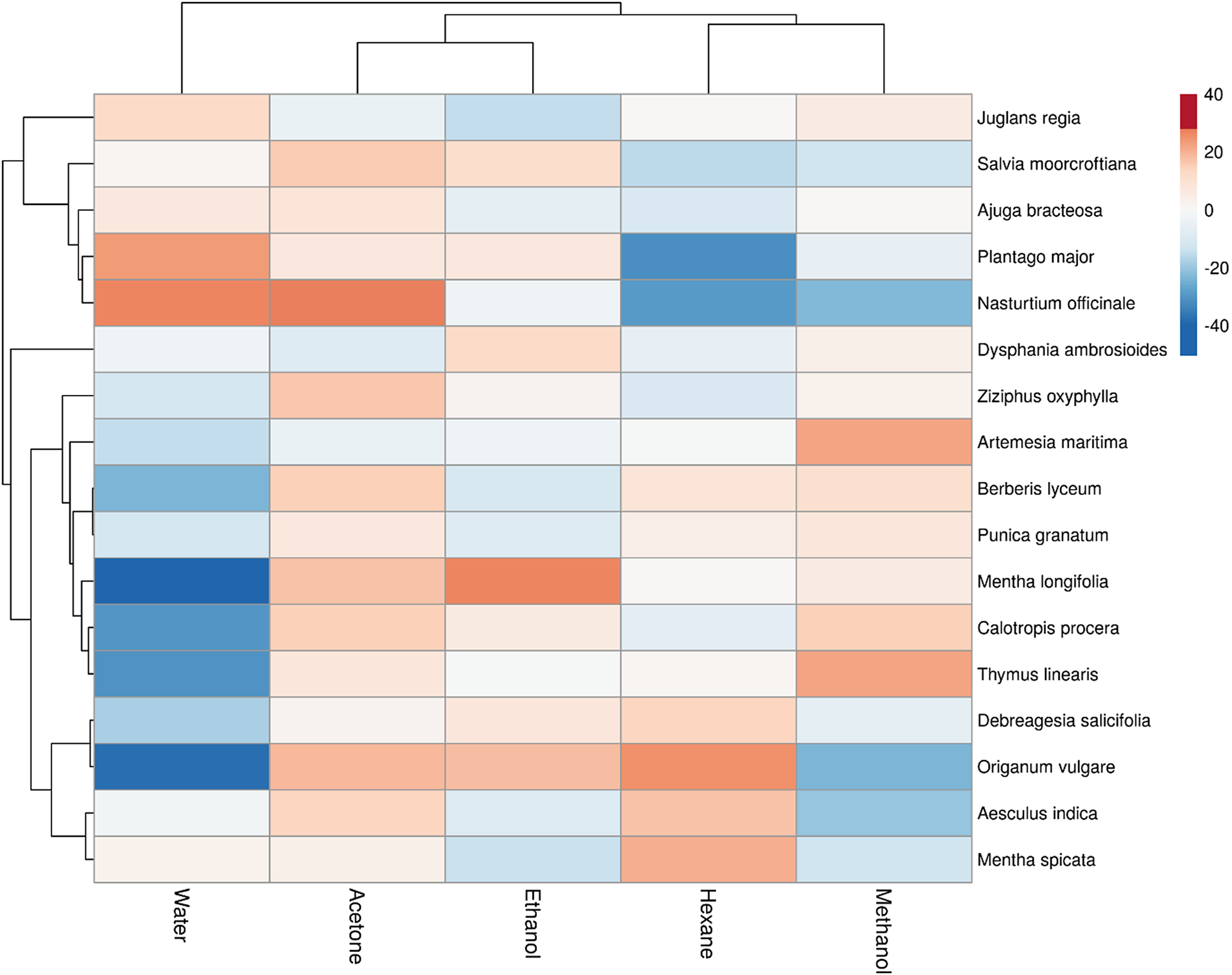
Figure 1. Long description
A heatmap with hierarchical clustering dendrograms on the top and left sides.
* X-axis (Solvents): From left to right, the columns are Water, Acetone, Ethanol, Hexane, and Methanol. A dendrogram at the top groups Water and Acetone together, and Hexane and Methanol together, with Ethanol as a separate branch.
* Y-axis (Plant Species): 17 species are listed on the right, including Juglans regia, Salvia moorcroftiana, Ajuga bracteosa, Plantago major, Nasturtium officinale, Dysphania ambrosioides, Ziziphus oxyphylla, Artemesia maritima, Berberis lyceum, Punica granatum, Mentha longifolia, Calotropis procera, Thymus linearis, Debreagesia salicifolia, Origanum vulgare, Aesculus indica, and Mentha spicata. A complex dendrogram on the left clusters these species based on activity patterns.
* Color Scale: A vertical legend on the far right ranges from minus 40 (dark blue, low/no reduction in motility) to 0 (white) to 40 (dark red, high anthelmintic activity).
* Data Trends:
* Nasturtium officinale shows strong positive activity (red) in Water and Acetone but negative activity (blue) in Hexane and Methanol.
* Plantago major shows strong negative activity (dark blue) in Hexane.
* Mentha longifolia shows strong negative activity in Water but positive activity in Ethanol.
* Origanum vulgare shows strong negative activity in Water and Methanol but positive activity in Hexane.
* Most other extracts show neutral (white) to moderate (light orange or light blue) activity across the solvents.
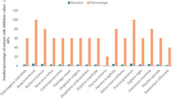
The overall anthelmintic potential was summarised by calculating the proportion of plant extracts prepared in five different solvents that exhibited significant anthelmintic activity (≥40% inhibition). The proportion of active extracts prepared in five different solvents was calculated to compare the overall activity pattern among plant species. The results presented in Figure 2 clearly indicate that the highest proportion of active extracts (100%) was observed in A. bracteosa and P. granatum, followed by B. lyceum, M. longifolia, and A. maritima, with an overall 80% proportion of active extracts, while the least active proportion was observed in extracts of T. linearis.
Overall anthelmintic potential of selected medicinal plants against C. elegans.
Note: Results were pooled and presented as numbers and percentages, using a ≥40% reduction in C. elegans motility as the threshold for active extracts. The analysis includes all extracts prepared from the selected medicinal plants using five different solvents (acetone, ethanol, hexane, methanol, and water), with an inhibition threshold of ≥40%. The X-axis represents the names of the plants, while the Y-axis represents the number and percentage of plant extracts showing ≥40% reduction in C. elegans motility, calculated as the proportion of active extracts for each plant relative to the total number of extracts tested for that plant.
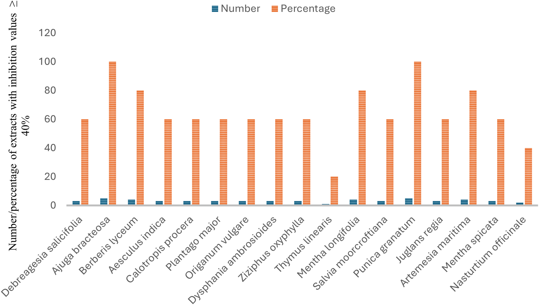
Figure 2. Long description
A grouped bar chart with a legend at the top center indicating blue solid bars for Number and orange striped bars for Percentage. The Y-axis is labeled Number forward slash percentage of extracts with inhibition values greater than or equal to 40 percent, with a scale from 0 to 120. The X-axis lists 17 plant species. From left to right, the data pairs (Number, Percentage) are:
* Debreagesia salicifolia: 3, 60
* Ajuga bracteosa: 5, 100
* Berberis lyceum: 4, 80
* Aesculus indica: 3, 60
* Calotropis procera: 3, 60
* Plantago major: 3, 60
* Origanum vulgare: 3, 60
* Dysphania ambrosioides: 3, 60
* Ziziphus oxyphylla: 3, 60
* Thymus linearis: 1, 20
* Mentha longifolia: 4, 80
* Salvia moorcroftiana: 3, 60
* Punica granatum: 5, 100
* Juglans regia: 3, 60
* Artemesia maritima: 4, 80
* Mentha spicata: 3, 60
* Nasturtium officinale: 2, 40
Ajuga bracteosa and Punica granatum show the highest potential with 100 percent of extracts being active, while Thymus linearis shows the lowest at 20 percent.
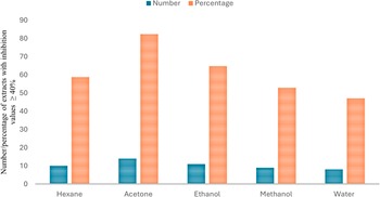
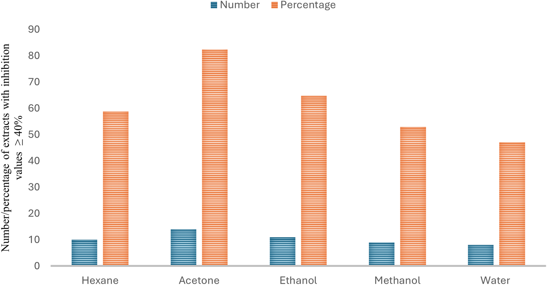
The solvent-wise distribution of anthelmintic activity showed differences in the percentage of plant extracts exhibiting significant activity (≥40% inhibition). The solvent-wise distribution of active extracts was evaluated to compare the activity obtained with different extraction solvents. As shown in Figure 3, the highest anthelmintic activity was observed in plant extracts prepared in acetone (82%), followed by ethanol (65%), hexane (59%), and methanol (53%), while the least activity was shown by extracts prepared in water (47%). However, chi-square analysis showed no statistically significant difference in the distribution of active extracts among solvents (p > 0.05).
Solvent-wise distribution of anthelmintic activity against C. elegans across all tested plant extracts.
Note: Results were pooled and presented as numbers and percentages, using a ≥40% reduction in C. elegans motility as the threshold for active extracts. The extracts were prepared using five different solvents: acetone, ethanol, hexane, methanol, and water. The X-axis represents the extraction solvents used, while the Y-axis represents the number and percentage of plant extracts showing ≥40% reduction in C. elegans motility, calculated as the proportion of active extracts obtained with each solvent relative to the total number of extracts prepared using that solvent.
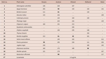
Based on the overall activity patterns observed in Figures 1–3, the most active extracts were further evaluated by determining their IC50 values (Table 2). Plant extracts showing anthelmintic activity with more than 40% inhibition of C. elegans motility were included in this analysis, whereas less active extracts are indicated by ‘–’, for which IC50 values were not calculated. The majority of the extracts exhibited moderate activity, with IC50 values above 100 μg/mL, whereas some showed potent activity, with IC50 values below 100 μg/mL. The most potent extracts with the lowest IC50 values include acetone extract of B. lyceum (81 μg/mL), M. longifolia (81 μg/mL), A. bracteosa (90 μg/mL), N. officinale (98 μg/mL), ethanol extract of M. longifolia (65 μg/mL), methanol extract of A. maritima (66 μg/mL), and hexane extract of P. granatum (99 μg/mL).
IC50 values of the plant extracts showing ≥40% reduction in C. elegans motility (anthelmintic activity) from the tested panel of 85 extracts prepared from 17 plants using 5 different solvents (μg/mL)
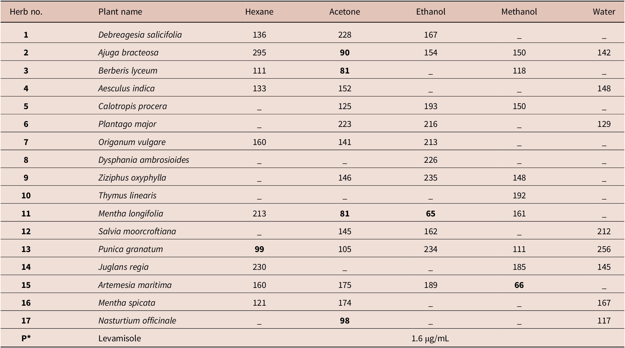
Table 2. Long description
The table lists 17 plants and their corresponding IC50 values in micrograms per milliliter for different solvent extracts.
* Column headers: Herb number, Plant name, Hexane, Acetone, Ethanol, Methanol, Water.
* Row 1: Debreagesia salicifolia; Hexane 136, Acetone 228, Ethanol 167.
* Row 2: Ajuga bracteosa; Hexane 295, Acetone 90, Ethanol 154, Methanol 150, Water 142.
* Row 3: Berberis lyceum; Hexane 111, Acetone 81, Methanol 118.
* Row 4: Aesculus indica; Hexane 133, Acetone 152, Water 148.
* Row 5: Calotropis procera; Acetone 125, Ethanol 193, Methanol 150.
* Row 6: Plantago major; Acetone 223, Ethanol 216, Water 129.
* Row 7: Origanum vulgare; Hexane 160, Acetone 141, Ethanol 213.
* Row 8: Dysphania ambrosioides; Ethanol 226.
* Row 9: Ziziphus oxyphylla; Acetone 146, Ethanol 235, Methanol 148.
* Row 10: Thymus linearis; Methanol 192.
* Row 11: Mentha longifolia; Hexane 213, Acetone 81, Ethanol 65, Methanol 161.
* Row 12: Salvia moorcroftiana; Acetone 145, Ethanol 162, Water 212.
* Row 13: Punica granatum; Hexane 99, Acetone 105, Ethanol 234, Methanol 111, Water 256.
* Row 14: Juglans regia; Hexane 230, Methanol 185, Water 145.
* Row 15: Artemesia maritima; Hexane 160, Acetone 175, Ethanol 189, Methanol 66.
* Row 16: Mentha spicata; Hexane 121, Acetone 174, Water 167.
* Row 17: Nasturtium officinale; Acetone 98, Water 117.
* Final Row: Positive control Levamisole with an I C sub 50 of 1.6 micrograms per milliliter.
Values below 100 are highlighted as potent, including Ajuga bracteosa (Acetone 90), Berberis lyceum (Acetone 81), Mentha longifolia (Acetone 81, Ethanol 65), Punica granatum (Hexane 99), Artemesia maritima (Methanol 66), and Nasturtium officinale (Acetone 98).
Note: ‘–’ indicates extracts that were tested but showed <40% inhibition, and IC50 values were therefore not determined.
Bold values indicate potent anthelmintic activity with IC50 values below 100 μg/mL.
P*: Positive control (levamisole, final concentration = 12 μg/mL).
Phytochemical analysis
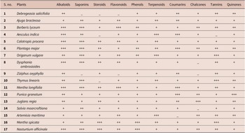
The phytochemical analysis of the acetone extracts of 17 selected medicinal plants from the Swat area of Pakistan (Table 3) revealed the presence of secondary metabolites, including alkaloids, saponins, steroids, flavonoids, phenols, terpenoids, coumarins, chalcones, tannins, and quinones. All tested extracts contained most of the tested phytoconstituents. The extracts of A. bracteosa, B. lyceum, C. procera, P. major, O. vulgare, M. longifolia, P. granatum, J. regia, M. spicata, and N. officinale contained all aforementioned types of secondary metabolites. D. ambrosioides and S. moorcroftiana contained all tested phytoconstituents except chalcones. T. linearis and A. maritima contained all tested secondary metabolites except steroids and coumarins, respectively. A. indica lacks steroids and tannins, while Z. oxyphylla lacks saponins, flavonoids, and chalcones among the tested secondary metabolites. Similarly, D. salicifolia contained all tested phytoconstituents except saponins, steroids, and flavonoids. Alkaloids were detected in high concentration in the majority of the extracts, while terpenoids, phenols, and quinones were also detected in medium or low concentration in all tested plant extracts. Coumarins and tannins were found in all plants except A. maritima and A. indica, respectively. Flavonoids and saponins were not detected in D. salicifolia and Z. oxyphylla only. Steroids were absent in D. salicifolia, A. indica, and T. linearis. Chalcones were not present in D. ambrosioides, Z. oxyphylla, and S. moorcroftiana.
Qualitative phytochemical analysis of acetone extracts from selected medicinal plants of Swat, Pakistan
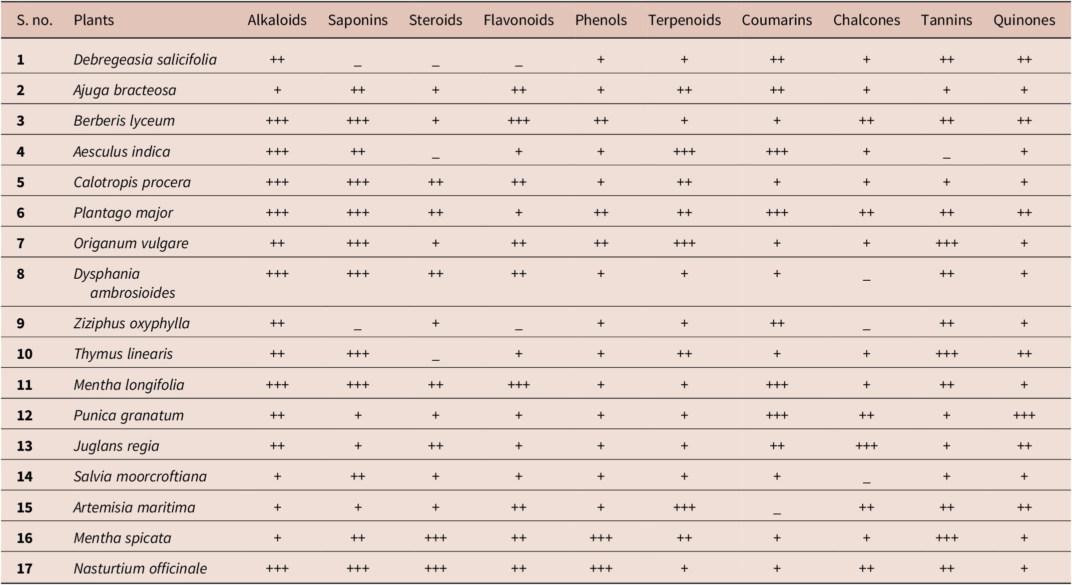
Table 3. Long description
A table with 12 columns: S. no., Plants, Alkaloids, Saponins, Steroids, Flavonoids, Phenols, Terpenoids, Coumarins, Chalcones, Tannins, and Quinones. The presence of phytochemicals is indicated by plus symbols: +++ for very strong, ++ for strong, + for weak, and an underscore for absence.
1. Debregeasia salicifolia: Alkaloids ++, Saponins _, Steroids _, Flavonoids _, Phenols +, Terpenoids +, Coumarins ++, Chalcones +, Tannins ++, Quinones ++.
2. Ajuga bracteosa: Alkaloids +, Saponins ++, Steroids +, Flavonoids ++, Phenols +, Terpenoids ++, Coumarins ++, Chalcones +, Tannins +, Quinones +.
3. Berberis lyceum: Alkaloids +++, Saponins +++, Steroids +, Flavonoids +++, Phenols ++, Terpenoids +, Coumarins +, Chalcones ++, Tannins ++, Quinones ++.
4. Aesculus indica: Alkaloids +++, Saponins ++, Steroids _, Flavonoids +, Phenols +, Terpenoids +++, Coumarins +++, Chalcones +, Tannins _, Quinones +.
5. Calotropis procera: Alkaloids +++, Saponins +++, Steroids ++, Flavonoids ++, Phenols +, Terpenoids ++, Coumarins +, Chalcones +, Tannins +, Quinones +.
6. Plantago major: Alkaloids +++, Saponins +++, Steroids ++, Flavonoids +, Phenols ++, Terpenoids ++, Coumarins +++, Chalcones ++, Tannins ++, Quinones ++.
7. Origanum vulgare: Alkaloids ++, Saponins +++, Steroids +, Flavonoids ++, Phenols ++, Terpenoids +++, Coumarins +, Chalcones +, Tannins +++, Quinones +.
8. Dysphania ambrosioides: Alkaloids +++, Saponins +++, Steroids ++, Flavonoids ++, Phenols +, Terpenoids +, Coumarins +, Chalcones _, Tannins ++, Quinones +.
9. Ziziphus oxyphylla: Alkaloids ++, Saponins _, Steroids +, Flavonoids _, Phenols +, Terpenoids +, Coumarins ++, Chalcones _, Tannins ++, Quinones +.
10. Thymus linearis: Alkaloids ++, Saponins +++, Steroids _, Flavonoids +, Phenols +, Terpenoids ++, Coumarins +, Chalcones +, Tannins +++, Quinones ++.
11. Mentha longifolia: Alkaloids +++, Saponins +++, Steroids ++, Flavonoids +++, Phenols +, Terpenoids +, Coumarins +++, Chalcones +, Tannins ++, Quinones +.
12. Punica granatum: Alkaloids ++, Saponins +, Steroids +, Flavonoids +, Phenols +, Terpenoids +, Coumarins +++, Chalcones ++, Tannins +, Quinones +++.
13. Juglans regia: Alkaloids ++, Saponins +, Steroids ++, Flavonoids +, Phenols +, Terpenoids +, Coumarins ++, Chalcones +++, Tannins +, Quinones ++.
14. Salvia moorcroftiana: Alkaloids +, Saponins ++, Steroids +, Flavonoids +, Phenols +, Terpenoids +, Coumarins +, Chalcones _, Tannins +, Quinones +.
15. Artemisia maritima: Alkaloids +, Saponins +, Steroids +, Flavonoids ++, Phenols +, Terpenoids +++, Coumarins _, Chalcones ++, Tannins ++, Quinones ++.
16. Mentha spicata: Alkaloids +, Saponins ++, Steroids +++, Flavonoids ++, Phenols +++, Terpenoids ++, Coumarins +, Chalcones +, Tannins +++, Quinones +.
17. Nasturtium officinale: Alkaloids +++, Saponins +++, Steroids +++, Flavonoids ++, Phenols +++, Terpenoids +, Coumarins +, Chalcones ++, Tannins ++, Quinones +.
+++ = very strong positive reaction; ++ = strong positive reaction; + = weak positive reaction; – = absence of detectable phytochemical reaction.
Thin layer chromatographic profiling
TLC profiling of the acetone extracts of selected medicinal plants was performed to separate and visualise the chemical constituents of the active acetone extracts. The calculated retention factor (Rf) of the plant extracts, along with the distance travelled and the number of observed phytochemical constituents, is shown in Supplementary Table S2. Unlike the preliminary qualitative phytochemical screening, TLC provided chromatographic fingerprints that allowed visual comparison of the chemical composition of the active extracts. The chromatographic profiles revealed differences in the number, position, color, migration distance, and Rf values of the separated bands among the tested plant extracts. Some extracts showed similar fingerprints and Rf values when TLC was performed using two different proportions of the selected mobile phases.
Discussion
Most communities in northern Pakistan, including the Swat region, rely on traditional medicinal plants to meet their primary health needs. This indigenous knowledge helps in the search for new drug candidates, and scientific research can play a crucial role in preserving and validating this medicinal knowledge (Wagner and Ulrich-Merzenich Reference Wagner and Ulrich-Merzenich2009). In the present study, the anthelmintic potential, preliminary phytochemical screening, and TLC profiling of the selected medicinal plants from the Swat region of Pakistan were evaluated. The results demonstrated variations in anthelmintic activity among plant species and extraction solvents, with acetone extracts showing comparatively higher activity than the other solvent extracts. The phytochemical screening and TLC profiling further revealed the presence of diverse secondary metabolites that may contribute to the observed biological activity. These findings provide scientific support for the traditional use of these medicinal plants and are discussed in relation to previous studies.
Anthelmintic activity
Caenorhabditis elegans has gained recognition as a useful model organism for preliminary anthelmintic screening and drug discovery due to its biological relevance and experimental advantages (Panda et al. Reference Panda, Padhi, Leyssen, Liu, Neyts and Luyten2017). The present study demonstrated significant variations in anthelmintic activity among extracts of the selected medicinal plants, with several extracts showing notable inhibition of C. elegans motility. The observed activity suggests that these plants may contain bioactive constituents contributing to their anthelmintic properties. Among the tested extracts, A. bracteosa, P. granatum, B. lyceum, M. longifolia, and A. maritima exhibited comparatively higher activity, suggesting that these species may represent promising sources of anthelmintic compounds. The differences in activity among plant extracts may be associated with variations in their phytochemical composition and the ability of extraction solvents to recover different classes of bioactive metabolites.
Despite the lack of statistical significance, acetone extracts consistently exhibited a higher proportion of anthelmintic activity than other solvents. This trend suggests that acetone may efficiently extract certain bioactive compounds of intermediate polarity, potentially contributing to the observed activity. However, further fractionation and bioassay-guided studies are required to confirm these observations. Solvent polarity greatly influences the solubility and extraction of compounds from plant material, which directly affects their bioactivities (Lezoul et al. Reference Lezoul, Belkadi, Habibi and Guillén2020). Acetone and ethanol are capable of extracting a broad range of moderately polar phytochemicals, which may contribute to the higher bioactivity observed against C. elegans compared with non-polar (hexane) and highly polar (water and methanol) extracts (Lee et al. Reference Lee, Jayakody, Kim, Jeong, Choi, Kim, Seo, Azimi, Hyun and Ryu2024).
The anthelmintic effects of A. maritima (Irum et al. Reference Irum, Ahmed, Mukhtar, Mushtaq, Mirza, Donskow-Łysoniewska, Qayyum and Simsek2015), J. regia (Das et al. Reference Das, Mehta and Gupta2011; Kale et al. Reference Kale, Gaikwada and Kamble2011), M. longifolia (Ahmad et al. Reference Ahmad, Tanveer and Zargar2013), C. procera (Iqbal et al. Reference Iqbal, Lateef, Jabbar, Muhammad and Khan2005; Mamat et al. Reference Mamat, Lame, Adeline, Yvette, Herman, Roger and Dieudonné2023), B. lyceum (Shah et al. Reference Shah, Sherwani, Sualeh, Kanwal, Khan and Kazmi2014), P. major (Turel et al. Reference Turel, Ayaz and Dinç2013), D. ambrosioides (Zamilpa et al. Reference Zamilpa, García-Alanís, López-Arellano, Hernández-Velázquez, Valladares-Cisneros, Salinas-Sánchez and Mendoza-de Gives2019), and Origanum vulgare (Štrbac et al. Reference Štrbac, Krnjajić, Maurelli, Stojanović, Simin, Orčić, Ratajac, Petrović, Knežević and Cringoli2022) have been reported before against various helminths. In contrast, the anthelmintic activity of A. indica, A. bracteosa, D. salicifolia, N. officinale, Z. oxyphylla, T. linearis, S. moorcroftiana, and M. spicata is reported here for the first time. Although the anthelmintic activity of some plants against certain parasitic worms has already been reported, we report the anthelmintic effects against C. elegans for the first time, thereby addressing a previously unexplored aspect of their anthelmintic potential and providing a basis for further detailed (e.g., mechanistic) studies. Advanced chemical and pharmacological techniques have facilitated the isolation and characterisation of novel bioactive compounds from medicinal plants, highlighting their importance as sources of potential therapeutic agents (Alamzeb et al. Reference Alamzeb, Ali, Mamoon-Ur-Rashid, Khan, Ihsanullah, Adnan, Omer, Ullah, Ali and Setzer2021).
The plant extracts with the lowest IC50 values, such as A. maritima, M. longifolia, B. lyceum, A. bracteosa, and P. granatum, need to be studied further using a bioassay-guided approach to isolate their anthelmintic compounds, which may serve as potential therapeutic agents. Therefore, bioassay-guided isolation and identification of active compounds from the promising plants, supported by chemical characterisation (Hu et al. Reference Hu, Tekin, Hu, Yaghoobi, Khan, Ghosh, Panda, Huang and Luyten2023), along with exploration of their mechanisms of action, structure–activity relationships, resistance evolution, and in vivo validation, are recommended for further studies.
Phytochemical analysis
The phytochemical composition of medicinal plants provides important insights into their potential biological activities and may help explain the observed anthelmintic effects. The present study revealed the presence of diverse secondary metabolites, including alkaloids, phenols, terpenoids, and quinones in all tested plant extracts, while other phytoconstituents were detected at varying concentrations. These findings suggest that the tested plants possess a broad range of bioactive compounds that may contribute to their biological activities. Together with the observed anthelmintic activity, these results support the potential of these plants as promising candidates for the development of new anthelmintic agents.
The present study revealed the presence of alkaloids, phenols, terpenoids, and quinones in all tested plant extracts, with other secondary metabolites at high concentrations in some extracts. These findings provide insights into the possible contribution of phytoconstituents to the biological activities of these plant materials and their potential therapeutic applications (Silva et al. Reference Silva, Nascimento da Silva, Martins da Fonseca, De Araujo, Correia, Cavalcanti and Lima2016).
The secondary metabolites of plants protect them against biotic and abiotic stresses, but humans have secondarily converted these phytoconstituents into medicines to combat various pathogenic microorganisms, thereby aiding the treatment of corresponding infections (Hemeg et al. Reference Hemeg, Moussa, Ibrahim, Dawoud, Alhaji, Mubarak, Kabli, Alsubki, Tawfik and Marouf2020). The antipathogenic properties of the plants are based on the concentration and interactions of the secondary metabolites present in various parts of the plants (Dzotam et al. Reference Dzotam, Touani and Kuete2015).
Alkaloids are among the most important secondary metabolites, often possessing potent antimicrobial, antiparasitic, and anti-HIV activities (Nugraha et al. Reference Nugraha, Achmad and Sitompul2019). Phenolic compounds are typically antiinflammatory (Persinos and Quimby Reference Persinos and Quimby1967). Terpenoids often have antiinflammatory, anticancer, antiviral, and antibacterial activity (Bhattacharya et al. Reference Bhattacharya, Maity, Pramanick, Hazra and Choudhury2016). Quinones also possess physiological and therapeutic effects (El-Najjar et al. Reference El-Najjar, Gali-Muhtasib, Ketola, Vuorela, Urtti and Vuorela2011). Flavonoids typically possess antioxidant and antitumour activity, while tannins produce antitumor, antibacterial, and antiviral effects (Adil et al. Reference Adil, Filimban, Ambrin, Quddoos, Sher and Naseer2024). Coumarins often have anticancer, anticoagulant, and antiinflammatory effects (Xu et al. Reference Xu, Zhao, Y-L and Zhang2015). Saponins boost the immune system and protect the body against various microbial infections (Prohp and Onoagbe Reference Prohp and Onoagbe2012). Steroids help women maintain their hormonal balance during pregnancy or breastfeeding (Ajiboye et al. Reference Ajiboye, Ibukun, Edobor, Ojo and Onikanni2013). Chalcones may possess antiparasitic, anthelmintic, antimicrobial, antiviral, anticancer, antioxidative, and immunosuppressive effects (Yerragunta et al. Reference Yerragunta, Suman, Anusha, Patil and Samhitha2013).
A number of phytochemical studies have revealed that biological activities, including antiparasitic, antiinflammatory, antimicrobial, anti-dyspepsia, and antihypertensive activities, are attributed to the presence of various secondary metabolites such as alkaloids, triterpenoids, flavonoids, and other phytochemical groups in the respective plants (Hu et al. Reference Hu, Hu, Peng, Ghosh, Khan, Sun and Luyten2021). The secondary metabolites detected in these plants may play an important role in treating helminthic and parasitic infections; thus, their isolation and purification should be of interest for drug development.
Thin layer chromatographic profiling
Thin Layer Chromatographic (TLC) profiling provides a comparative approach for evaluating the chemical complexity of plant extracts and supports the interpretation of preliminary phytochemical findings. Although TLC does not identify individual compounds, it provides a rapid comparative assessment of extract complexity and serves as a valuable first step for selecting extracts for bioassay-guided fractionation and compound isolation (Bladt, Reference Bladt2009; Harborne Reference Harborne1998). Various techniques can be used to select plants of therapeutic interest, but TLC is comparatively simple, flexible, and rapid, allowing many samples to be profiled in parallel, with results that can be used to carry out further investigative work (Panda et al. Reference Panda, Das, Leyssen, Neyts and Luyten2018).
Separation on a TLC plate is based on polarity; some compounds will strongly bind to the adsorbent surface, while others will move, and these differences characterise the test samples (Kagan and Flythe Reference Kagan and Flythe2014). Rf values below 0.5 indicate the presence of non-polar compounds, while values above 0.5 indicate polar compounds (Gomathi et al. Reference Gomathi, Kalaiselvi, Ravikumar, Sophia, Gopalakrishnan and Uma2012). Our study also indicates that these plant extracts possess both polar and non-polar compounds. The wide range of Rf values observed in the active acetone extracts further supports the presence of chemically diverse metabolites with different polarities, consistent with the qualitative phytochemical screening results.
Similar TLC fingerprints observed for some extracts may reflect the presence of related classes of phytochemicals, whereas distinct banding patterns suggest differences in chemical composition that may contribute to the variation in anthelmintic activity observed among the extracts (Bladt Reference Bladt2009). Conversely, extracts showing distinct Rf values and banding patterns are likely to contain different phytochemical constituents or different relative abundances of these constituents, which may partially explain the differences in their biological activities.
This complementary information strengthens the selection of the most active extracts for future purification and identification of the compounds responsible for the observed anthelmintic activity. TLC fingerprints can also be used to determine bioactive phytoconstituents directly by exposing the test microorganism to the chromatograms via bioautography (Marston Reference Marston2011). TLC fingerprinting has been approved by the WHO for the identification and authentication of plant materials (Zahiruddin et al. Reference Zahiruddin, Parveen, Khan, Parveen and Ahmad2021).
Our anthelmintic tests also support the ethnomedicinal use of these plants by traditional knowledge holders. Therefore, further studies are recommended to isolate and purify the compounds responsible for the bioactivities against specific pathogens.
The present study concludes that the selected traditional medicinal plants of the Swat region of Pakistan possess promising anthelmintic activity against C. elegans. Among the tested plants, A. maritima, M. longifolia, B. lyceum, A. bracteosa, and P. granatum exhibited the most potent activities, supporting their traditional ethnomedicinal uses. The selected plants contain various groups of phytoconstituents that may contribute to their bioactivities against different pathogens. Furthermore, TLC profiling supported the phytochemical variability and chemical diversity among the active acetone extracts, providing preliminary fingerprints for future bioassay-guided isolation studies. This study further highlights the importance of traditional medicinal plants as potential sources of novel anthelmintic drug candidates. Therefore, the most active plants are recommended for further phytochemical isolation, purification, and mechanistic studies using bioassay-guided approaches.
Supplementary material
The supplementary material for this article can be found at http://doi.org/10.1017/S0022149X26102016.
Acknowledgements
The authors are thankful to the authorities of the University of Swat, Pakistan, and KU Leuven, Belgium, for providing facilities to conduct this research. The authors are also thankful to all those who helped directly or indirectly during this research work.
Financial support
This work was supported indirectly by a doctoral fellowship grant to the author, Ajmal Khan, from the Higher Education Commission of Pakistan, the University of Swat, Pakistan, and KU Leuven, Belgium. Ajmal Khan and Sujogya Kumar Panda were also supported in part by CELSA Grant 23/014. All other authors largely supported themselves.
Competing interest
The authors declare none.
Ethical standard
Not applicable.
Artificial intelligence (AI) statement
Grammarly AI was used for grammatical corrections, rephrasing the sentence structure, and improving the readability of the manuscript. All revisions were carefully reviewed, edited, and verified by the authors. The authors take full responsibility for the final content of the manuscript.