

Introduction
The effect of long-distance running on vascular volumes has been of interest for researchers over decadesReference Dixon, Collop and Batt1–Reference de Paz, Villa, Lopez and Gonzalez-Gallego3. Long-distance running is associated with a wide range of significant changes in haematological parameters, and intravascular haemolysis has been suggested as an important reason for the alteration of vascular volumes in these runners. Prolonged running has been associated with a significant destruction of red blood cells (RBC), with RBC turnover being substantially higher in runners compared with untrained controlsReference Weight, Klein, Noakes and Jacobs4. Cr-labelled RBC survived for only 74 days in individuals running up to 130 km week− 1 compared with 114 days in untrained subjectsReference Weight, Klein, Noakes and Jacobs4. In contrast, one study reported increased RBC destruction after intense physical trainingReference Schmidt, Maassen, Trost and Boning5. These research reports provide strong evidence that RBC lifespan is reduced in endurance runners.
Foot impact force during running has been suggested as one of the important causes of haemolysis in runnersReference Miller, Pate and Burgess6, Reference Telford, Sly, Hahn, Cunningham, Bryant and Smith7. Many researchers have proposed that mechanical damage to RBC occurs in the tiny capillaries of the foot during the footstrike phase of runningReference Weight, Klein, Noakes and Jacobs4, Reference Falsetti, Burke, Feld, Frederick and Ratering8, Reference Poortmans and Haralambie9. However, intravascular haemolysis has also been reported in sports in which foot impact does not occur. Reports on haemolysis have been documented in cyclistsReference Telford, Sly, Hahn, Cunningham, Bryant and Smith7, Reference Dufaux, Hoederath, Streitberger, Hollmann and Assmann10, rowersReference Dufaux, Hoederath, Streitberger, Hollmann and Assmann10, skatersReference Roberts and Smith11 and swimmersReference Selby and Eichner12–Reference Pizza, Flynn, Boone, Rodriguez-Zayas and Andres14, suggesting that mechanisms other than footstrike can also lead to haemolysis in athletes. It was postulated that after strenuous exercise erythrocytes are more susceptible to stressReference Senturk, Gunduz, Kuru, Kocer, Ozakaya and Yesilkaya15, whether of the mechanical, oxidative or osmotic typeReference Bonilla, Narvaez and Chuaire16, resulting in increased reactive oxygen species production, oxidative stress and tissue damageReference Chevion, Moran, Heled, Shani, Regev and Abbou17, and, consequently, in haemolysis. Incidence of ‘hand strike’ haemolysis has also been documentedReference Martinez, Villa, Aguilo, Tur and Pons18.
Therefore, the purpose of our study was to analyse intravascular haemolysis in athletes during running and compare it with cycling. We speculated that foot impact force during running would result in increased breakdown of tiny capillaries in the foot. Specifically, we hypothesized that haemolysis occurs more during running than during cycling.
Methods
Participants
A total of 20 male long-distance runners participated in this study. We calculated the sample size of the study using sample size software (nMaster 1.0; Christian Medical College, Vellore, India). A sample size calculator estimated a sample size of five per group for the given data (effect size 1.73). We used 10 samples per group to increase the power of the study (power 0.53). We screened all the participants using a physical activity readiness questionnaire. Female runners and athletes under regular Fe supplementation were excluded from the study. The experimental protocol was approved by the Institutional Ethics Committee of the Department of Sports Medicine and Physiotherapy. All participants gave written informed consent after the design and the risks of the study had been described to them. A further condition was that no intense training should be carried out 24 h before testing.
Grouping of participants
We randomly assigned the participants into two groups (using online research randomizer software version 4.0), with 10 athletes in each group. Grouping of athletes was as follows: running (group 1, n = 10) and cycling (group 2, n = 10).
Experimental protocol
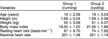
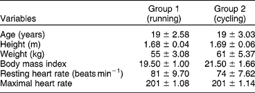
Initially, each athlete reported to the exercise physiology laboratory before the actual running and cycling protocols. The physical characteristics of all athletes were measured (Table 1) and the target heart rate (THR, 60–70%) was calculated, which was set as the intensity for both the running and cycling protocols (THR was calculated according to age and maximal heart rate (HRmax) (HRmax = 220 − age)). All athletes wore medium-weight training shoes of a similar type, with all shoes in good condition.
Physical and physiological characteristics of all participants (means±SD)
The running protocol (group 1) consisted of 5 min of jogging as a warm-up at 40–50% of the THR, after which the athletes ran for a period of 1 h at the calculated THR (60–70%). The cycling protocol (group 2) consisted of an initial warm-up of 5 min of cycling, after which the athletes were allowed to cycle for a period of 1 h in the cycle ergometer at 40–50% of the THR. The speed and resistance required to achieve the THR (60–70%) was found within 5 min of the test, after which the athletes sustained their speed to maintain their exercise intensity. The cycle ergometer was placed in the exercise therapy laboratory, where there was no air conditioning available and the windows were open for free air circulation to avoid gross temperature variations compared with the running trial.
The aim of running or cycling around the THR (60–70%) during the 1-h period for each mode of exercise on an individual basis was to closely approximate potential oxidative and metabolic stress in the RBC.
Heart rate monitoring
Heart rate was measured at regular intervals (every 15 min), after which adjustments were made, if necessary, to the speed. Heart rate was monitored every 15 min, to ensure that the athletes ran at the intensity around their calculated THR. Heart rate was monitored by the athlete and the investigator using a polar heart rate monitor (PC Coach Light, version 3.0.3; Biometrics, Inc., Boulder, CO, USA).
To verify whether haemolysis found after running and cycling was caused by exercise and not due to temporal variation or damage during blood collection, blood samples were collected and analysed from a non-exercising group over the same time period as that of the exercise group. Two participants per day (n = 10) [mean age (years) 20 ± 2.78 (SD), height (m) 1.68 ± 0.05 (SD), weight (kg) 67 ± 6.08 (SD), body mass index 23.5 ± 1.31 (SD) and resting heart rate 77 ± 3.05 (SD)], who were moderately active and had similar anthropometric characteristics to athletes, were recruited.
Blood sample collection
Blood samples were collected before and immediately after the running and cycling protocols. At each sample point, blood was collected via venepuncture of the antecubital vein, with athletes in the sitting position. To control the plasma volume shift due to postural change, sampling before and after the running and cycling trials was carried out in an identical fashion, with athletes in the sitting position. Extreme care was taken during the collection of blood samples to avoid the possibility of haemolysis. Two millilitres of blood were collected in an EDTA tube for haemoglobin (Hb) estimation. Two millilitres of blood were transferred into a serum tube and allowed to clot at room temperature. The blood samples were stored in the ice box and immediately transferred to the laboratory (situated 200 m from the exercise laboratory) and refrigerated. The serum-clotted samples were centrifuged at 3000 rpm for 10 min. The refrigerated blood samples were evaluated within 24 h of collection. Blood parameters used to measure haemolysis were unconjugated bilirubin (UBR), lactate dehydrogenase (LDH), serum ferritin (FER) and Hb. Blood samples were analysed using a minitechno-biochemistry analyser (Lamicoat International, under license of I.S.E S.R.L, Rome, Italy, 2007) for the estimation of LDH, UBR and Hb, and using an ELISA-APR microplate reader (Logotech-ISE Group, Rome, Italy, 2007) for the estimation of FER.
Blood samples from two runners were rejected as these were clotted, and blood measurements of one runner were not included as he was not able to complete the 1 h of running. In total, measurements from seven runners, 10 cyclists and 10 non-exercising group participants were considered for data analysis.
Statistical analysis involved a comparison between the profiles of repeated measurements obtained from the running and cycling trials. A paired t-test was used to evaluate the amount of haemolysis between the pre- and post-run. Non-parametric data evaluation was carried out for the cycling and non-exercising group participants, as the observed value did not satisfy the criteria for parametric test evaluation (mean ≥ 3 SD). The level of intra-group significance in total for the cycling and non-exercising group participants was evaluated using the Wilcoxon signed-rank test. The Mann–Whitney U-test was performed to evaluate the significance of haemolysis between the running and cycling group participants. The significance level was set at P ≤ 0.05 and all statistical analyses were carried out using SPSS statistical software (version 17.0).
Results
Paired t-testing revealed a significant difference in the mean values of UBR (t = 4.509, P < 0.01) and LDH (t = 2.497, P <0.05) post-run (group 1), indicating the occurrence of haemolysis in these athletes (Table 2). No significant differences were observed in any of the haematological variables (UBR, LDH, Hb and FER) before and after cycling (P>0.05), indicating insignificant haemolysis occurrence in the cycling group participants.
Haematological variables of all group participants (means±SD)

* P < 0.05. ** P < 0.01.
However, we did observe a significant variation in mean difference values (between the pre- and post-tests) for the haematological variables when compared between the running and cycling groups (UBR: Z = 3.035; P < 0.001, LDH: Z = 2.641; P < 0.01, Hb: Z = 2.446; P < 0.05; FER: Z = 0.683; P = 0.536). Consequently, we assumed that running was the main reason for these significant changes observed, as we previously observed insignificant changes in the cycling group.
The investigation of temporal variation in the blood samples taken from the non-exercising group (n = 10) revealed no significant differences (P>0.05) in any haemolytic variable, indicating that the previously demonstrated significant changes over this time period were exercise-related.
Discussion
The aim of this experimental study was to evaluate intravascular haemolysis during running and compare it with cycling. The main finding of this study was that the participants demonstrated a significant amount of haemolysis during running when compared with cycling. As foot impact is the important factor of relevance differentiating between running and cycling, the results observed in our study strongly suggest that footstrike is the mechanism responsible for greater haemolysis during running. The results are consistent with previous studies indicating the occurrence of haemolysis after prolonged runningReference Dressendorfer, Wade and Frederick2, Reference Telford, Sly, Hahn, Cunningham, Bryant and Smith7.
Researchers have noted increases in bilirubin and LDH to be the indicators of increased intravascular haemolysisReference Deitrick19, Reference Wu, Chen, Shee, Chang, Huang and Yang20, which is consistent with our study findings. However, acute effects of haemolysis on Hb and FER did not conform to previous studies. Hb values tended to decrease significantly (P < 0.05), whereas FER values revealed no significance. Even though studies have reported a significant decrease in Hb following runningReference Dressendorfer, Wade and Frederick2, Reference Deitrick19, the observed effects in these studies were due to prolonged running for more than a week and not an acute response to running.
Red cell haemolysis has been reported after various forms of exercise, whereas the foot impact force during running has been suggested to be an important mechanism causing haemolysisReference Miller, Pate and Burgess6, Reference Telford, Sly, Hahn, Cunningham, Bryant and Smith7. At the time of contact between the foot and the ground, impact forces and pronation forces place a large stress on structures of the lower extremityReference Spurgeon21–Reference Nigg and Morlock23, whereas the exposure to repeated impact loading was linked to the development of runners' injuriesReference Aguinaldo and Mahar24. Previous researches suggested the role of shoe insoles and midsoles in the reduction of impact forces on the foot during runningReference Cook, Kester, Brunet and Haddad25, Reference Fredrick, Clarke, Hamil and Fredrick26 and hence haemolysisReference Dressendorfer, Wade and Frederick2, Reference Hardin and Hamill27. These reports concur with the findings from our study, proving that mechanical trauma during footstrike could be the major cause of haemolysis during running.
Our results indicate insignificant changes in haematological parameters in the non-exercising group participants, thus eliminating the role of environmental temperature, blood sample collection and blood sample handling in haemolysis. Even though participants who cycled (group 2) did not demonstrate any significant haemolytic changes in our study, previous study results indicate that activities that do not involve footstrike can also result in haemolysisReference Selby and Eichner12, Reference Bula, Ziobro and Sutylo28. The reasons for haemolysis in these studies were mainly attributed to oxidative stressReference Senturk, Gunduz, Kuru, Kocer, Ozakaya and Yesilkaya15, Reference Smith29, perturbation of osmotic homeostasisReference Telford, Sly, Hahn, Cunningham, Bryant and Smith7 and compression of larger muscle groups on capillaries during exerciseReference Telford, Sly, Hahn, Cunningham, Bryant and Smith7, Reference Stenner, Gianoli, Biasioli, Piccinini, Delbello and Bussani30. The reason for the insignificant degree of haemolysis during cycling in our study may be decreased exercise intensity (THR 60–70%) compared with previous studies (maximal heart rate, MHR, 75–95%)Reference Telford, Sly, Hahn, Cunningham, Bryant and Smith7, Reference Peter, Brian, Carmel, Grant, Erwin and Dorine31. This indicates that training at low intensities can itself result in greater haemolysis in runners as observed in our study, which shows the significance of this present study. A recent study has reported greater haemolysis at higher intensities compared with lower intensities during runningReference Peter, Brian, Carmel, Grant, Erwin and Dorine31, which signifies the importance of training intensity on intravascular haemolysis.
Our study involved a grass surface, which, on inspection, revealed an uneven surface with bumps not readily visible and grass erosion on some areas along the course of the track. Surface impact absorption is an important factor that may contribute to haemolysisReference Peter, Brian, Carmel, Grant, Erwin and Dorine31, Reference Schwellnus, Penfold, Cilliers, Kuyl and van der Heever32. During running, the transient force during the impact phase of the ground reaction force can be up to three times that of body weightReference Aguinaldo and Mahar24. These ground reaction forces can be higher on hard surfacesReference Stiles and Dixon33, Reference Feehery34, such as asphalt, compared with softer surfaces, but can increase significantly on an uneven surfaceReference Grimmer, Ernst, Gunther and Blickhan35. As the foot impact force is considered as being an important cause of intravascular haemolysis during runningReference Miller, Pate and Burgess6, Reference Telford, Sly, Hahn, Cunningham, Bryant and Smith7, we assumed that the unevenness of the grass surface could have resulted in decreased, uneven impact force absorption, consequently resulting in increased foot impact force during running, which could have increased the degree of haemolysis observed in the present study. On the other hand, Indian athletes can be more prone to this problem as some still run barefoot or on irregular or uneven surfaces. A recent study reported an increased incidence of injuries on grass surfaces and attributed their findings to inadequate maintenance of the grass in terms of quality and texture in Indian settingsReference Kaur, Yadav and Sandhu36, which supports the fact that Indian athletes are more prone to this phenomenon.
Practical applications
Intravascular haemolysis is thought to be a physiological adaptation to exerciseReference Robinson, Cristancho and Boning37, Reference Schumacher, Schmid, Grathwohl, Bultermann and Berg38. Adaptations include beneficial effects such as dilutional pseudo-anaemiaReference Selby and Eichner12 and increased red cell massReference Smith, Martin, Telford and Ballas39, which could benefit the cardiorespiratory endurance adaptations of erythrocythaemia and bring about an optimal haematological state for performanceReference Deitrick19. However, prolonged strenuous training may disturb this conservative mechanism and repeated episodes of haemolysis over time may significantly impact runners. Although intravascular haemolysis may not lead to sports anaemia or true iron-deficiency anaemia, it can result in an iron-deficiency statusReference Dufaux, Hoederath, Streitberger, Hollmann and Assmann10, Reference Hunding, Jordal and Paulev40, Reference Ehn, Carlmark and Hoglund41, which may hinder these runners in achieving maximal aerobic powerReference Green, Carter, Grant, Tupling, Coates and Ali42 and impact their performanceReference Rowland, Black and Kelleher13, Reference Rogers, Goodman, Mitchell and Hattingh43.
Conclusion
Our study findings indicate that intravascular haemolysis occurs significantly during prolonged running when compared with cycling, and we conclude that mechanical trauma due to footstrike is the major cause of intravascular haemolysis during running. Further research is needed regarding the impact force characteristics of shoes and surfaces on haemolysis, as well as the repeated effect of haemolysis over a longer period of training, and their relationship to performance.