INTRODUCTION
Toxoplasma gondii is a widespread protozoan in warm-blooded animals including humans and birds (Tenter et al. Reference Tenter, Heckeroth and Weiss2000). Felids are the definitive hosts and shed oocysts after infection while almost all warm-blooded animals and humans are intermediate hosts and develop tissue cysts in several organs, especially in neural and muscular tissue (Tenter et al. Reference Tenter, Heckeroth and Weiss2000; Dubey, Reference Dubey2010a ).
The main risk factor for humans to become infected with T. gondii is the consumption of rare or undercooked meat (Cook et al. Reference Cook, Gilbert, Buffolano, Zufferey, Petersen, Jenum, Foulon, Semprini and Dunn2000). Typically, poultry meat is consumed well done. But as part of a changing lifestyle more and more poultry is consumed as raw sausages or ham. According to Dubey (Reference Dubey1997) and Pott et al. (Reference Pott, Koethe, Bangoura, Zöller, Daugschies, Straubinger, Fehlhaber and Ludewig2012) tissue cysts are still viable after brief salting or curing and there is a risk of infection with T. gondii by consumption of fermented raw sausages.
An average conventional fattening cycle in turkey husbandry lasts up to 16 weeks in female turkeys and 22 weeks in male turkeys (Krautwald-Junghanns et al. Reference Krautwald-Junghanns, Ellerich, Bohme, Cramer, DellaVolpe, Mitterer-Istyagin, Ludewig, Fehlhaber, Schuster, Berk, Aldehoff, Fulhorst, Kruse, Dressel, Noack and Bartels2009). A short fattening period takes 9–12 weeks for both sexes. Usually, chickens are fattened for 35–42 days in conventional husbandry whereas organic fattening lasts up to 10 weeks. Livestock kept in free-range husbandries has a significantly increased risk of infection with T. gondii (Kijlstra et al. Reference Kijlstra, Meerburg and Mul2004; Maksimov et al. Reference Maksimov, Buschtons, Herrmann, Conraths, Gorlich, Tenter, Dubey, Nagel-Kohl, Thoms, Bötcher, Kühne and Schares2011) compared with livestock raised indoors. Fowl is considered to be one of the most important intermediate hosts in the life cycle of T. gondii (Dubey, Reference Dubey2010b ), but little is known about the duration of persistence of T. gondii in tissue of poultry. Zöller et al. (Reference Zöller, Koethe, Ludewig, Pott, Daugschies, Straubinger, Fehlhaber and Bangoura2013) demonstrated a persistence of T. gondii in turkeys over a period of 12 weeks but to our knowledge the persistence of T. gondii in chicken has not been investigated. There is little information as to whether chickens are able to eliminate a T. gondii infection, as has been observed for some of the other intermediate host species (Beverley et al. Reference Beverley, Henry, Hunter and Brown1977).
The aim of this study was to investigate the duration of persistence of T. gondii in turkeys and chickens and to estimate the risk of finding T. gondii-positive meat and organs at the time of slaughtering according to the duration of conventional fattening cycles. Additionally, the distribution of T. gondii in tissues depending on time of slaughtering was analysed.
MATERIALS AND METHODS
Parasites
For infection of poultry the T. gondii type III strain NED (Howe and Sibley, Reference Howe and Sibley1995) was used. Tachyzoites for infection of the animals were grown in VERO cells cultivated with IMDM medium (PAA, Pasching, Austria), 5% foetal calf serum, 1% penicillin/streptomycin and 1% amphotericin B as described before (Zintl et al. Reference Zintl, Halova, Mulcahy, O'Donovan, Markey and DeWaal2009) at 37 °C, 5% CO2. To harvest the parasites, supernatant of cell culture was centrifuged at 2000×g for 5 min. The pellet was resolved in 1 mL PBS solution and tachyzoites were counted using a Neubauer chamber.
Animals and husbandry
In vivo trials were performed at the Institute of Parasitology, University Leipzig, Germany. Conventionally reared and commercially available turkeys (BUT B.I.G. 6, n = 30) and chickens (ISA JA 757, n = 18) were kept on a bedding consisting of wood shavings. Turkeys were fed ad libitum with poultry feed for pet birds without anticoccidials (deuka Wild-und Ziergeflügelfutter, Deutsche Tiernahrung Cremer, Duesseldorf, Germany) because no conventional starter and grower feed for turkeys without anticoccidials was available. The feed was enriched with powdered milk and supplements (Korvimin ZVT+Reptil, WDT, Garbsen, Germany) to obtain the recommended nutritional values for turkeys. Chickens received conventional starter and grower feed for chickens without anticoccidials ad libitum. Animals had free access to water.
Infections and study design
After a raising period of 8 weeks turkeys were divided into five groups. Four groups were infected and slaughtered at 4, 8, 12 or 16 weeks post-infection (p.i.), and one group remained uninfected as negative control. Regarding chickens, 1-week-old chicks were divided into three groups, two of which were infected and slaughtered 5 or 10 weeks p.i., to simulate the conventional and organic fattening time line, and the remaining group again served as uninfected negative control.
Animals were infected each with 1×10⁶ tachyzoites of the T. gondii NED strain in approximately 0·5 mL of a sterile isotonic saline solution (B. Braun Melsungen AG, Melsungen, Germany) intravenously into the wing vein (Vena cutanea ulnaris).
Serological examinations
Blood samples were drawn from the wing vein once before and weekly after infection for T. gondii antibody screening (IgY fraction in both bird species) and stored at −20 °C until testing. Sera of turkeys were analysed with a kinetic ELISA (KELA) as previously described (Koethe et al. Reference Koethe, Pott, Ludewig, Bangoura, Zöller, Daugschies, Tenter, Spekker, Bittame, Mercier, Fehlhaber and Straubinger2011). Briefly, microtitre plates were coated with 0·13 μg GRA7/GRA8 antigen mix (1:1) in coating buffer containing 0·1 m NaHCO3 buffer (pH 9·6), 1·69 mm SDS and 1·9% 2-mercaptoethanol and subsequently frozen until use. Coated plates were thawed and washed (4×100 μL well−1 PBS-Tween 0·05%) and blocked with 200 μL well−1 5% skim milk for 1 h at 37 °C. After washing, 100 μL well−1 1:50 diluted sera were incubated for 1 h at 37 °C and after another washing 100 μL well−1 1:1000 diluted detection antibody (goat-anti-turkey IgY-HRP) was incubated for 30 min at ambient temperature. After a last washing step, 100 μL well−1 TMB substrate was added and after 105 s plates were read on an ELISA reader (SpectraMax 340PC384, Molecular Devices, Germany). Kinetic development of signal was recorded by reading every 55 s for, in total, four times. The resulting slope of increasing signal was used by the reader software (SoftMax Pro v5, Molecular Devices) for calculating KELA values. Four standard sera were included in every plate to reduce plate-to-plate variances by applying linear regression-based recalculation of obtained values.
Chicken sera were shipped to the Friedrich-Loeffler-Institut, Wusterhausen and processed by an immunofluorescent antibody test (IFAT) as described by Maksimov et al. (Reference Maksimov, Buschtons, Herrmann, Conraths, Gorlich, Tenter, Dubey, Nagel-Kohl, Thoms, Bötcher, Kühne and Schares2011) but with minor modifications. Ten μL of a suspension of cell culture-derived T. gondii RH strain tachyzoites (5×10⁶ mL−1) in PBS were used to sensitize IFAT slide wells. Slides were air-dried and stored frozen at −20 °C until used. The slides were fixed with ice-cold acetone for 10 min and then incubated in PBS for 10 min. Chicken sera were titrated in PBS in two-fold steps starting at a dilution of 1:25. Anti-chicken IgY (H+L) produced in goat and coupled to FITC [Rockland Immunochemicals Inc., Gilbertsville, USA] diluted 1:50 in PBS, 0·2% Evans Blue was used to detect primary antibodies. The slides were examined using an Axiovert fluorescence microscope (AHBT3, Olympus, Hamburg, Germany). Only complete peripheral fluorescence of the tachyzoite was considered specific. A titre of 100 was used as the positive cut-off titre.
Sample processing
Slaughtering of the animals was performed after anaesthesia with ketamine (25 mg kg−1) and xylazine (1 mg kg−1) intramuscularly. The animals were dissected and tissue samples from 16 different locations were taken including edible parts (heart, breast muscle, drumstick, thigh muscle, liver, gizzard) that may be purchased for human consumption and non-edible parts (glandular stomach, pancreas, spleen, kidneys, brain, retina, gonads, intestine, lung, bone marrow). Directly after slaughtering or after storage at −20 °C, samples were homogenized with commercial household blenders (La Moulinette, Tefal Groupe SEB, Offenbach, Germany) or mortar and pestles, except muscles where approximately 300 g sample material from different locations was taken. All homogenized samples were stored in microcentrifuge tubes at −20 °C until further processing.
DNA extraction was performed from 25 mg tissue per sample with the QIAamp DNA Mini Kit® (Qiagen, Hilden, Germany) following the manufacturers instructions. After no more than three samples from infected birds at least one sample of the negative control group was handled. This order was kept up through the whole processing including PCR and gel electrophoresis as control of possible contamination. Subsequently, DNA content of samples was measured using a spectrophotometer (Nanodrop 2000c, Thermo Scientific, Waltham, MA, USA). 80–200 ng template DNA was used per PCR reaction. A positive control consisting of DNA from cell culture-derived ME49 tachyzoites and a negative control (aliquot of currently used DNA elution buffer) were carried along in every PCR batch. The PCR reaction was performed as a direct PCR followed by a nested PCR as described by Zöller et al. (Reference Zöller, Koethe, Ludewig, Pott, Daugschies, Straubinger, Fehlhaber and Bangoura2013). The mastermix for a direct PCR reaction with 25 μL contained 0·5 U GoTaq® Flexi Polymerase and 1×GoTaq® Flexi Buffer (Promega GmbH, Mannheim, Germany), 200 μ m of each dNTP (Fermentas, St. Leon Rot, Germany), 3 mm MgCl2, 0·4 μ m of each primer Tg1 (5′-AAA AAT GTG GGA ATG AAA GAG-3′) and Tg2 (5′-ACG AAT CAA CGG AAC TGT AAT-3′). For nested PCR the same assembled mastermix was used with the primers Tgnested1 (5′-CGC TAA TGT GTT TGC ATA GG-3′) and Tgnested2 (5′-GGC ACG TCT CTT GTT CTT CT-3′), 1·5 mm MgCl2 and 2·5 μL of the direct PCR product as template. Both PCRs were performed in the iCycler® Thermal Cycler (Bio-Rad, Hercules, CA, USA) or the M×3000P®Real-Time PCR System (Stratagene/Agilent Technologies, La Jolla, USA) under the following cycling conditions: preheating (2 min at 94 °C), amplification for 35 cycles (direct PCR) or 25 cycles (nested PCR) of 1 min at 94 °C, 40 s at 55 °C and 1 min at 72 °C and one final extension step of 5 min at 72 °C.
Approximately 10 μL of the nested PCR products were examined with gel electrophoresis on a 1·5% agarose gel, which was stained with ethidium bromide. DNA bands were visualized with UV-light in positive samples at 375 bp fragment length.
Statistics
Statistical analysis was carried out with the IBM SPSS statistics Version 20 software package (IBM, New York, USA).
The Kolmogorov–Smirnov test was performed to test data for normal distribution. All data were non-normally distributed and were therefore further analysed by Kruskal–Wallis test and Friedman test. The Spearman rank correlation test was performed to explore possible correlations. Group differences and correlations with P values of less than 0·05 were defined as statistically significant.
RESULTS
Experimental infections of turkeys
None of the turkeys showed symptoms of clinical toxoplasmosis throughout the entire experiment. Two of the 24 infected turkeys had to be euthanized due to leg and wing problems at 13 and 14 weeks p.i., respectively.
Serology
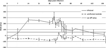
All infected animals showed a seroconversion (see Fig. 1, Table S1). Eleven turkeys already had a detectable antibody level at 1 week p.i. The remaining 13 animals seroconverted by 2 weeks p.i. At 9weeks p.i. antibody levels of the 12 remaining animals were decreasing and until slaughtering the levels of 3 animals fell below the cut-off. No seroconversion of the uninfected control was detected over the entire period of the experiment.
Mean antibody levels of infected and uninfected turkeys determined in a kinetic ELISA (error bars indicate s.d.).

Direct parasite detection (PCR)
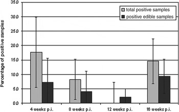
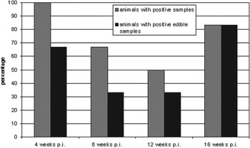
Sixty-one of 384 tissue samples (15·9%) contained detectable parasite DNA. At every time of slaughtering positive samples were found. There were no significant differences between the proportions of positive findings in infected turkey slaughtered at different time points p.i. (P>0·05; see Fig. 2). In 30 edible parts (7·8%) T. gondii was observed. Again no significant differences were observed when the proportion of positive findings in turkeys slaughtered at different time points after infection was compared (P>0·05). Twenty of the 24 infected turkeys (including the two deceased animals) showed T. gondii DNA in at least one sampled part, 14 of these in at least one edible part. Positive findings were observed distributed evenly over the examination time points (see Fig. 3).
Mean proportion of positive samples per turkey stratified for the length of the period between infection and slaughter (4, 8, 12 weeks p.i., n = 96; group 16 weeks p.i., n = 64, error bars indicate s.d.).

Number of positive tested turkeys depending on time of slaughtering separately by animals positive in at least one organ (grey) and animals positive in at least one edible organ (black).

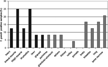
All parts except the kidneys tested positive at least once (see Fig. 4, Table S1). Most frequently, breast (29·2%) and thigh muscles (29·2%) tested positive followed by drumsticks, intestine, bone marrow (25% each) and brain (20·8%) though no body part was statistically significantly more often DNA positive than other parts (P>0·05). Correlation of T. gondii detectability in different muscular tissues was tested and a statistically significant correlation between thigh and breast muscle positivity could be found (Spearman rho = 0·542, P = 0·009).
Positive organ samples over all infected turkeys (n = 24) plotted by edible organs (black) and other organs (grey).

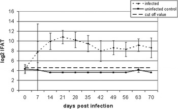
Mean antibody level of infected and uninfected chickens in IFAT. Titres are provided as log2 values of the IFAT titres (error bars indicate s.d.).

Experimental infections of chickens
None of the chickens showed signs of clinical toxoplasmosis.
Serology
Every infected chicken seroconverted during the study (see Fig. 5, Table S2). Eight chickens of the infected group showed a seroconversion at 1 week p.i. The other four chickens had a detectable antibody titre at 2 weeks p.i. Antibody titres of all chickens stayed on a high level until slaughtering, apart from one animal where the titre dropped below the cut-off at 4 weeks p.i. The uninfected negative control did not seroconvert over the entire experiment.
Direct parasite detection (PCR)
Over both examination time points, a total of 4 out of 192 (2·1%) samples from infected chickens tested positive for T. gondii DNA by PCR (see Table S2). Two of the four positive parts were edible parts (1·0% of all organs). The positive samples were: heart, retina, pancreas and drumstick of four different positive chickens (33·3% of all infected chickens). Three positive tested parts (retina, pancreas, drumstick) were found at 5 weeks p.i. One positive part (heart) belonged to a chicken of the 10 weeks p.i. group.
DISCUSSION
None of the infected birds showed symptoms related to clinical toxoplasmosis. This is in accord with observations from previous studies in chickens and turkeys (Biancifiori et al. Reference Biancifiori, Rondini, Grelloni and Frescura1986; Dubey et al. Reference Dubey, Ruff, Camargo, Shen, Wilkins, Kwok and Thulliez1993b ; Kaneto et al. Reference Kaneto, Costa, Paulillo, Moraes, Murakami and Meireles1997; Sedlak et al. Reference Sedlak, Literak, Vitula and Benaak2000; Chumpolbanchorn et al. Reference Chumpolbanchorn, Anankeatikul, Ratanasak, Wiengcharoen, Thompson and Sukthana2009; Zöller et al. Reference Zöller, Koethe, Ludewig, Pott, Daugschies, Straubinger, Fehlhaber and Bangoura2013) where subclinical toxoplasmosis in experimentally infected birds was seen. However, Dubey et al. (Reference Dubey, Ruff, Camargo, Shen, Wilkins, Kwok and Thulliez1993b ) described cases of death due to acute toxoplasmosis in chickens after inoculation of a high dose (100 000 oocysts) of the T. gondii GT1 strain. Fatal toxoplasmosis has been described in naturally infected wild turkeys (Howerth and Rodenroth, Reference Howerth and Rodenroth1985; Quist et al. Reference Quist, Dubey, Luttrell and Davidson1995). Differences in infectivity and pathogenicity in avian toxoplasmosis could be explained by the use or occurrence of T. gondii strains of variable virulence and infective doses. Nonetheless, our study confirms that gallinaceous birds have a high tolerance with respect to high infection doses of T. gondii.
We detected measurable antibody levels as soon as day 7 after infection in both species which matches several previous reports (Dubey et al. Reference Dubey, Camargo, Ruff, Wilkins, Shen, Kwok and Thulliez1993a , Reference Dubey, Ruff, Camargo, Shen, Wilkins, Kwok and Thulliez b ; Sedlak et al. Reference Sedlak, Literak, Vitula and Benaak2000; Chumpolbanchorn et al. Reference Chumpolbanchorn, Anankeatikul, Ratanasak, Wiengcharoen, Thompson and Sukthana2009; Zöller et al. Reference Zöller, Koethe, Ludewig, Pott, Daugschies, Straubinger, Fehlhaber and Bangoura2013). Also the observed decrease of antibody levels near or beyond the cut-off was described before (Dubey et al. Reference Dubey, Camargo, Ruff, Wilkins, Shen, Kwok and Thulliez1993a ; Sedlak et al. Reference Sedlak, Literak, Vitula and Benaak2000; Martinez-Carrasco et al. Reference Martinez-Carrasco, Ortiz, Bernabe, Ruiz De Ybanez, Garijo and Alonso2004; Vitaliano et al. Reference Vitaliano, Mineo, Andre, Machado, Mineo and Werther2010) but did not coincide with elimination of the parasite burden since parasite DNA was still detected in turkeys at 12 and 16 weeks p.i. at unaltered levels (seen as number of positive tested tissues) compared with earlier time points. Therefore, seroprevalence studies probably under-report the presence of T. gondii in turkey flocks since antibody levels may fall beyond the detection limit about 9 weeks after infection (depending on the detection method). Tissue stage persistence in animals used for meat production has been studied previously. Dubey et al. (Reference Dubey, Murrell and Fayer1984) and Dubey (Reference Dubey1988) found a persistence of T. gondii in meat of pigs at least 171–875 days after infection with oocysts. In beef cattle tissue cysts persisted up to 1191 days after infection with T. gondii oocysts (Dubey and Thulliez, Reference Dubey and Thulliez1993). In the present study, T. gondii was detected by PCR, thus the type of stages present in the tissues cannot be further described. The aim of the study was to determine the ability of the parasite to persist in a range of different tissues over a certain period of time equivalent to common fattening cycles. We demonstrated the persistence of T. gondii in turkeys up to 16 weeks after infection, i.e. over a whole fattening cycle, assuming infection during the first weeks of life. This fact indicates that an infection of turkeys with T. gondii possibly leads to T. gondii-positive edible tissues and a potential risk for infection of the consumer cannot be excluded. The close correlation found between T. gondii presence in thigh and breast muscle indicates that often several muscles are parasitized simultaneously in affected turkeys.
Though Dubey et al. (Reference Dubey, Camargo, Ruff, Wilkins, Shen, Kwok and Thulliez1993a ) found no infectious tissue cysts in brain and liver of turkeys, we found liver and brain of the intravenously infected turkeys to be positive for T. gondii DNA as had been shown previously in oocysts-infected turkeys by Bangoura et al. (Reference Bangoura, Zöller, Koethe, Ludewig, Pott, Fehlhaber, Straubinger and Daugschies2013).
The infection of poultry with tachyzoites was practiced before in other studies (Drobeck et al. Reference Drobeck, Manwell, Bernstein and Dillon1953; Kaneto et al. Reference Kaneto, Costa, Paulillo, Moraes, Murakami and Meireles1997) to describe effects of the parasite in an in vivo infection model, though this is not the natural way of infection. We have chosen this route of infection in the present trial to benefit from in vitro propagation of T. gondii instead of in vivo passages to gather infection material since previous studies revealed no significant differences between turkeys infected by the intravenous route or the oral route, regarding the number of positive tested (edible) tissue samples (Bangoura et al. Reference Bangoura, Zöller, Koethe, Ludewig, Pott, Fehlhaber, Straubinger and Daugschies2013).
The infection of broiler chickens revealed only a small number of positive samples (4 of 192), but seroconversion of all chickens showed the validity of the infection. Our results indicate a low concentration of T. gondii in the organs tested. Similar results in infected chickens were found by Sedlak et al. (Reference Sedlak, Literak, Vitula and Benaak2000). We consider that the differences in positive findings between chicken and turkey are a result of the high resistance of the former to T. gondii (Bickford and Saunders, Reference Bickford and Saunders1966; Biancifiori et al. Reference Biancifiori, Rondini, Grelloni and Frescura1986; Dubey et al. Reference Dubey, Ruff, Camargo, Shen, Wilkins, Kwok and Thulliez1993b ). In chickens a decrease of antibody levels was seen starting 4 weeks after infection which is in line with the findings in turkeys. Thus seroprevalence studies may in fact underestimate the true prevalence of T. gondii in chickens in the field.
However, in our study the detection limit of T. gondii DNA is high due to limited amount of sample material we could use for DNA extraction and the following PCR. Thus, in this study the number of T. gondii-positive samples is presumably underestimated in both host species. In further studies this problem could be avoided by using a DNA extraction method for large sample volumes as described by Opsteegh et al. (Reference Opsteegh, Langelaar, Sprong, den Hartog, De Craeye, Bokken, Ajzenberg, Kijlstra and van der Giessen2010).
In turkeys, a high proportion of tissues was found positive for T. gondii DNA though, as stated above, methodological limitations to sensitivity were present. This comprised also edible tissues such as breast and thigh muscle. Therefore, poultry and particularly turkey could be shown to represent potential intermediate hosts for T. gondii with persistence in various organs and tissues. Thus a risk of transmission of T. gondii by raw or undercooked poultry meat, especially turkey meat, to the consumer even after early infection at the beginning of a fattening cycle cannot be excluded by the present studies. However, the presence of T. gondii stages in poultry tissues as detected cannot be linked directly to infectivity by the study design chosen and has not been tested so far.
SUPPLEMENTARY MATERIAL
To view supplementary material for this article, please visit http://dx.doi.org/S003118201400078X followed by article doi number.
ACKNOWLEDGEMENTS
The authors thank the zoonosis network Toxonet02 and the members of the Institute of Parasitology, University Leipzig, for practical support. The work was funded by the German Ministry for Education and Research (BMBF) within the network Toxonet02 (grant 01KI1002C). The experiment was conducted in compliance with current European and German legal requirements.