Introduction
Endoscopic cricopharyngeal myotomy was first described in 1994.Reference Halvorson and Kuhn1 This surgery has subsequently been reported to be a safe and effective replacement for transcervical cricopharyngeal myotomy.Reference Pitman and Weissbrod2 Transcervical cricopharyngeal myotomy improves cricopharyngeal dysfunction, but is associated with higher surgical morbidity and complication rates than endoscopic cricopharyngeal myotomy. Despite the benefits of endoscopic cricopharyngeal myotomy, the surgery is not yet widely performed. The reasons include: surgeons’ concerns about performing a new technique and lack of familiarity with inside-out anatomy; morbidity associated with the procedure, especially mediastinitis; and the inability to confirm intra-operatively whether an adequate incision has been made.Reference Pitman and Weissbrod2
Another problem relates to the lack of cricopharyngeal exposure. A systematic review reported that the rate of conversion to open surgery was 7.7 per cent in patients undergoing internal pharyngoesophageal myotomy for Zenker's diverticulum.Reference Aiolfi, Scolari, Saino and Bonavina3 Previous studies have excluded patients with trismus and neck disease that prevented neck extension. Thus, there has been no evaluation of which cases will require conversion to transcervical cricopharyngeal myotomy. Some of these ‘contraindication’ cases may actually be suitable for endoscopic cricopharyngeal myotomy.
In order to overcome these issues, we performed endoscopic cricopharyngeal myotomy using fluoroscopic radiography to monitor the cricopharyngeal muscle. This paper describes the method, safety and effectiveness of intra-operative fluoroscopic radiography during endoscopic cricopharyngeal myotomy.
Materials and methods
Between April 2013 and March 2017, 15 consecutive patients with idiopathic cricopharyngeal dysfunction and cricopharyngeal stenosis secondary to post-radiation fibrosis were identified. Diagnosis was based on medical history, endoscopic examination, and videofluoroscopy of the pharynx and oesophagus. Approval from the Tottori University Institutional Review Board (number: 1688) was obtained to perform endoscopic cricopharyngeal myotomy.
For this study, we selected patients with the following characteristics: (1) aged 20 years or older; (2) a diagnosis of cricopharyngeal dysfunction due to neurological, iatrogenic or inflammatory causes; (3) oesophageal introitus obstruction due to pharyngeal contractile dysfunction; and (4) a performance status score of 0 or 1. Exclusion criteria included poor mouth opening, brachygnathia and poor neck extension.
The criterion for discontinuing surgery was poor pharyngeal opening that precluded adequate exposure of the oesophageal introitus for treatment of pharyngeal stenosis. Furthermore, if achieving haemostasis was difficult, endoscopic cricopharyngeal myotomy was changed to transcervical cricopharyngeal myotomy.
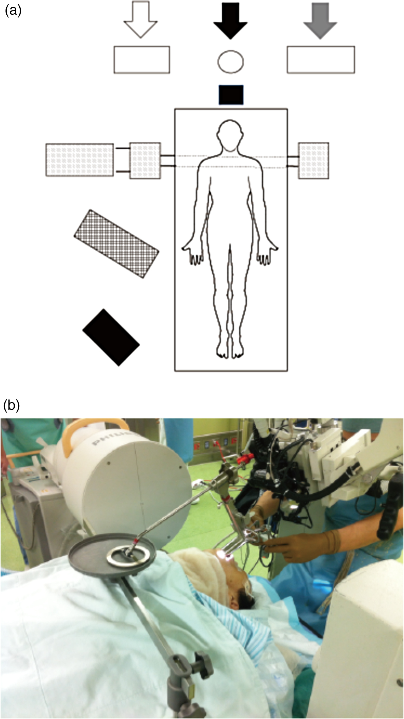
The endoscopic cricopharyngeal myotomy procedure was performed under general anaesthesia with oral intubation. A nasopharyngeal tube was inserted into the oesophagus to help identify the oesophageal introitus and guide placement of the diverticuloscope. We used peri-operative X-ray fluoroscopic radiography (Philips, Amsterdam, Netherlands) to check the position and direction of the diverticuloscope and forceps. The X-ray source was positioned on the patient's right side, and the C-arm was located under the patient's bed. The operating theatre layout is shown in Figure 1.
Schematic diagram of the operating theatre during endoscopic cricopharyngeal myotomy. The operating theatre consists of: a surgical bed, fluoroscopy machine (dotted area), C-arm (dotted area), microscope (grey arrow), carbon dioxide laser (white arrow), fluoroscopy monitor (crosshatched area) and anaesthesia apparatus (black area). The surgeon was positioned cephalad to the patient (black arrow). The fluoroscopy X-ray generator was positioned at the side of the patient's neck. The C-arm was positioned under the surgical bed.

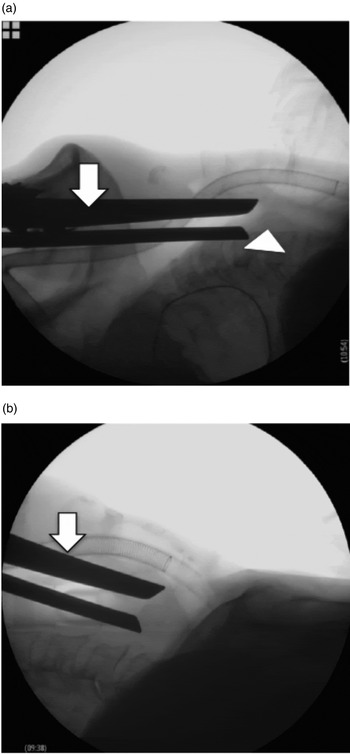
A Weerda diverticuloscope (Karl Storz, Tuttlingen, Germany) was placed in the post-cricoid area. When the larynx was lifted with the diverticuloscope, the cricopharyngeal muscle was stretched and became visible, bulging posteriorly like the cricopharyngeal bar. The surgeon could see the position and direction of the diverticuloscope and identified the cricopharyngeal bar. With fluoroscopic radiography, the surgeon could make sure that the surgical field was expanded at the correct position and that the cricopharyngeal muscle was sufficiently exposed, as shown in Figure 2. If the surgeon could not insert the Weerda diverticuloscope into the post-cricoid area through the oesophageal introitus, and could not see the cricopharyngeal muscle, the endoscopic cricopharyngeal myotomy was changed to transcervical cricopharyngeal myotomy.
Intra-operative fluoroscopy images during endoscopic cricopharyngeal myotomy. Lateral-view fluoroscopy showed the position of the Weerda diverticuloscope (arrow) and cricopharyngeal muscle (arrowhead) (a), and indicated whether the diverticuloscope (arrow) was inserted parallel to the oesophagus (b). In this case, it was not inserted parallel to the oesophagus.

A transmucosal vertical incision was made with a carbon dioxide (CO2) laser (Lumenis CO2 Laser 30C; Lumenis, Dreieich, Germany) set to a 6–10 W continuous wave mode and equipped with a micromanipulator connected to the microscope. The muscle was cut transmucosally in the midline up to the buccopharyngeal fascia. If necessary, the surgeon could simultaneously see fluoroscopic views. The cricopharyngeal muscle was cut as laterally as possible. Myotomy was performed without mucosal repair. The extent of the inferior incision was confirmed fluoroscopically using forceps.
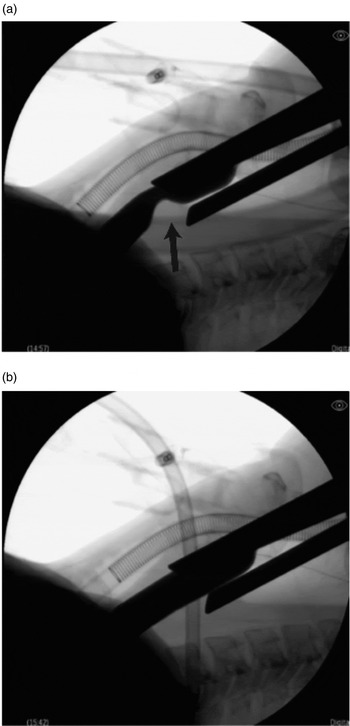
After the resection of cricopharyngeal muscle, we inserted a balloon dilatation catheter (Boston Scientific, Marlborough, Massachusetts, USA) through the mouth and performed the balloon dilation with 10 ml barium. If the resection was inadequate, fluorography showed the partial balloon filling defect, as shown in Figure 3, and we performed the additional cricopharyngeal resection. Finally, we confirmed no balloon filling defect.
Intra-operative fluoroscopy image with balloon dilatation catheter. Lateral-view fluoroscopy showed the X-ray shadow of the balloon dilatation catheter positioned at the cricopharyngeal muscle. Following inadequate resection of cricopharyngeal muscle, fluorography showed the partial balloon filling defect (arrow) (a). After the additional cricopharyngeal resection, fluoroscopy showed no balloon filling defect (b).

Feeding tubes (nasopharyngeal gastric tubes) were placed in all patients following the myotomy, under endoscopic sight in the operative field. Patients received prophylactic antibiotic treatment for 48 hours. On post-operative day 4, swallowing function and the possible presence of a pharyngeal fistula were examined using videofluoroscopy. If a pharyngeal fistula and aspiration were visible, oral intake was delayed and the patient was re-examined 3 days later. Patients were discharged when the nasogastric feeding tube could be removed and oral intake was sufficient.
Before surgery and two months after surgery, changes in swallowing function were evaluated with the penetration aspiration scale using videofluoroscopy. Before surgery and 12 months after surgery, changes in feeding condition were evaluated with the functional outcome swallowing scale.
Statistical analysis
All values are presented as means ± standard deviation. For statistical analyses, the Wilcoxon signed-rank test was used to evaluate the differences in: functional outcome swallowing scale scores before and 12 months after surgery, and penetration aspiration scale scores before and 2 months after surgery.
Results
Patients and surgical procedures
Between April 2013 and March 2017, 15 consecutive patients with idiopathic cricopharyngeal muscle dysfunction and post-radiation fibrosis were identified.
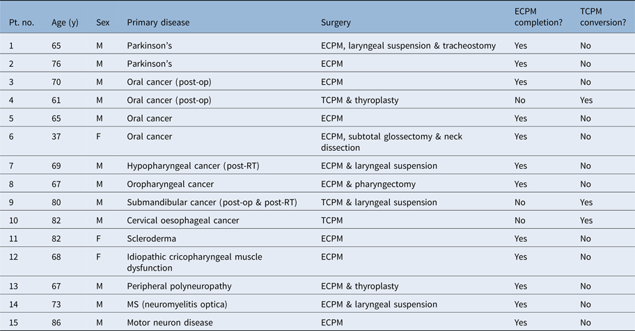
The average age of the patients was 66.7 ± 13.1 years; 12 patients were male and 3 were female. Regarding performance status, one patient had a score of 0, nine had a score of 1 and five had a score of 2. The primary diseases resulting in cricopharyngeal dysfunction are shown in Table 1. In this study, two patients had Parkinson's disease, two had post-operative oral cancer, two had oral cancer, one had hypopharyngeal cancer (post-radiotherapy), one had oropharyngeal cancer, one had submandibular cancer (post-operation and post-radiation), one had cervical oesophageal cancer, one had scleroderma, one had idiopathic cricopharyngeal muscle dysfunction, one had peripheral polyneuropathy, one had multiple sclerosis and one had motor neuron disease. Four patients had been diagnosed with cervical spondylosis.
Patient characteristics and feasibility of endoscopic cricopharyngeal myotomy

Pt. no. = patient number; y = years; ECPM = endoscopic cricopharyngeal myotomy; TCPM = transcervical cricopharyngeal myotomy; M = male; post-op = post-operation; F = female; RT = radiotherapy; MS = multiple sclerosis
Intra-operative fluoroscopic radiography was able to show the Weerda diverticuloscope positioned in the post-cricoid area near the cricopharyngeal muscle, and resulted in sufficient exposure to permit the muscle's resection. When the surgeon could not insert the Weerda diverticuloscope through the oesophageal introitus into the post-cricoid area, or could not see the cricopharyngeal muscle, the endoscopic cricopharyngeal myotomy was abandoned and changed to transcervical cricopharyngeal myotomy.
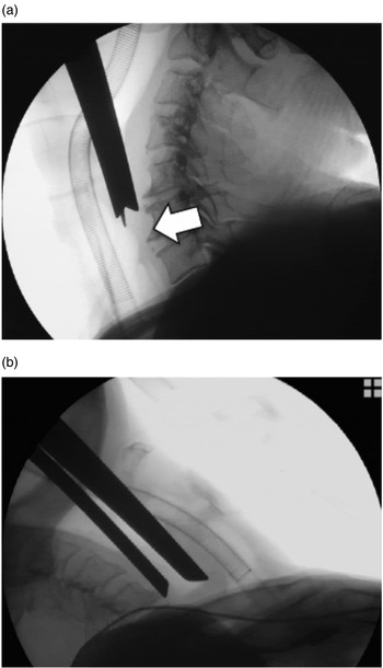
In three patients, intra-operative conversion to transcervical cricopharyngeal myotomy was required because of inadequate exposure of the affected area. Regarding the first converted patient, who required wide resection of the oral floor, we found it difficult to lift the patient's larynx with the diverticuloscope. The X-ray fluoroscope showed that the diverticuloscope was situated cephalad to the cricopharyngeal muscle. It was impossible to expose the cricopharyngeal bar (Figure 4a). With regard to the second converted patient, who underwent ion beam therapy for submandibular cancer (adenocystic carcinoma), we found it difficult to insert the diverticuloscope through the oesophageal entrance due to stenosis. The X-ray fluoroscope showed that the position of the diverticuloscope was located at the hypopharynx (Figure 4b). The third patient was converted for the same reason as the second patient.
Intra-operative fluoroscopy findings in a case of transcervical cricopharyngeal myotomy conversion. Lateral-view fluoroscopy showed that the Weerda diverticuloscope could not be inserted into the post-cricoid area through the oesophageal introitus (arrow) (a), and the cricopharyngeal muscle could not be seen (b).

In 12 patients, including 4 with cervical spondylosis, we could identify the cricopharyngeal muscle, confirm sufficient resection, and complete the endoscopic cricopharyngeal myotomy procedure with no peri-operative complications. In four patients with low pharyngeal pressure and laryngoptosis, we performed concurrent laryngeal suspension (three cases between the larynx and mandible, and one case between the hyoid bone and larynx). In 3 out of 12 patients who underwent endoscopic cricopharyngeal myotomy, we could see the cricopharyngeal bar on fluoroscopy, despite its not being visible with the microscope. Therefore, the endoscopic cricopharyngeal myotomy procedure could be completed in these patients.
Peri-operative outcomes
There were no intra-operative complications. Excluding laryngeal elevation, no tracheostomies were performed intra-operatively or post-operatively. The mean blood loss in all patients who underwent endoscopic cricopharyngeal myotomy was 5.7 ± 2.4 ml. No patients required a blood transfusion. The average operation time for the endoscopic cricopharyngeal myotomy procedure, including fluorography setup, was 91 ± 10 minutes.
No intra-operative or peri-operative fatalities occurred, nor was there any catastrophic haemorrhage or emergent airway compromise. None of the patients experienced serious adverse events that required hospitalisation or intervention, or that were considered life-threatening.
Swallowing outcomes
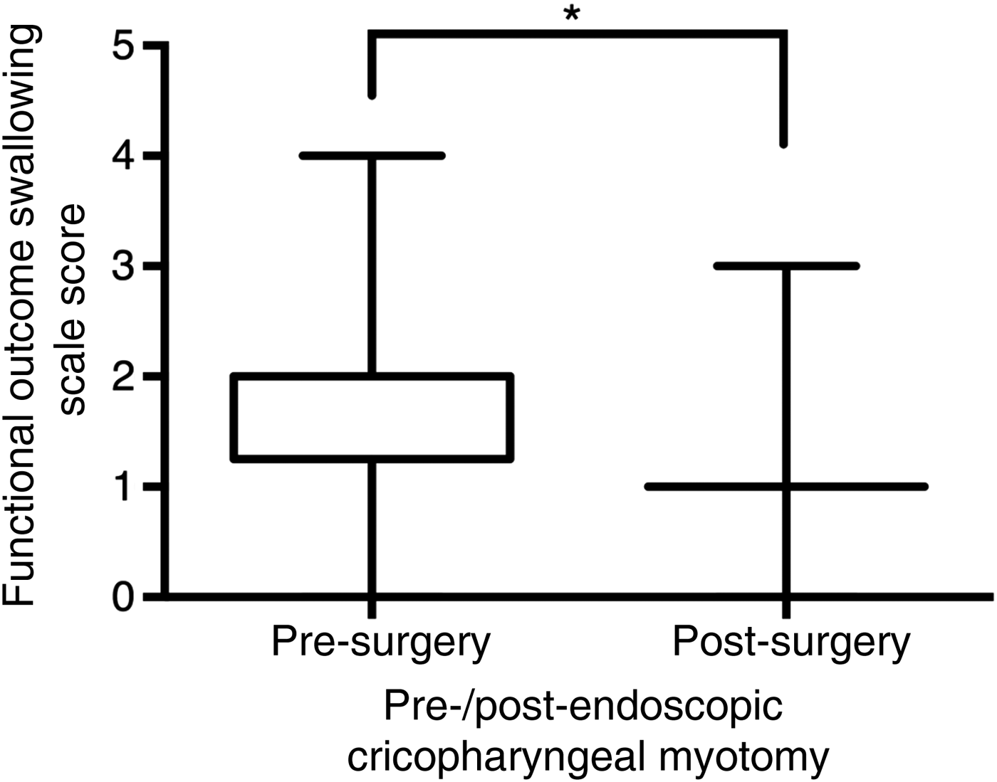
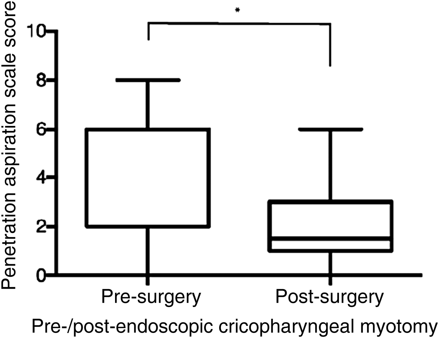
The mean functional outcome swallowing scale score of the endoscopic cricopharyngeal myotomy patients was 2.38 ± 0.27 pre-operatively and 1.30 ± 0.26 post-operatively, which reflected a significant difference (Figure 5). The mean penetration aspiration scale score of the endoscopic cricopharyngeal myotomy patients (excluding those who underwent endoscopic cricopharyngeal myotomy with pharyngectomy) improved from 4.7 ± 0.79 pre-operatively to 2.2 ± 0.66 post-operatively (Figure 6). At three years post-operatively, only one patient required a feeding tube for oral feeding difficulty.
Functional outcome swallowing scale scores were lower after endoscopic cricopharyngeal myotomy than before (*p = 0.027).

Penetration aspiration scale scores were lower after endoscopic cricopharyngeal myotomy than before (*p = 0.031).

Discussion
Endoscopic cricopharyngeal myotomy is a new and less invasive technique for transecting the cricopharyngeal muscle endoscopically and transmucosally, using a CO2 laser. It is performed in patients with defective relaxation of the upper oesophageal sphincter, usually due to cricopharyngeal dysfunction. Traditionally, cricopharyngeal myotomy has been performed via an external transcervical approach. Compared to transcervical cricopharyngeal myotomy, endoscopic cricopharyngeal myotomy has several advantages: a shorter operation time, less morbidity and scar-free surgery.Reference Takes, van den Hoogen and Marres4, Reference Dauer, Salassa, Luga and Kasperbauer5 However, there are several drawbacks to endoscopic cricopharyngeal myotomy. The surgeon may have concerns about feasibility, especially regarding endoluminal exposure of the cricopharyngeal muscle for the initiation of endoscopic cricopharyngeal myotomy. In addition, endoscopic cricopharyngeal myotomy requires sufficient muscle resection to obtain long-lasting relief from dysphagia. Thus, the surgeon must confirm the extent of resection. For this purpose, it is ideal to have a vertical view as well as a horizontal view. In order to address these issues, we introduced intra-operative X-ray fluoroscopy to endoscopic cricopharyngeal myotomy. This allowed us to confirm the position of the cricopharyngeal muscle and the Weerda diverticuloscope.
Three of 15 patients (20 per cent) required intra-operative conversion from endoscopic cricopharyngeal myotomy to transcervical cricopharyngeal myotomy because the cricopharyngeal bar could not be adequately exposed and the oesophageal introitus could not be opened due to stenosis. A systematic review reported a conversion rate of 7.7 per cent in patients undergoing internal pharyngoesophageal myotomy for cervical diverticulum.Reference Aiolfi, Scolari, Saino and Bonavina3 There was a small number of conversions to transcervical cricopharyngeal myotomy. However, in this study, none of the patients with a history of cervical spondylosis required conversion to transcervical cricopharyngeal myotomy.
Thus, the feasibility of endoscopic cricopharyngeal myotomy can be determined intra-operatively. Intra-operative fluoroscopy from the lateral side provides surgeons with information about the direction and position of the Weerda diverticuloscope, and the exposure of the surgical field. This informs feasibility decisions, especially in cases of difficult endoluminal exposure of the cricopharyngeal muscle, and, in turn, increases safety. The surgeon can see the cricopharyngeal bar if the Weerda diverticuloscope is positioned correctly. In this study, there were three patients in whom we could not see the cricopharyngeal bar using the microscope; however, we could confirm cricopharyngeal muscle by reference to the position of cricoid cartilage, and subsequently complete endoscopic cricopharyngeal myotomy.
Using the functional outcome swallowing scale, we demonstrated that swallowing outcomes improved following endoscopic cricopharyngeal myotomy. Two other studies showed similar outcomes, though their patient selection was different. Bergeron and Chhetri evaluated swallowing ability after endoscopic cricopharyngeal myotomy and found that it improved significantly, with functional outcome swallowing scale scores decreasing from 2.6 to 1.6.Reference Bergeron and Chhetri6 Hoesseini et al. reported that 85 per cent of patients experienced subjective relief of symptoms after endoscopic cricopharyngeal myotomy.Reference Hoesseini, Honings, Taus-Mohamedradja, van den Hoogen, Marres and van den Broek7
One problem with endoscopic cricopharyngeal myotomy is that there is no way to confirm the extent of resection intra-operatively because of the narrow working space and a view from only one direction. We used fluoroscopy and a balloon dilatation catheter with barium intra-operatively. They provided information on whether the cricopharyngeal muscle was sufficiently resected and the cricopharyngeal bar was completely relieved, via a lateral X-ray image. Thus, intra-operative fluoroscopy increased the effectiveness and success of endoscopic cricopharyngeal myotomy. Indeed, despite this study's small number of patients, the post-operative swallowing function outcomes were comparable to those of previous reports.
Only one patient required a feeding tube three years after endoscopic cricopharyngeal myotomy. His swallowing dysfunction was thought to be due to recurrent stenosis resulting from a history of chemoradiotherapy. Bachy et al. reported that 13 per cent of patients underwent a second endoscopic cricopharyngeal myotomy because of symptom recurrence.Reference Bachy, Matar, Remacle, Jamart and Lawson8 Hoesseini et al. showed that 26 per cent of patients underwent repeat laser surgery, with a mean interval of 16 months.Reference Hoesseini, Honings, Taus-Mohamedradja, van den Hoogen, Marres and van den Broek7
Another problem with endoscopic cricopharyngeal myotomy is recurrent swallowing dysfunction. This did not occur in this study, except in the one patient mentioned above who had previously received chemoradiotherapy. The lower rate of recurrent swallowing dysfunction compared to previous reports may have been a result of the contribution of intra-operative fluoroscopy in confirming the extent of resection.
In this study, there were no intra-operative or post-operative complications. In a series of 205 patients, Brouillette et al. reported that 2 patients (1 per cent) developed fistulas and 11 (5.3 per cent) had wound infections.Reference Brouillette, Martel, Chen and Duranceau9 Another study reported that two patients experienced intra-operative oesophageal perforation and one patient developed a fistula.Reference Lindgren and Ekberg10 As our study included a small number of cases, it was difficult to draw firm conclusions from the results; nevertheless, the findings indicate that intra-operative fluoroscopy might help the surgeon confirm the depth of the buccopharyngeal fascia, which could reduce the complication rate.
• Fluoroscopic radiography was utilised during endoscopic cricopharyngeal myotomy to improve feasibility issues
• Peri-operative fluoroscopic radiography can show the diverticuloscope's position and extent of incision
• The additional use of a balloon catheter was helpful in checking whether resection was sufficient
• Intra-operative fluoroscopic radiography during endoscopic cricopharyngeal myotomy may improve swallowing function and reduce peri-operative complications
It should be noted that intra-operative fluoroscopy has several disadvantages. First, it requires additional setup time. The surgeons have to adjust the position of the surgical bed and the C-arm of the fluoroscopy unit, in co-operation with the anaesthesiologists and operating theatre nurses. However, with practice and repetition, setup times will become shorter. Second, the surgical staff and patient might be exposed to radiation; the surgical staff will require radiation protection suits during the surgery.
One limitation of this study relates to its single-arm design, namely the lack of patients who did not undergo fluoroscopy. Further studies will be needed to conclusively demonstrate the effectiveness of intra-operative fluorography.
Intra-operative fluoroscopy might be helpful for confirming the extent of resection, increasing surgical effectiveness, and reducing morbidity and complications in patients undergoing endoscopic cricopharyngeal myotomy.
Competing interests
None declared