Loeys–Dietz syndrome is an autosomal dominant connective tissue disorder known to cause life-threatening cardiovascular and intracerebral incidents in paediatric patients. Reference Patel, Alejo and Crawford1 This genetic disorder is characterised by heterozygous mutations in transforming growth factor β receptors 1 and 2, which manifests as an overproduction of collagen, loss of elastin content, and disruption of elastic fibres in the vasculature tree. Reference Cleuziou, Eichinger, Schreiber and Lange2 In addition to the classical triad of generalised arterial tortuosity, hypertelorism, and a bifid uvula or cleft palate, patients are also predisposed to aneurysm formation and arterial dissection and rupture. Reference Patel, Alejo and Crawford1,Reference Liu, Fraser, Zhou, Cameron, Vricella and Hibino3 Compared to other CTDs, LDS presents in a more aggressive manner and is associated with earlier morbidity and mortality due to the earlier occurrence of catastrophic cardiovascular events Reference Liu, Fraser, Zhou, Cameron, Vricella and Hibino3,Reference Everitt, Pinto, Hawkins, Mitchell, Kouretas and Yetman4 Aortic dissections and aneurysms have been reported in patients as young as 3–6 months of age, with surgical intervention often required. Reference Williams, Loeys and Nwakanma5–Reference Wisniewski, Singer, Kehl, Nawrocki, Januszewska and Malec7
Case
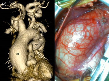
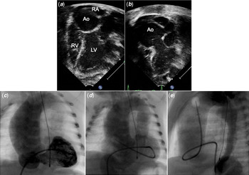
We report a 36-day-old, 3.3 kg term male diagnosed with LDS through immediate postnatal genetic testing who presented with a large aortic root aneurysm (31 mm, z score +10) that was surgically reconstructed 5 days later. CT of the chest (Fig 1a) revealed severe dilation of the aortic root, ascending aorta, and epiaortic vessels. Echocardiogram showed a massively dilated aortic root causing leftward displacement of the heart with significant compression of the right atrium and right ventricle, leading to a low end-diastolic volume of the right ventricle and marked right-to-left atrial shunting across a patent foramen ovale (Fig 2a and b). Prior to being transferred to our care, the patient had suffered multiple systemic desaturations and was discharged to hospice care after being deemed inoperable. To investigate the cause of the patient’s unstable haemodynamics, an angiogram was performed and revealed that the ascending aorta and transverse aortic arch measured 31 mm and 21 mm in diameter, respectively, (z score +17.7, +8.4, respectively). Additional findings include a left main coronary artery aneurysm, type B aortic dissection flap, and a tortuous abdominal aorta (Fig 2c, d and e).
(a) CT 3D reconstruction of the great arteries performed shortly after birth showing severe dilation of the aortic root and ascending aorta, tortuous head and neck vessels, as well as dilated main PA and PDA. (b) Intraoperative image showing severe dilation of aortic root engulfing the entire surgical field. Ao=Aorta; PA=Pulmonary Artery; PDA=Patent Ductus Arteriosus.

(a) Apical four-chamber view. The aorta significantly compresses the RA. Note that the RV is under filled. (b) Apical three-chamber view. The aortic root is massively dilated. Ao=Aorta; LV=Left Ventricle; RA=Right Atrium; RV=Right Ventricle. (c) AP projection of LV angiogram showing massively dilated aortic root and ascending aorta in comparison to LV size. (d) AP projection of aortic root angiogram showing the dilated aortic root and ascending aorta, left coronary artery aneurysm, and tortuous abdominal aorta. (e) Lateral projection of an ascending aorta angiogram showing a large ductal ampulla, type B aortic dissection in the descending aorta, and tortuous abdominal aorta. AP=Anteroposterior; LV=Left Ventricle.

The surgical plan included excision of the ascending aorta and hemiarch with remodelling of the sinotubular junction and non-coronary sinus (asymmetrically dilated). Upon opening the sternum, the aortic root and ascending aorta were found to encompass the entire surgical field (Fig 1b). The aortic valve was bicuspid with typical right–left coronary cusp fusion, with a healed localised dissection in the descending aorta. A 14 mm aortic homograft was used to replace the hemiarch and ascending aorta. Distal arch reconstruction showed an intramural haematoma had closed the false lumen of the type B dissection, and small clots were aspirated. The proximal end of the graft was then anastomosed to the aortic root just above the STJ. Exploration into the right atrium revealed a small PFO, which was left open due to a relatively small right ventricle.
Stable haemodynamics and full saturations were obtained in recovery. The patient was discharged home on room air, losartan, furosemide, and aspirin on post-operative day 10 in stable condition. Discharge echocardiogram showed a 14 mm aortic root, normal biventricular systolic function, and a predominately right-to-left shunt across a small PFO. Post-operative observation revealed an excellent contour of the hemiarch reconstruction without obstruction.
Comment
Loeys–Dietz syndrome is a connective tissue disorder known to cause aggressive aortopathy and is associated with high incidences of aortic dissection and aneurysmal rupture in paediatric patients. Reference Liu, Fraser, Zhou, Cameron, Vricella and Hibino3 LDS differs from similar CTDs by its earlier onset of catastrophic cardiovascular events that often occur at smaller aortic diameters. Although patients with LDS typically present with cardiovascular incidents starting in their teenage years, aneurysm rupture and dissection have been reported in infants as young as 3–6 months with aortic diameters less than 4.0 cm. Reference Williams, Loeys and Nwakanma5–Reference Wisniewski, Singer, Kehl, Nawrocki, Januszewska and Malec7
Due to the disease’s malignant phenotype and high mortality, prophylactic surgical intervention is recommended at smaller aortic diameters compared to patients with other CTDs. Reference Cleuziou, Eichinger, Schreiber and Lange2,Reference Everitt, Pinto, Hawkins, Mitchell, Kouretas and Yetman4,Reference Williams, Loeys and Nwakanma5 Studies have shown favourable outcomes in LDS patients undergoing surgical intervention in childhood with low morbidity and mortality. Reference Everitt, Pinto, Hawkins, Mitchell, Kouretas and Yetman4 However, LDS patients see higher rates of reoperation Reference Everitt, Pinto, Hawkins, Mitchell, Kouretas and Yetman4 due to the occurrence of both anastomotic or root pseudoaneurysms at the prior site of intervention, and de novo aneurysms at different sites of the arterial vasculature, highlighting the aggressive nature of this disease. These observations underscore the importance of keen, longitudinal surveillance to ensure timely surgical intervention. Reference Williams, Loeys and Nwakanma5
While absolute structural diameters may guide surgical decision-making, treatment strategies become complicated when patients present at exceptionally young ages. It is exceedingly rare for LDS to present for cardiovascular intervention in early infancy, with only a handful of cases reported in the literature. Reference Jacques, Grosse-Wortmann, Hickey, Chitayat, Van Arsdell and Bradley6,Reference Wisniewski, Singer, Kehl, Nawrocki, Januszewska and Malec7 We believe this to be the first case of an infant with LDS requiring aortic aneurysm repair in the neonatal period. In this case, the presenting pathophysiology was particularly impressive, resembling that of tricuspid atresia with restrictive pulmonary blood flow. This case confirms that these patients should not be deemed inoperable solely based on the diagnosis of LDS with a vascular presentation in the neonatal period.
Acknowledgements
None.
Financial support
This research received no specific grant from any funding agency, commercial, or not-for-profit sectors.
Conflict of interest
None.
Ethical standards
This report does not include any human or animal experimentation.