Implications
The study of the morphology and function of the mammary gland in production animals has been traditionally restricted to samples obtained from necropsy procedures. Current research has focused on obtaining in vivo samples; however, the potential adverse effects on milk production and udder health must be reported and further studied.
Introduction
The study of the morphology and function of the mammary gland in dairy animals has been traditionally restricted to samples obtained from necropsy procedures. Even though non-invasive methodologies such as ultrasonography and endoscopy have been developed, in recent years, research has focused on obtaining in vivo mammary tissue samples for histological and biochemical evaluation. This has been done despite difficulties in the procedure and possible adverse effects on milk production deriving from elasticity and vascularization of lactating mammary tissue and the possibility of hemorrhage and mastitis (Farr et al., Reference Farr, Stelwagen, Cate, Molenaar, McFadden and Davis1996).
Mammary gland biopsy is a technique used in both research and clinical diagnosis. On large animals, two biopsy methods are commonly reported. The first involves surgically obtaining large tissue fragments (0.75–1 g), resulting in complications such as external hemorrhage and formation of fistulas (Farr et al., Reference Farr, Stelwagen, Cate, Molenaar, McFadden and Davis1996). The second technique uses needles and biopsy pistols (automatic needle jab system), being capable of extracting small fragments (20–25 mg) from a small incision, minimizing hemorrhage (Ollier et al., Reference Ollier, Leroux, De la Foye, Bernard, Rouel and Chilliard2009; Soberón et al., Reference Soberón, Lukas, Van Amburgh, Capuco, Galton and Overton2010). VanKlompenberg et al. (Reference VanKlompenberg, McMicking and Hovey2012) described a biopsy technique that extracts tissue fragments of ~100 mg via a vacuum system followed by a saline lavage of the biopsied site, with or without vasoconstrictors. This method could potentially reduce complications after biopsy.
Greater milking frequency per day increases milk production in several species. It has also been associated with a decrease in somatic cell count (SCC) (Smith et al., Reference Smith, Ely, Graves and Gilson2002; Dahl et al., Reference Dahl, Wallace, Shanks and Lueking2004; Wright et al., Reference Wright, Wall and McFadden2013), an indicator of mammary gland health. However, other authors did not observe such effect (Wall and McFadden, Reference Wall, Bond and McFadden2007; Shields et al., Reference Shields, Rezamand, Sevier, Seo, Price and McGuire2011). No previous study has evaluated the effect of milking frequency on the udder health after biopsy. The objectives of the present study were (a) to describe and evaluate the adaptation of the use of a biopsy needle to assess bovine mammary tissue, (b) to determine the main occurrences related to milk yield and udder health after the procedure, and (c) to evaluate the effect of increased milking frequency on such occurrences.
Material and methods
Animals, management and sampling
The Federal University of Minas Gerais ethical committee (CETEA/UFMG n. 58/2011) approved all the experimental procedures. The study was conducted at the Experimental Farm of the Agricultural Research Company of Minas Gerais (EPAMIG), located in Felixlândia, Minas Gerais, Brazil, between June and December of 2013. Biopsies were performed at early lactation in 16 F1 Holstein×Zebu multiparous cows (third and greater lactations), homogenously distributed into two groups according to parity, mean milk production and SCC of the previous lactation, genetic composition, and calving date. The groups were: (1) 2×: cows milked twice/day (n=8) and (2) 4×: cows milked four times/day (n=8), from two to 21 days postpartum. After day 21 of lactation, both groups were milked twice/day until 28 days in milk (DIM). Milkings started on day 2 of lactation. Milking times were 0600 and 1400 h for the 2× group. Cows in the 4× group were also milked at 0930 and 1530 h. All animals were fed the same diet ad libitum and kept in the same location during the experimental period. Milk yield was measured with Milk Meter-Mark V collectors (α-Laval Agri; Jaguariúna-SP, Brazil) on days where the milk from the biopsied quarter was normal at visual inspection. Milk samples (pooled from successive a.m. and p.m. milkings) were collected 1 day before biopsy and daily after biopsy, starting at the day when no visible contamination by blood was seen on the milk. Percentage of fat, protein and lactose, and SCC were measured in each milk sample using spectroscopy and flow cytometry (Bentley Combi System 2300 and Bentley Instruments Incorporated, Chaska, USA, respectively) at the Laboratory of Milk Analysis and Quality at the Federal University of Minas Gerais Veterinary School.
Feed intake behavior was assessed by visual observation immediately after the biopsy. BW and body condition score (BCS) were assessed 1 day before and 7 days after each biopsy (respectively at days 1, 6, 13, 20 and 27 postpartum, and at 7, 14, 21, 28 and 35 days postpartum) by a single evaluator. The BCS was scored on a 1 to 5 point scale based on inspection of the pelvis and tail head, ribs, and back according to Edmonson et al. (Reference Edmonson, Lean, Weaver, Farver and Webster1989).
Udder health was assessed daily until 60 DIM by visual examination of the milk for blood, milk clots, and blood clots, by California Mastitis Test (CMT), and by monitoring clinical signs of mastitis (redness, increased udder temperature, pain on palpation, and edema). The CMT was scored according to McDougall et al. (Reference McDougall, Supré, De Vliegher, Haesebrouck, Hussein, Clausen and Prosser2010) as follows: Score 0 or negative: no reaction between the reagent and milk; trace: suspicious reaction; score 1 or +: weak positive reaction; score 2 or ++: positive reaction; and score 3 or++ +: strong positive reaction.
Tissue sampling and analysis
Mammary biopsies were performed in the medial region of the mammary quarter in eight cows from each group on days 2, 7, 14, 21, and 28 postpartum. Biopsies were performed immediately after the 0600 h milking for 2× cows, and after the 0930 h milking for 4× cows. For each animal, one fragment of secretory tissue was collected from only one mammary quarter at a time, alternating between the four mammary quarters per collection day. Only one mammary quarter was biopsied twice. First, second, third and fourth biopsy samples were collected from, respectively, right rear quarter, left rear quarter, right front quarter, and left front quarter. The right rear quarter was sampled again at the fifth biopsy. A total of 80 biopsy samples were collected.
Biocore II needles comprising a 12-gauge cannula and a 12-cm perforating monoface mandrel were used (MMdiagnostika; São Paulo-SP, Brazil). After milking, the cows were restrained in a squeeze chute and sedated with 1.5 ml of 1% acepromazine administered intramuscularly (0.03 mg/kg, Acepran; VETNIL Ind. and Com. of Veterinary Products Ltd., Louveira-SP, Brazil). Cows were not fasted before the biopsy and remained standing throughout the procedure. To allow adequate visibility of the biopsy site and increase safety, the rear limb ipsilateral to the biopsied quarter was suspended and restrained on the chute. The area to be biopsied (20 cm2 without visible subcutaneous blood vessels) was marked, trichotomized, and cleansed aseptically using 10% povidone-iodine and 70% alcohol. Skin and mammary tissue were locally anesthetized with 2% lidocaine hydrochloride without vasoconstrictors (20 mg/ml, Xylestesin; Cristália–Pharmaceutical Products Ltd., Itapira-SP, Brazil) administered subcutaneously (1 ml) and into the superficial intramammary tissue (2.5 ml). A small skin incision was made with a 40×16 needle to introduce the biopsy needle. The biopsy needle and mandrel were introduced through the skin incision ~1.5 cm into the mammary parenchyma. Subsequently, the mandrel was manually positioned toward the parenchyma, rotated 360°, and then fixed; next, the cannula was pushed, completely covering the mandrel, and the needle was removed from the mammary gland.
If mammary tissue was not retrieved, the needle was reintroduced, and the procedure repeated until an appropriate amount of tissue was obtained. After the procedure, a silver sulfadiazine healing spray (Bactrovet Prata AM; Konig, Avellaneda-BS, Argentina) was applied onto the incision site. Neither intramammary drugs nor intravenous antibiotics were administered in order to evaluate mastitis incidence without the use of preventive measures. If mastitis was diagnosed, intramammary gentamicin sulfate 150 mg (Mastifin; Ourofino Group, Cravinhos-SP, Brazil) was administered for 3 consecutive days.
Histological procedures
Tissue samples were fixed in formalin (10% neutral and buffered) for 8 h and transferred to 70% alcohol. Samples were then serially dehydrated in alcohol (80% and 90% for 1 h, and 100% for 2 h), clarified in xylene (5 min), infiltrated with liquid paraffin (56–58°C for 5 min), and embedded in paraffin blocks. The blocks were cut into 4-μm-thick slices (RM 2255 Leica Microtome), mounted on slides, stained with hematoxylin-eosin (HE) and covered with cover slips using mounting medium (Etellan).
The slides were observed under a microscope. A 4× objective lens was used to randomly select the fields to be photographed. Once a field was selected, a 40× objective lens was used to the capture the highest number of images with a Q Color S Olympus digital camera coupled to an Olympus BX-43 Microscope Image Pro Plus software version 7.0 was used for image capturing. Images were analyzed for histological evaluation of mammary tissue.
Statistical analyses
A χ 2 test was used to analyze the association between biopsy-related occurrences and milking frequency. Comparison between duration of occurrences and milking frequency was analyzed with a random design. BW and SCC were standardized with a logarithmic function and the duration in days of presence of blood clots in the milk with a root function. BW, milk composition and SCC were evaluated by comparing means before and after biopsy for dependent samples using paired t test (P<0.05). The BCS was analyzed using the Wilcoxon test, with a 95% confidence interval. Descriptive analyses were used for the morphological results, CMT, and animal health evaluation. Regression analyses were conducted to estimate individual milk production for the days when measuring was not done due to the presence of blood in the milk. Comparison of estimated milk production throughout the days was analyzed through random units, a unit being the animal. The Student–Newman–Keuls test was used to compare mean differences in milk production. Analyses were conducted with SAS 9.1 (SAS Institute Inc., Cary, NC, USA).
Results and discussion
Evaluation of biopsy samples
Biopsies were easily obtained in various mammary sites using a rapid procedure lasting no more than 20 min from animal restraint to the end of the procedure. Using the biopsy needle, a core of mammary tissue of ~6×3 mm was obtained at each biopsy. The biopsy technique does not allow the removal of tissue fragments from precise locations, therefore it is unknown which mammary gland region is being sampled. In total, 12 samples showed no secretory mammary tissue. Two were identified as mammary gland cistern tissue, and 10 were fibrous tissue only, likely from the gland cistern.
Histological evaluation revealed that samples were obtained with minimal tissue damage. The fragments produced an average of 10 fields at 40× magnification. Microscopic structures were clearly visible on histology, and the samples contained predominantly healthy alveolar secretory tissue. Compared to the method described by Farr et al. (Reference Farr, Stelwagen, Cate, Molenaar, McFadden and Davis1996) and used in other studies (Hale et al., Reference Hale, Capuco and Erdman2003; Miller et al., Reference Miller, Delbecchi, Petitclerc, Wagner, Talbot and Lacasse2006; Annen et al., Reference Annen, Fitzgerald, Gentry, McGuire, Capuco, Baumgard and Collier2007), one disadvantage of the present biopsy method is that only small tissue fragments can be collected. However, multiple samples can be collected from a single incision by directing the biopsy needle toward a different location.
There were no evident changes in mammary morphology as a result of milking 2× or 4× per day at any of the evaluated time points. Biopsies (day 21 and 23 postpartum) of Holstein cows milked two or four times per day for the first 21 DIM yielded similar results (Wall et al., Reference Wall and McFadden2013).
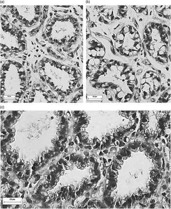
On the 2nd day of lactation, it was observed for both groups that alveoli were comprised of heterogeneous cell populations, most of these having the nuclei located near the basal region, occupying much of the intracellular area, also with presence of small quantities of small cytoplasmic vacuoles (Figure 1a). According to Akers et al. (Reference Akers, Capuco and Keys1977, Reference Akers, Heald, Bibb and McGilliard2006) and Annen et al. (Reference Annen, Fitzgerald, Gentry, McGuire, Capuco, Baumgard and Collier2007), these cells can be classified as immature, poorly differentiated or resting cells, respectively. Some alveoli presenting cells with large vacuoles in the cytoplasm occupying most of the intracellular space, characteristic of fat globules, which result in nucleus of irregular shape and polarity yet to be established (Figure 1b) were also observed. These alveoli were observed at 20 DIM in cows milked continuously during the dry period and defined by Annen et al. (Reference Annen, Fitzgerald, Gentry, McGuire, Capuco, Baumgard and Collier2007) as engorged alveoli. Holst et al. (Reference Holst, Hurley and Nelson1987) observed similar characteristics in mammary alveoli during the dry period. Thus, the engorged alveoli observed at 2 DIM are likely to be entering the differentiation stage. These morphologic characteristics have been previously described in Holstein cows at day 2 of lactation (Akers et al., Reference Akers, Capuco and Keys1977).
Micrographs of (a) alveoli with poorly differentiated cells at day 2 after calving, (b) engorged alveoli at day 2 after calving, and (c) alveoli with poorly differentiated cells at day 7 after calving, from mammary tissue samples obtained from a single cow milked twice/day until 28 days in milk (DIM).

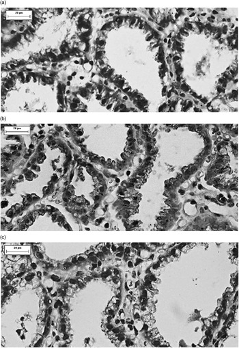
From 7 DIM, the nucleus of alveolar cells began to show basal position and the proportion of the cell area occupied by the nucleus diminished over DIM (Figures 1c and 2), resulting in a more evident cytoplasm. According to Akers et al. (Reference Akers, Capuco and Keys1977) the smaller nuclear : cytoplasm ratio was attributed to an increase in the cytoplasmic volume. Annen et al. (Reference Annen, Fitzgerald, Gentry, McGuire, Capuco, Baumgard and Collier2007) reported the presence of resting alveoli in day 1 and day 7 postpartum, suggesting that not all the mammary epithelium cells are fully differentiated postpartum. Different from that observed by Annen et al. (Reference Annen, Fitzgerald, Gentry, McGuire, Capuco, Baumgard and Collier2007), in the present study these cells with morphology suggestive of repose were not visualized at 7 DIM, suggesting a more rapid cell differentiation in animals used in this experiment when compared to the Holstein cows.
Micrographs of alveoli and cells of mammary tissue sample obtained at (a) 14, (b) 21, and (c) 28 days after calving from cows milked two or four times/day until 21 days in milk (DIM) and twice/day from 22 to 28 DIM.

The proportion of interalveolar connective tissue appeared to decrease throughout the period studied while the alveolar luminal area increased, although these changes were not quantitated (Figure 2). The increased interalveolar space was observed mainly at 2 DIM. Interalveolar space was filled with loose connective tissue, but a large amount of cells was also found in these areas. These cells consisted mainly of fibroblasts, mononuclear cells (lymphocytes, monocytes and macrophages) and plasmocytes. Mononuclear cells and plasmocytes were also observed migrating into the alveolar lumen. The greater cellularity in mammary stroma and lumen in early lactation can result from the physiological process of colostrogenesis is which begins at the end of the dry period.
Udder health
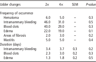
After biopsy, cows were released form the chute and moved to their pens. All animals began feeding immediately, indicating a fast post-biopsy recovery. There was no difference between number of milkings per day (2× or 4×) in the frequency of hematomas, intramammary bleeding, blood clot, edema, fibrosis, mastitis, intramammary hemorrhage duration and presence of blood clots in the milk and edema (P>0.05; Table 1). In addition, post-biopsy SCC was not different between 2× and 4× groups once blood contamination was no longer visible in the milk (2×=305.8×1000 cells/ml and 4×=445.2×1000 cells/ml; P>0.05). Although SCC was not different between 2× and 4× groups, observed counts are indicative of inflammation following biopsy, once the SCC threshold for mammary gland inflammation is above 250×1000 cells/ml (Smith, Reference Smith1996). Furthermore, it was expected that the 4× group would have faster recovery of biopsied gland health (shorter duration of high SCC and of observed alterations), due to more frequent removal of milk from the gland, with consequent removal of blood and clot. However, this effect was not observed, demonstrating that recovery of the mammary gland after biopsy may be related to intrinsic factors of the animal. Similarly, Leininger et al. (Reference Leininger, Roberson, Elvinger, Ward and Akers2003) did not observe an effect of increased frequency of milk removal in SCC of cows with experimentally induced Escherichia coli mastitis.
Effect of milking two (2× or four (4×) times per day during the first 21 days of lactation, followed by twice a day milking until 28 days of lactation on frequency of occurrence of hematoma, intramammary bleeding, blood clots, edema, areas of fibrosis, mastitis and duration in days of intramammary bleeding, blood clots and edema of F1 Holstein×Zebu cow after the biopsy procedure

Biopsy sites healed without any infections. Subcutaneous hematomas were observed in 11 of the 80 biopsies (14%) and were reabsorbed within 1 day. According to Farr et al. (Reference Farr, Stelwagen, Cate, Molenaar, McFadden and Davis1996), surgical biopsy causes intense external hemorrhage and subcutaneous hematomas persisting up to six days. This outcome was not observed in any of the present subjects, demonstrating an advantage of the biopsy needle procedure compared to the surgical methodology.
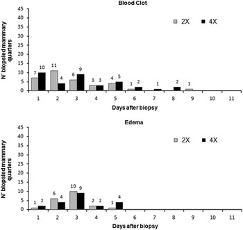
Most of the biopsied mammary quarters (96%) showed intramammary bleeding, and only three quarters showed no visible blood contamination in the milk. Out of 80 biopsies, 25 quarters showed visible blood in the milk for 2 days, 27 for 4 days, 19 for 6 days. Only six quarters had blood contamination persisting over 6 days (Figure 3). VanKlompenberg et al. (Reference VanKlompenberg, McMicking and Hovey2012) performed the biopsy technique using a vacuum system and observed blood residue or blood clot in the milk for no more than 7 days, 1 day longer than most of the biopsies in the present study.
Effect of milking two (2×) or four (4×) times per day for the first 21 days of lactation, followed by milking two times per day until 28 days of lactation on frequency of milk with blood, milk with blood clots, or edema by number of biopsied quarters and days after biopsy.

In addition to blood, blood clots were also observed in the milk for 69 biopsies (87%) persisting for no more than 6 days; only five quarters had blood clots persisting for more than 6 days (Figure 2). Blood clots were manually removed during and after milking. According to Farr et al. (Reference Farr, Stelwagen, Cate, Molenaar, McFadden and Davis1996), knowledge of mammary gland vascularization is important to choose the best biopsy site in order to avoid arteries and veins of large caliber, thus reducing the incidence of internal hemorrhage. After the biopsy, 43 mammary quarters (54%) had edema that resolved in 3 days; however, the edema persisted for more than 3 days in quarters positive for mastitis. No difference was observed between front and rear quarters as to duration of edema and milk contamination with blood or blood clots. Small visible or palpable fibrotic lesions were observed at the biopsy site in five biopsied quarters (6%), which resolved spontaneously between 15 to 20 days. Marx and Caruolo (Reference Marx and Caruolo1963), who performed surgical biopsies, reported similar findings.
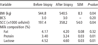
Although BCS was not affected by biopsy (P>0.05), BW at the day previous to biopsy (PAB=544.8 kg) and 1 week after biopsy (PPB=540.5 kg) were different (P<0.05; Table 2). This loss of body mass should be interpreted cautiously as it likely reflects the body reserve mobilization that occurs during early lactation due to negative energy balance, rather than the biopsy procedure.
Effect of biopsy procedure on BW, milk composition, milk somatic cell count (SCC), and body condition score (BCS) of F1 Holstein×Zebu milking cows before and after the biopsy procedure

Before biopsy, all mammary quarters tested negative on CMT (score 0). One day after the biopsy, 75% of the glands (n=60) had a positive CMT reaction (trace: 0% [0]; score 1: 40% [24]; score 2: 43% [26]; and score 3: 17% [10]) that persisted an average of 5.2 days and a maximum of 12 days in cases diagnosed with clinical mastitis. At 60 days of lactation, only three biopsied quarters showed CMT alterations, suggesting that the biopsy procedure may cause a transitory SCC increase. The SCC of post-biopsy milk (SCC=358.2×1000 cells/ml) was higher than that in milk collected prior to the procedure (SCC=197.4×1000 cells/ml; P<0.05; Table 2). The SCC decreased to values smaller than pre-biopsy values after no more than 11 days, which is 5 days after the end of visible abnormalities in the milk, except in the mammary quarters positive for clinical mastitis.
Among the 80 biopsies, 10 mammary quarters (12%) were diagnosed and treated for clinical mastitis, nine recovered quickly, and one quarter was lost due to mastitis-related complications. These results indicate that the biopsy procedure may cause an inflammatory reaction due to physical trauma caused by the needle. Most studies using biopsy needle or gun, or surgical methods report an absent (Annen et al., Reference Annen, Fitzgerald, Gentry, McGuire, Capuco, Baumgard and Collier2007) or low mastitis incidence (Farr et al., Reference Farr, Stelwagen, Cate, Molenaar, McFadden and Davis1996; Sorensen et al., Reference Sorensen, Nørgaard, Theil, Vestergaard and Sejrsen2006). However, this may be due to the use of prophylactic antibiotics, either systemic or intramammary, pre and post-biopsy, or other factors such as installations and milking management since mastitis incidence is associated with multiple factors. Marx and Caruolo (Reference Marx and Caruolo1963) reported mastitis incidence of 14% after needle biopsy without concurrent prophylactic antibiotics, which is similar to the present finding. Thus, the use of prophylactic antibiotics could minimize the risk of mammary infection post-biopsy.
Production and milk composition
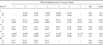
In the biopsies performed on days 2 postpartum, milk yield was reduced only on the day following the biopsy and rapidly went back to pre-biopsy values on the day after biopsy as a result of milking 2× or 4× per day. For biopsies performed on day 7 of lactation, there was no effect on milk yield of cows milked 2× per day. Cows milked 4× per day, however, had greater milk production at day 5 post-biopsy performed at 7 DIM (P<0.05). Regarding biopsies done on days 14 and 28 of lactation, no difference was found on the estimation of milk production on the days following the biopsy, independent of milking frequency (P>0.05; Table 3). Biopsies performed at 21 DIM were associated with reduced milk yield at day 1 v. day 4 post-biopsy on cows milked 2× per day. Cows milked 4× per day had greater milk yield at the day after biopsy performed at 21 DIM, which was coincidental with peak lactation for these cows. Thus, there was a decline in milk yield between day 1 and day 5 post-biopsy. This decline in milk yield was also coincidental with the switch from milking 4× to 2× per day, which might have contributed to the reduction in post-biopsy milk yield. These results demonstrate that alterations in milk production after biopsy are reversible and have limited duration.
Effect of biopsy procedure on days 2, 7, 14, 21 and 28 of lactation on estimated mean milk production (kg/day) of F1 Holstein×Zebu cows milked (2×) or (4×) times per day one day before biopsy (−1), on the day of biopsy (0) and 5 consecutive days after biopsy (1, 2, 3, 4, 5)

Days PP=days postpartum.
a,cValues within a row with different superscripts differ significantly at P<0.05.
According to Farr et al. (Reference Farr, Stelwagen, Cate, Molenaar, McFadden and Davis1996), milk production in the biopsied gland during the intermediary lactation phase is significantly reduced when compared with the pre-biopsy period, recovering within 6.5 days post-biopsy. Annen et al. (Reference Annen, Fitzgerald, Gentry, McGuire, Capuco, Baumgard and Collier2007) showed that daily milk production in biopsied animals was not influenced by the biopsy procedure. However, it is important to state that the progressive increase in milk production until the peak of lactation may mask potentially negative effects on production, which may explain the minimal or absent effects observed in the present study and by Annen et al. (Reference Annen, Fitzgerald, Gentry, McGuire, Capuco, Baumgard and Collier2007).
Percentages of fat, protein and lactose in milk were not affected by milking frequency during the evaluated period (2 to 30 DIM) (P>0.05). Pre and post-biopsy milk samples did not show any difference in fat percentage (P>0.05), but pre-biopsy samples had greater percentage of protein and smaller percentage of lactose compared to post-biopsy (P<0.05; Table 2). According to Tsioulpas et al. (Reference Tsioulpas, Grandison and Lewis2007), the physiological composition of milk is characterized by a high percentage of protein on the first day of lactation that gradually declines over 30 days, whereas the percentage of lactose, which is initially low, increases over the next 60 days of lactation. Thus, because the biopsies were performed in the first 28 days of lactation, the observed changes in milk composition may be related to physiological changes in milk composition occurring during this period instead of being a consequence of the biopsy.
Elevated SCC may result in alterations in milk composition, with a low percentage of solids because of reduced synthetic activity of the mammary tissue (Harmon, Reference Harmon1994). However, despite the post-biopsy increase in SCC (Table 2), this could only explain the post-biopsy reduction in protein percentage, being incompatible with post-biopsy increase in lactose percentage.
Conclusions
The method for mammary gland biopsy using only a biopsy needle can be conducted and repeated on the same animal using a quick and efficient procedure when it comes to morphology and gland function assessment, without large negative effects to production, milk composition and animal health. In addition, increased milking frequency did not minimize the negative impact of biopsy procedures on udder health.
Acknowledgments
This research was funded by FAPEMIG (Fundação de Amparo à Pesquisa do Estado de Minas Gerais; Minas Gerais, Brazil; CVZ APQ 0845/11). The authors are also thankful the Pró-Reitoria de Pesquisa da Universidade Federal de Minas Gerais and EPAMIG farm in Felixlândia for providing the facilities and labour necessary to execute this study.