Introduction
Prostate cancer accounts for more than 200,000 new cancer cases and 32,000 deaths annually in the United States.Reference Jemal, Siegel, Xu and Ward 1 For patients with localised prostate cancer, radiation therapy (RT) remains a common and effective treatment modality. In radiotherapy, a localised and sufficient dose is delivered to the target (such as the prostate) to achieve maximum tumour control while dose to surrounding normal tissues such as the rectum and the bladder is minimised to avoid complications. Technological advancements over the past decade have resulted in the introduction of various planning and delivery techniques that maximise the dose of radiation delivered to the prostate while sparing organs at risk (OAR). Intensity-modulated radiation therapy (IMRT) has replaced three-dimensional conformal RT for prostate cancer because of its ability to produce more conformal dose distributions and reduce normal tissue dose.Reference Amin and Konski 2 – Reference Plowman 5 The most common method for IMRT delivery for prostate cancer involves multiple fixed gantry positions with modulation achieved through variable multileaf collimator (MLC) positions or segments. A more recent IMRT technique, known as volumetric-modulated arc therapy (VMAT), involves a continuous irradiation at a constant or variable dose rate when the gantry rotates around the patient using one or more arcs.Reference Shaffer, Morris and Moiseenko 3 , Reference Otto 6 – Reference Wolff, Stieler and Welzel 12
In general, IMRT techniques (static or rotational) provide a highly individualised dose distribution designed to fit tightly around the prostate and avoiding as best as possible any critical organ. The use of IMRT in the treatment of prostate cancer has led to a substantial increase in dose conformity and, therefore, has provided opportunity for dose escalation to the prostate that has resulted in improved biochemical control without an increase in late bladder or rectal toxicity.Reference Leibel, Fuks and Zelefsky 13 , Reference de Crevoisier, Tucker and Dong 14 In a study by Zietman et al.,Reference Zietman, DeSilvio and Slater 15 patients were randomised to receive external beam radiation to a total dose of either 70·2 Gy (conventional dose) or 79·2 Gy (high dose) using a combination of conformal photon and proton beams. They determined that patients with clinically localised prostate cancer have a lower risk of biochemical failure if they receive high dose rather than conventional dose conformal radiation without any associated increase in acute or late urinary or rectal morbidity.Reference Zietman, DeSilvio and Slater 15
Several articles have also reported on the dosimetric benefits of rotational therapy versus fixed-beam IMRT.Reference Palma, Vollans and James 7 , Reference Tsai, Wu, Chao, Tsai and Cheng 11 , Reference Wolff, Stieler and Welzel 12 , Reference Guckenberger, Richter, Krieger, Wilbert, Baier and Flentje 16 – Reference Dearnaley, Sydes and Graham 18 However, those reports had a planning focus with limited number of patient datasets (cohorts of patients). In general, VMAT provides an efficient delivery technique with reduced beam-on time and monitor units compared to static-field IMRT.Reference Shaffer, Morris and Moiseenko 3 , Reference Davidson, Blake, Batchelar, Cheung and Mah 8 – Reference Wolff, Stieler and Welzel 12 However, there is no conclusive agreement on whether VMAT delivers lower doses to the OAR (such as bladder, rectum and femoral heads).Reference Davidson, Blake, Batchelar, Cheung and Mah 8 , Reference Fontenot, King, Johnson, Wood, Price and Lo 10 , Reference Wolff, Stieler and Welzel 12 , Reference Guckenberger, Richter, Krieger, Wilbert, Baier and Flentje 16 Attempts have been made by various researchers to compare single-arc versus double-arc VMAT technique,Reference Wolff, Stieler and Welzel 12 , Reference Guckenberger, Richter, Krieger, Wilbert, Baier and Flentje 16 , Reference Yoo, Wu, Lee and Yin 19 but results vary between the different studies.
In order to conduct a dosimetric evaluation of our implementation of VMAT technique for prostate treatment, we have undertaken to retrospectively perform a comprehensive analysis of VMAT plans for 300 patients treated over a period of 2 years. To improve the standard of care for RT patients, there is a need to define a consistent treatment pathway for all patients with the capacity to reduce any significant variations in acceptable treatment plans. Whereas the criteria for an acceptable plan within a clinical trial are usually well-defined, consistent criteria for an acceptable treatment plan in terms of target coverage and dose to OAR are lacking outside of clinical trials. There is, therefore, a growing need for the development of evidence-based institutional plan criteria to ensure effective generation of optimal treatment plans. This is likely to help improve confidence in treatment planners or dosimetrists, reduce variation in treatment plans and improve overall workflow while ultimately enabling efficient and optimal patient care. The purpose of this paper is to report on the dosimetric review of 300 patients treated over a 2-year period in an effort to evaluate the effectiveness of previously adopted plan optimisation and acceptability criteria and consider any changes necessary.
Methods
Study design
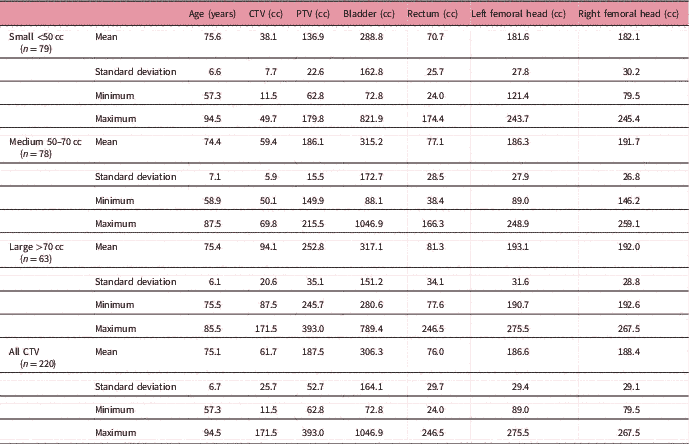
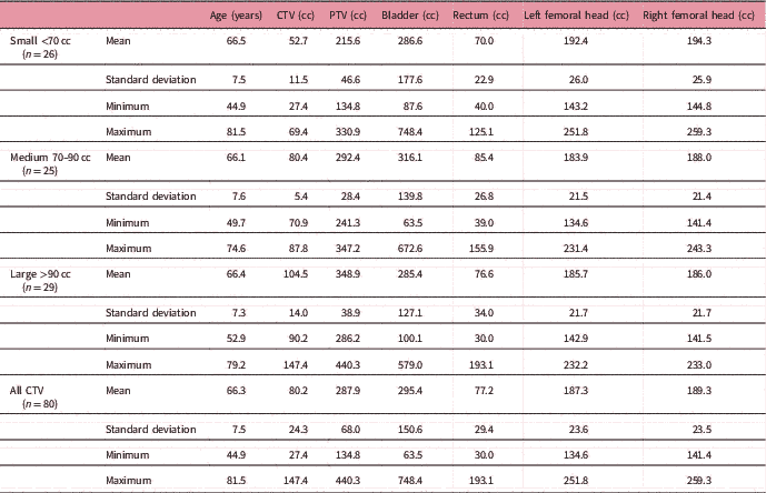
We retrospectively reviewed the treatment plans for 300 prostate cancer patients treated at our cancer centre, with the VMAT technique via RapidArc (Varian Medical Systems, Palo Alto, CA, USA). In all, 220 patients treated with a prescription of 78 Gy in 39 fractions and 80 patients with a prescription of 66 Gy in 33 fractions were considered in this study. All patient plans were delivered with a 6 MV photon beam. A summary of the patient characteristic and dosimetric parameters are provided in Tables 1a and 1b.
Summary of the statistical analysis of age, target volumes (CTV and PTV) and OAR volumes (bladder, rectum, left and right femoral head) for the patients receiving 78 Gy in 39 fractions

Abbreviations: CTV, clinical target volume; PTV, planning target volume; OAR, organs at risk.
Summary of the statistical analysis of age, target volumes (CTV and PTV) and OAR volumes (bladder, rectum, left and right femoral head) for the patients receiving 66 Gy in 33 fractions

Abbreviations: CTV, clinical target volume; PTV, planning target volume; OAR, organs at risk.
Treatment simulation and planning
As part of our institutional standard treatment protocol for prostate RT, all the patients were positioned supine with both arms on their chest and a kneefix placed under their knees. Computed tomography (CT) scanning was based on our institutional ‘prostate’ scan protocol, with a slice thickness of 0·3 cm, full bladder and empty rectum. The scan data were exported to the virtual simulator where the laser origin was marked at the location of the ball-bearings. This point was used for marking setup reference on the patient. The patient was tattooed at the anterior, right lateral and left lateral setup points. In some cases, additional tattoos were placed inferiorly for levelling if the patient’s anatomy required a more stable setup point.
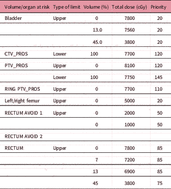
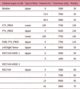
In some patients, a T2-weighted magnetic resonance imaging (MRI) scan was used as part of the planning process. In such cases, the CT and MRI images were co-registered with one another within the Eclipse (Varian Medical Systems, Palo Alto, CA, USA) treatment planning system (TPS) and used to better delineate the prostate volume. Contouring of all structures was standardised based on our institutional guidelines. The clinical target volume (CTV) was contoured to encompass the prostate or the prostate bed. Margins of 0·7 cm posteriorly and 1·0 cm radially in other directions were applied to the CTV to create the planning target volume (PTV). Portions of the seminal vesicles were automatically included within the PTV. OARs were contoured accordingly for bladder from dome to base; rectum starting from the sigmoid flexure to the bottom of the ischial tuberosities for a length of up to 13 cm; and femoral heads to include the lesser trochanter. Additional structures were created to aid the VMAT optimisation process. The details of such structures are outlined in Table 2. To account for the attenuation properties of the couch used for treatment, the Varian Exact IGRT Couch Top model available in the Eclipse TPS was adopted and included in the dose optimisation and calculation.
Structures and objective parameters used for VMAT optimisation
Abbreviation: VMAT, volumetric-modulated arc therapy.
Radiation Therapy Oncology Group (RTOG) guidelinesReference Shaw, Kline and Gillin 20 , Reference Knoos, Kristensen and Nilsson 21 for conformity and homogeneity of dose within the PTV were adopted as part of our treatment planning goals. A summary of the treatment planning objectives is provided in Table 3. The plans were considered acceptable if at least 99% of the PTV received 95% of the prescribed dose and no more than 2% of the PTV volume received 105% of the prescribed dose. All plans were generated using the Eclipse external beam TPS, version 10·0 (Varian Medical Systems, Palo Alto, CA). The beam geometry for the VMAT plans consisted of two full arcs spanning 358° each and gantry angle of 180·1 to 179·9 in clockwise and 179·9 to 180·1 in counter-clockwise rotation. Collimator angles for each plan were typically up to 15° and 345°. VMAT plan optimisation was carried out by the progressive resolution optimiser, which considers the plan objectives for an increasing number of beam angles. The VMAT optimisation is performed in four resolution levels: Resolution level 1 contains five steps, level 2 has two steps, level 3 has two steps and level 4 has one step. The majority of the actual optimisation occurs in levels 1 and 2. Levels 3 and 4 focus on ensuring that an optimal fluence is obtained. As the optimisation progresses, the optimiser becomes more constrained but the resolution increases. The VMAT dose optimisation was carried out by the dose–volume optimiser and dose calculations were done using the anisotropic analytical algorithm. A summary of the plan optimisation objectives, including the normal tissue objective is included in Table 2. If a compromise had to be made between achieving the desired target coverage and limiting OAR dose–volume values below the stated limits, a violation of some OAR dose limits was permitted to maintain similar target coverage in all planning tasks. The choice of the OAR limit was left to the discretion of the radiation oncologist. All plans used 6 MV photons and a dose calculation grid size of 2·5 mm, which incorporated heterogeneity corrections. Treatment was delivered on Varian Clinac 2100iX Linac or Varian TrueBeam Linac, both with 120-leaf MLC and a daily cone-beam CT image guidance was used for setup verification and target localisation.
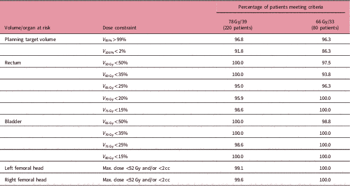
The planning objectives showing proportion of patients meeting each criterion

Data analysis
For all patients, statistical analysis of the cumulative dose–volume histograms (DVHs) and dosimetric parameters for the PTV and OARs were calculated and compared. The dose metrics evaluated include the mean and maximal doses to the PTV and the volume of the OARs exceeding each of the secondary dose constraints listed in Table 3.
Plan quality
The quality of a RT plan may be considered on the basis of how well the prescribed dose conforms to the target volume and to what extent the dose distribution varies within the target volume. In this study, the plan quality was quantitatively evaluated by calculating the homogeneity index (HI), uniformity index (UI) and the conformity index (CI) for each plan. The HI, CI and UI evaluate the dose homogeneity, conformity and uniformity, respectively, within the PTV and are calculated as:
 $${\rm HI}{\equals}{\rm }{{D_{2} {\minus}D_{{98}} } \over {D_{{{\rm PD}}} }}$$
 $${\rm UI}{\equals}{\rm }{{D_{5} } \over {D_{{95}} }}$$
 $${\rm CI}{\equals}{\rm }{{V_{{{\rm RI}}} } \over {{\rm TV}}}$$
where D 2, D 5, D 95 and D 98 are the doses received by 2, 5, 95 and 98% of the PTV, respectively. D PD is the prescribed dose, V RI is the volume of PTV covered by the reference isodose line, which in this case is the 95% isodose line, and TV is the target volume (which in this case is the PTV).
Dose to OAR
The doses to the OARs were assessed by determining the percentage volume (V) of an organ receiving n dose (V n ). To get a complete understanding of how VMAT planning impacts on dose delivered across the rectum, bladder and femoral head, V 40 Gy, V 50 Gy, V 60 Gy, V 65 Gy, V 70 Gy, V 75 Gy and V 80 Gy for both rectum and bladder, and the V 35 Gy, V 40 Gy, V 45 Gy for left and right femoral heads were assessed.
Results and Discussion
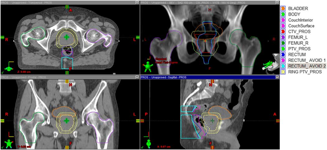
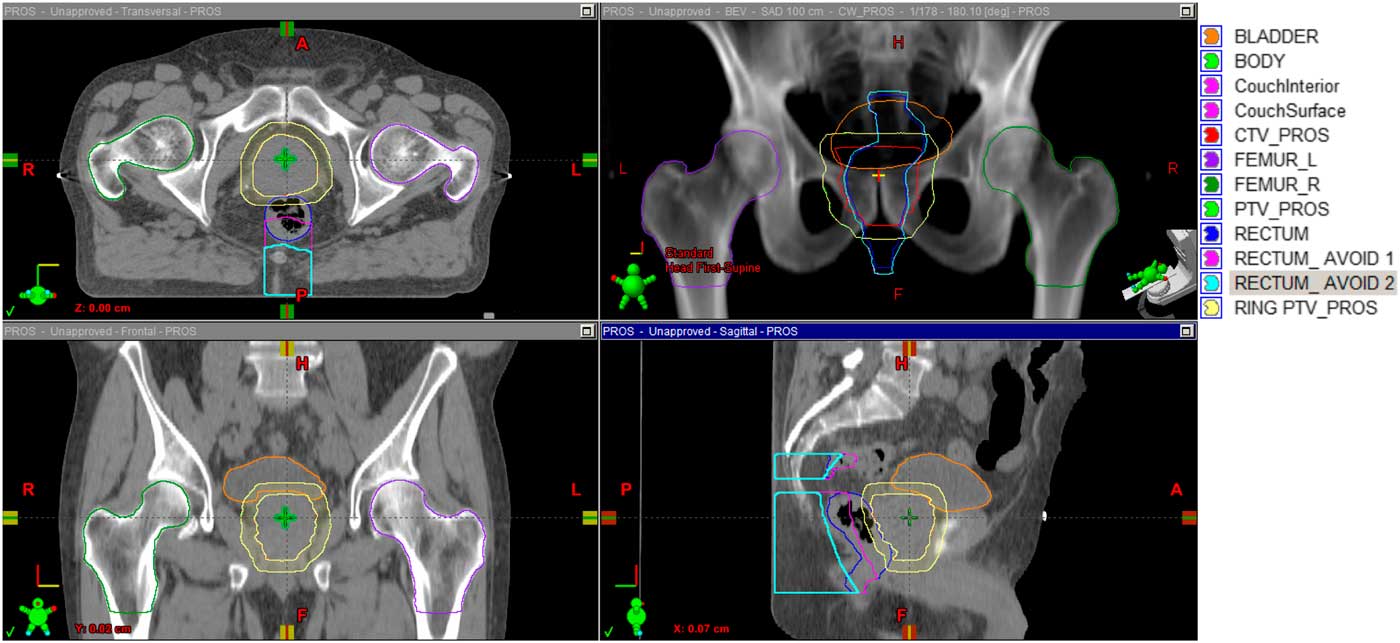
The dosimetry of VMAT plans for patients with prostate cancer treated at our centre over a period of 24 months were retrospectively reviewed. We stratified the patients into two cohorts; patients treated with a prescription dose of 78 Gy in 39 fractions as the primary radiation therapy treatment (XRT), and 66 Gy in 33 fractions as a post-op or salvage XRT. All patients were planned with the VMAT technique via RapidArc (Varian Medical Systems, Palo Alto, CA, USA). The quality of each patient plan was assessed by examining the extent to which each plan achieved the dosimetric objectives. This was qualitatively assessed by evaluating the DVH for the PTV and OAR. Figure 1 shows a typical segmentation of the CTV, PTV, rectum, femoral heads and bladder structures. For each treatment plan dose distribution, we determined the mean, standard deviation, minimum and maximum doses to the PTV, bladder, rectum, left and right femoral heads, and analysed the corresponding DVHs for all the structures. The dosimetric analysis was further stratified into three patient groups based on the target (CTV) volume for each prescription cohort of patients: small (<50 cc), medium (50≤x≤70 cc) and large (>70 cc) for patients receiving 78 Gy; and small (<70 cc), medium (70≤x≤90 cc) and large (>90 cc) for patients receiving 66 Gy.
Typical segmentation of the CTV, PTV, rectum, femoral heads and bladder structures. Abbreviations: CTV, clinical target volume; PTV, planning target volume.
Patient characteristics
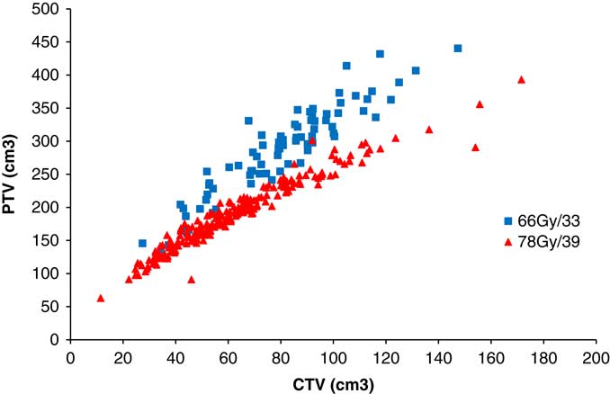
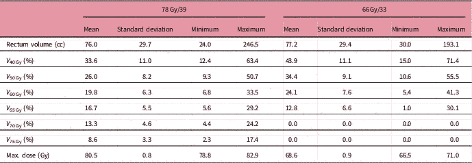
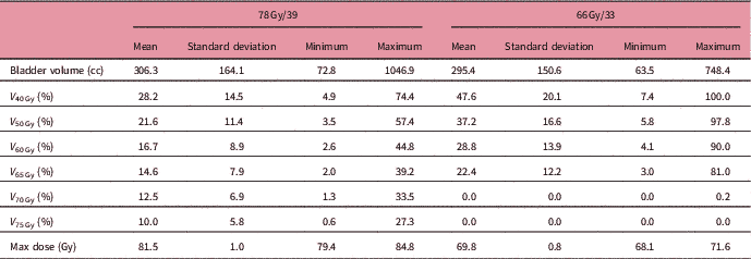
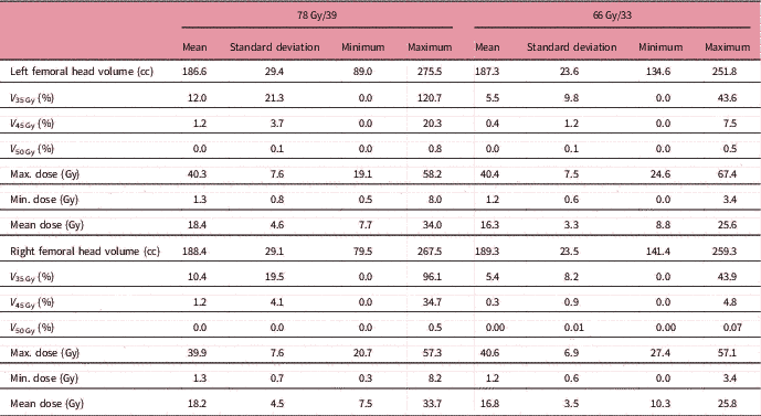
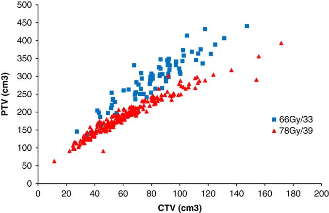
Table 1 shows a summary of the patient characteristics stratified into three groups by prostate size. For each patient, we determined the age, and volumes of the prostate (CTV), PTV, bladder, rectum and femoral heads. The mean patient age was 75·1 years (range: 57·3–94·5 years) and 66·3 years (range: 44·9–81·5 years) for patients prescribed with 78 Gy in 39 fractions and 66 Gy in 33 fractions, respectively. Of all the patients in this study, 220 (73·3%) patients were treated with a prescription of 78 Gy in 39 fractions, whereas 80 (26·7%) were treated to a prescription of 66 Gy in 33 fractions. There were significant differences in the target (CTV and PTV) volume between the patients treated with 78 and 66 Gy. The variations in target volume were independent of age. Figure 2 shows a correlation of the CTV with the PTV volume for both prescriptions. The average prostate (CTV) volumes for patients treated with 78 and 66 Gy were 61·7 cc (range, 11·5–171·5 cc) and 80·2 cc (range, 27·4–147·4 cc), respectively. Similarly, the average target (PTV) volumes for patients treated with 78 and 66 Gy were 187·5 cc (range, 62·8–393·0 cc) and 287·9 cc (range, 134·8–440·3 cc), respectively. The average volume of the bladder was 306·3 cc (range, 72·8–1046·9 cc) and 295·4 cc (range, 63·5–748·4 cc) for patients treated with 78 and 66 Gy, respectively. Similarly, the average volume of the rectum was also 76·0 cc (range, 24·0–246·5 cc) and 77·2 cc (range, 30·0–193·1 cc) for patients treated with 78 and 66 Gy, respectively. The average volumes of the left femoral head were 186·6 cc (range, 89·0–275·5 cc) and 187·3 cc (range, 134·6–251·8 cc) for patients treated with 78 and 66 Gy, respectively; and for the right femoral head were 188·4 cc (range, 79·5–267·5 cc) and 189·3 cc (range, 141·4–259·3 cc) for patients treated with 78 and 66 Gy, respectively.
A plot for the correlation between CTV and PTV for the two prescriptions, 78 Gy in 39 fractions and 66 Gy in 33 fractions. Abbreviations: CTV, clinical target volume; PTV, planning target volume.
Plan quality
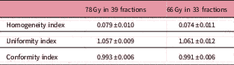
Plan quality was quantitatively assessed by calculating the HI, UI and the CI for the PTV for all patients. Table 4 shows the summary of the statistical analysis of the HI, UI and the CI for the PTV structures for all patients. Our results show improved dose conformity for prostate VMAT plans. The improved conformity is inherent to the arc delivery that delivers dose from up to 360°. Results show that all plans considered in this study demonstrated superior conformity to the PTV irrespective of the size of the prostate or the intended prescription.
Summary of the statistical analysis of the homogeneity index, uniformity index and the conformity index for the PTV

Abbreviation: PTV, planning target volume.
PTV dose analysis
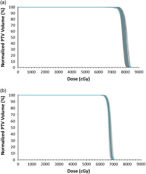
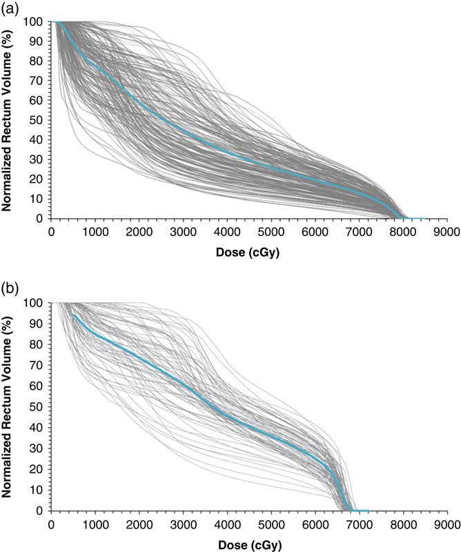
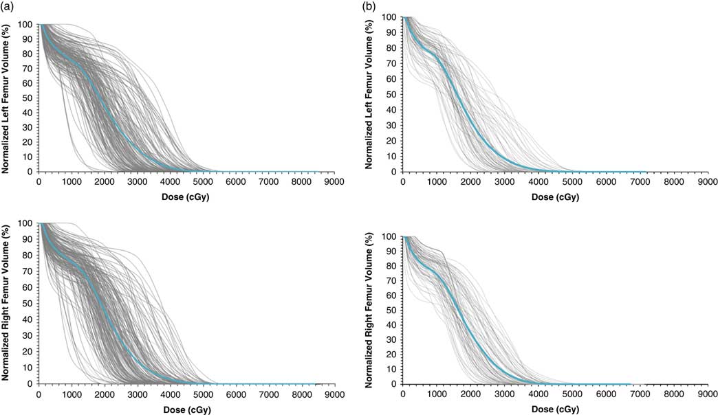
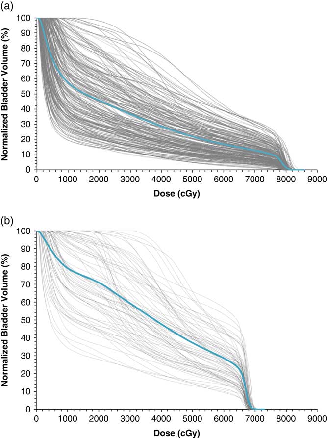
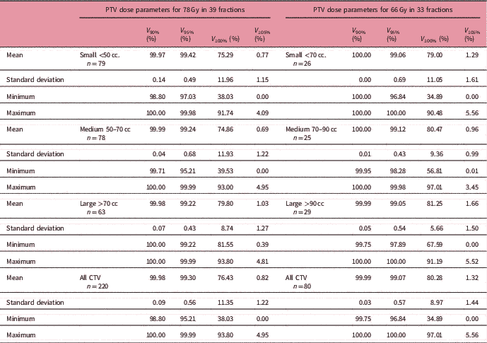
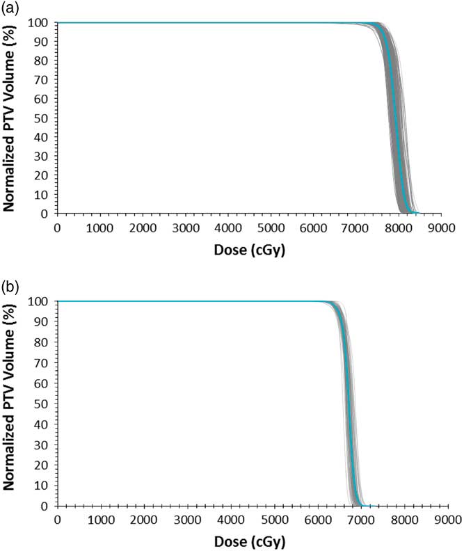
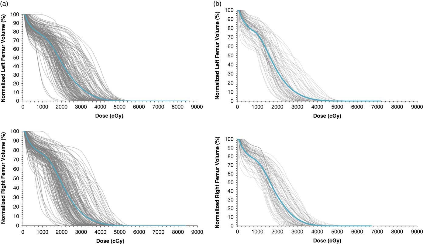
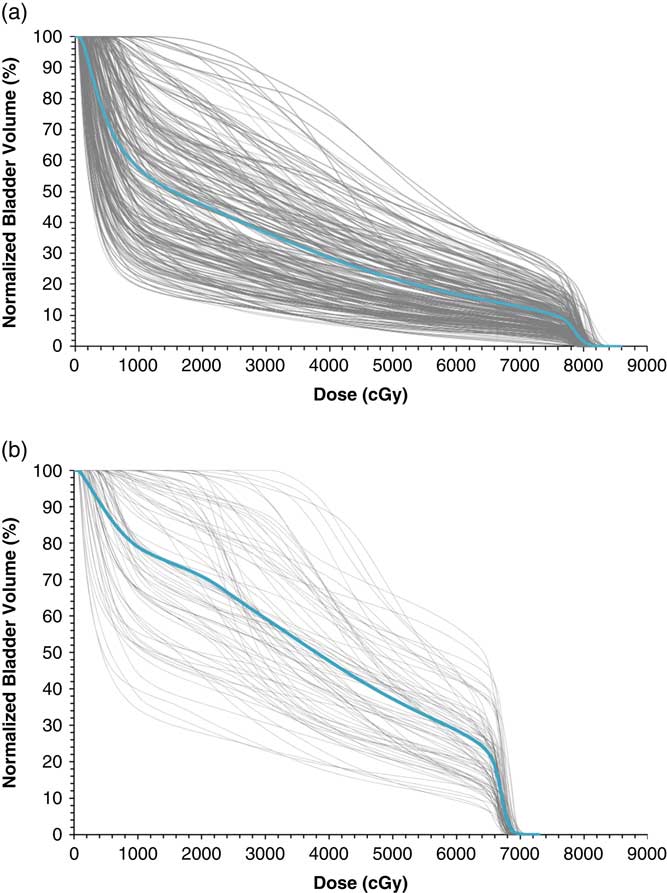
Figures 2–6 show DVH plots of the various structures for all the patients in each prescription group. Adequate CTV and PTV coverage was achieved for all patients reviewed. Target coverage and dose uniformity parameters such as the PTV doses: V 95%, V 100% and V 105% (PTV volume receiving 95, 100 and 105% of the prescribed dose, respectively) were calculated. Table 5 shows the dosimetric analysis of PTV with the patients grouped into those treated with 78 Gy in 39 fractions and those treated with 66 Gy in 33 fractions. Also shown in Table 5 are the dosimetric analyses, with patients for each prescription cohort further stratified into three groups (based on CTV volume). According to our current data, patients treated with a prescription of 78 Gy in 39 fractions, the mean V 90%, V 95%, V 100% and V 105% are 99·98% (range: 98·80–100·0%), 99·3% (range: 95·21–99·99%), 76·43% (range: 38·03–93·80%) and 0·82% (range: 0·0–4·95%). Similarly, for patients treated with 66 Gy in 33 fractions, the mean V 90%, V 95%, V 100% and V 105% are 99·99% (range: 99·75–100·0%), 99·07% (range: 96·84–100·0%), 80·28% (range: 34·89–97·01%) and 1·32% (range: 0·0–5·56%), respectively.
A plot of the dose–volume histograms (DVHs) of PTV for all patients at the prescription of (a) 78 Gy in 39 fractions and (b) 66 Gy in 33 fractions. Also shown in each plot is the mean DVHs for all patients. Abbreviation: PTV, planning target volume.
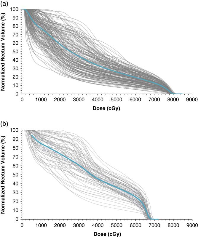
A plot of the dose–volume histograms (DVHs) of the rectum for all patients at the prescription of (a) 78 Gy in 39 fractions and (b) 66 Gy in 33 fractions. Also shown in each plot is the mean DVHs for all patients. Abbreviation: PTV, planning target volume.
A plot of the dose–volume histograms (DVHs) of the femoral head for all patients at the prescription of (a) 78 Gy in 39 fractions and (b) 66 Gy in 33 fractions. Also shown in each plot is the mean DVHs for all patients.
A plot of the dose–volume histograms (DVHs) of the bladder for all patients at the prescription of (a) 78 Gy in 39 fractions and (b) 66 Gy in 33 fractions. Also shown in each plot is the mean DVHs for all patients.
A summary of the statistical analysis of normalised PTV volume receiving 90%, 95%, 100% and 105% of prescribed dose for all patients

Abbreviations: PTV, planning target volume; CTV, clinical target volume.
OAR dose analysis
Doses to the OAR such as the rectum (and or rectal wall), bladder (and or bladder wall) and femoral heads have been compared. A number of studies have reported significantly better dose distributions and significant dose reduction to the OAR with VMAT compared to static-field IMRT.Reference Guckenberger, Richter, Krieger, Wilbert, Baier and Flentje 16 , Reference Khan, Jiang, Kiciak, Ur Rehman, Afzal and Chow 22 The rectum is the primary dose-limiting organ when treating prostate cancer by radiotherapy and therefore, significantly influences the treatment prescribed dose and the plan quality. The volume of rectum receiving ≥60 Gy is consistently associated with the risk of Grade ≥2 rectal toxicity or rectal bleeding. Late injuries are usually clinically manifested within 3–4 years after RT and may include stricture, diminished rectal compliance and decreasing storage capacity. These morbidities can be severe and can markedly affect the quality of life of patients.Reference Michalski, Gay, Jackson, Tucker and Deasy 23 For typical DVHs, the normal tissue complication probability models predict that following a conservative DVH constraint for rectum: V 50 Gy<50%, V 60 Gy <35%, V 65 Gy<25%, V 70 Gy<20% and V 75 Gy<15% should limit Grade ≥2 late rectal toxicity to <15% and the probability of Grade ≥3 late rectal toxicity to <10% for prescriptions up to 79·2 Gy in standard 1·8–2 Gy fractions.Reference Michalski, Gay, Jackson, Tucker and Deasy 23
To assess the extent of the dose delivered across each of the OAR, we determined the percentage volume (V) of each organ receiving n dose (V n ). For the rectum and bladder, the analysis included V 40 Gy, V 50 Gy, V 60 Gy, V 65 Gy, V 70 Gy and V 75 Gy (Tables 6a and 6b). Our data show rectal mean V 40 Gy, V 50 Gy, V 60 Gy, V 65 Gy, V 70 Gy and V 75 Gy of 33·6, 26·0, 19·8, 16·7, 13·3 and 8·6%, respectively, for the 78 Gy in 39 fractions cohort and 43·9, 34·4, 24·1, 12·8, 0·0 and 0·0%, respectively, for the 66 Gy in 33 fractions cohort (Table 6a). The corresponding bladder mean values for the V 40 Gy, V 50 Gy, V 60 Gy, V 65 Gy, V 70 Gy and V 75 Gy are 28·2, 21·6, 16·7, 14·6, 12·5 and 10·0%, respectively, for the 78 Gy in 39 fractions cohort and 47·6, 37·2, 28·8, 22·4, 0·0 and 0·0, respectively, for the 66 Gy in 33 fractions cohort (Table 6b). For each of the left and right femoral head, the maximum, minimum and mean doses, as well as the V 35 Gy, V 45 Gy and V 50 Gy were evaluated (Table 6c). The mean doses to the femoral heads are not significantly different for both cohorts of patients (Table 6c). The proportions of patients meeting our institutional OAR dosimetric criteria are summarised in Table 3. Our results indicate that at least 94% of all the patients considered in this study successfully met all the rectal dose constraints, and that at least 99% met all the bladder dose constraints, and that all patients met the femoral head constraints (Table 3).
A summary of the statistical analysis of the OAR volumetric doses for the rectum

Abbreviations: OAR, organs at risk.
A summary of the statistical analysis of the OAR volumetric doses for the bladder

Abbreviation: OAR, organs at risk.
A summary of the statistical analysis of the OAR volumetric doses for the left and right femoral head volumes

Abbreviation: OAR, organs at risk.
Conclusion
This study shows that highly conformal radiation dose distribution for the treatment of prostate cancer is achievable with VMAT technique. With the quality of plans achievable with this technique, it should be possible to safely dose escalate without compromising significantly on possible toxicity. Based on our promising initial experiences, we are confident about how VMAT technique has been established as a mode of a conformal radiation delivery system for localised prostate cancer at our institution. Results from this study provide evidence to support the adoption of such conformal technology for possible higher radiation doses in many other disease sites. Individual institutes are encouraged to evaluate and adopt the most suitable planning criteria tailored to their own needs and priority. We believe our experience reported here could help form the foundation for such endeavour, which will hopefully provide further improvement in the planning process aimed at providing an effective and efficient delivery of radiotherapy for the treatment of prostate cancer and similar sites. Further study to determine long-term results with VMAT and possible correlations with outcome is underway. The results of comparison of genitourinary and gastrointestinal toxicity assessed by the RTOG toxicity score administered by the physician between the treatment groups and a correlation with dose received will be presented in a subsequent manuscript.
Acknowledgement
The authors would like to acknowledge with much gratitude the financial support from the Kitchener-Waterloo Chapter of the TELUS Ride For Dad and the Prostate Cancer Fight Foundation for this study and Kinga Alliet for guidance on the planning process.