Introduction
Bacterial meningitis has been reported in children fitted with cochlear implants,Reference Loundon, Blanchard, Roger, Denoyelle and Garabedian1 and more recently in children with auditory brainstem implants.Reference Colletti, Shannon, Carner, Veronese and Colletti2 The presumptive diagnosis of meningitis is clinical, while definitive diagnosis relies on radiological investigation and lumbar puncture. After posterior fossa surgery, meningitis generally occurs following wound infection and cerebrospinal fluid (CSF) leakage.
We describe the case of a patient with Mondini's dysplasia whose clinical course was complicated by meningitis developing six years after auditory brainstem implantation. The aetiology was believed to be otogenic. An exploratory tympanotomy, performed after resolution of the infectious meningitis, demonstrated a stapes footplate malformation. This direct communication between middle and inner ear was the most likely route for the development of meningitis.
Case report
An eight-year-old boy presented to the emergency department with a three-day history of severe headache unresponsive to analgesia and progressive onset of neck stiffness. He also complained of phonophobia and photophobia. The patient had a previous history of meningitis at the age of 13 months, treated with antibiotic therapy.
On admission, the patient had a temperature of 38.5°C but essentially no other significant findings. His white blood cell count was 19 100/μl.
The patient also had a history of Mondini's dysplasia associated with profound, bilateral hearing loss. Six years earlier, he had undergone auditory brainstem implantation (Figure 1) on the right side, without any complications.
Intra-operative view of auditory brainstem implant insertion.

The scar from the implantation surgery showed no signs of infection, nor did the skin at the level of the auditory brainstem implant receiver.
On otoscopy, an opaque, bulging right tympanic membrane was observed, with normal findings on the left side.
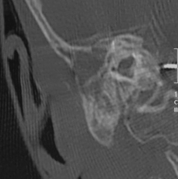
On the basis of the clinical symptoms and surgical history, cerebral and temporal bone computed tomography (CT) scans were performed to investigate the possibility of infection at the site of the auditory brainstem implantation surgery. This revealed opacity of the right mastoid cavity, but, surprisingly, no signs of infection at the previous retrosigmoid craniotomy site or around or below the auditory brainstem implant receiver (Figure 2).
High-resolution, coronal computed tomography scan of the right ear, showing the presence of fluid in the right middle ear and mastoid cavity, indicative of acute mastoiditis, in a child with Mondini's dysplasia.

A lumbar puncture was performed due to suspected meningitis, producing cloudy fluid under elevated pressure. The CSF contained 5070 cells/mm3, with a glucose concentration of 12 mg/dl (the plasma glucose was 72 mg/dl) and a protein concentration of 321 mg/dl.
Tympanocentesis was performed, with drainage of pus.
The child was commenced on intravenous antibiotics, including ceftriaxone (1500 mg every 12 hours) and vancomycin (450 mg every 6 hours).
The patient's vaccine status was up to date, and there were no identifiable risk factors except for the Mondini malformation.
Microbial cultures of blood, middle-ear aspirate and CSF were all positive for β-lactam-sensitive Streptococcus pneumoniae.
Improvement was achieved within 48 hours. The child was discharged with complete resolution of his meningitis and mastoid infection, documented on CT scan. Furthermore, his auditory brainstem implant was functional with good outcomes.
The child's quick recovery from meningitis and complete clearance of mastoid infection suggested that mastoidectomy with mastoid obliteration was unnecessary. Thus, an exploratory tympanotomy was scheduled for one month after complete resolution of disease.
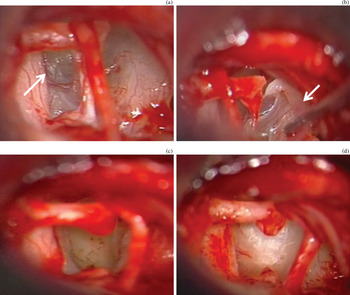
On exploration of the middle ear, the stapes superstructure appeared normal. However, the intercrural area contained a thin, bulging tissue structure (Figure 3a). Opening this structure released gushing fluid. The stapes was removed (Figure 3b and 3c), the vestibule obliterated with fascia and fibrin glue, and the defective oval window area further covered with fascia (Figure 3d).
Intra-operative images taken during exploratory tympanotomy. (a) The normal stapes footplate bone is partially absent, being partially replaced by a thin, bulging tissue structure (arrow). (b) Removal of the stapes; the stapes superstructure anatomy is seen to be normal (arrow). (c) The oval window area defect after removal of the stapes. (d) Repair of the oval window defect with fascia, after removal of the malformed stapes.

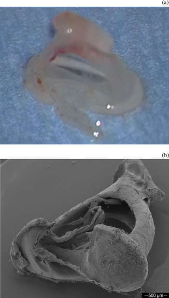
The defective stapes (Figure 4a) was examined under scanning electron microscopy (Figure 4b).
(a) The removed, malformed stapes. (b) Scanning electron micrograph of the removed stapes.

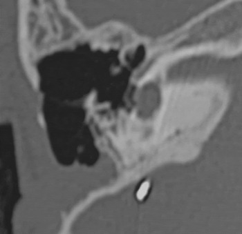
Computed tomography scans performed one year after the procedure showed no signs of middle-ear, mastoid or intracranial disease (Figure 5). No further complications were observed during two years of follow up.
High-resolution, coronal computed tomography scan of the right ear, performed one year after surgical repair of the malformed oval window area, showing no signs of middle-ear, mastoid or intracranial disease.

Discussion
In the general paediatric cochlear implant population, bacterial meningitis is encountered in less than 1 per cent of cases.Reference Loundon, Blanchard, Roger, Denoyelle and Garabedian1, Reference McJunkin and Jeyakumar3 It may occur as an early or delayed complication of surgery.Reference Loundon, Blanchard, Roger, Denoyelle and Garabedian1, Reference McJunkin and Jeyakumar3 The most likely causative event is generally an infection at the surgical site (especially over the cochleostomy when it is not properly sealed with muscle and fascia).Reference Loundon, Blanchard, Roger, Denoyelle and Garabedian1
Rarely, recurrent meningitis may be caused by malformation of the inner ear, with a resultant perilymphatic fistula.Reference Sennaroglu4–Reference Torkos, Czigner, Jarabin, Toth, Szamoskozi and Kiss7 Mondini's dysplasia is the most common temporal bone anomaly associated with CSF leakage in children. The meningitis associated with this malformation generally develops as a result of retrograde infection tracking from the middle ear directly to the CSF.
In the case of otogenic meningitis in children fitted with a cochlear implant, obliteration of the mastoid cavity, and even petrosectomy, are considered effective treatments.
However, there is currently very limited data on meningitis associated with paediatric auditory brainstem implantation surgery.Reference Colletti, Shannon, Carner, Veronese and Colletti2 Risk factors for the development of meningitis after posterior fossa surgery include wound infection, CSF leakage, prolonged surgery, long-term steroid use and diabetes.
The patient presented here demonstrates a case of meningitis not related to auditory brainstem implantation surgery. The source of infection was an ipsilateral mastoid bone infection associated with a rare malformation of the stapes footplate. The thin, bulging structure that had replaced the stapes footplate bone may have easily ruptured during previous episodes of middle-ear and/or mastoid infection, with subsequent perilymphatic contamination. A congenital defect in the oval window area, as described in our patient, can be related to other inner ear malformations such as Mondini's dysplasia, since all middle-ear structures are branchial in origin apart from the stapes footplate, which is derived from the otic capsule. Furthermore, a fistula of the stapes footplate may also be caused by a wide defect in the lamina cribrosa and modiolus, with resultant erosion due to continuous CSF pressure over the medial surface of the oval and round windows.Reference Sennaroglu4 In our patient, the close connection between the perilymphatic spaces of the inner ear and the intracranial subarachnoid space was the most likely route by which bacteria reached the meninges. In children with inner ear malformations, various congenital defects may play a role in increasing the risk of meningitis, such as absence of the bony wall between the internal auditory canal and the base of the malformed cochlear cavity,Reference McJunkin and Jeyakumar3, Reference Sennaroglu4 and a widely patent cochlear aqueduct. However, a pathological communication between the middle and inner earReference Sennaroglu4, Reference Torkos, Czigner, Jarabin, Toth, Szamoskozi and Kiss7, Reference Kaddour8 is the most likely ‘route of least resistance’ for such infection.
Patients with inner ear malformations, especially Mondini's dysplasia, need to be carefully evaluated pre-operatively to reduce or eliminate all anatomical conditions that may predispose to meningitis. Moreover, most of these conditions are bilateral; thus, it may be possible to observe a child fitted with a cochlear or auditory brainstem implant who presents with meningitis induced by infection in the contralateral malformed ear.Reference Sennaroglu4
It has previously been thought that pre-operative radiological investigations are unable to detect all possible cochlear abnormalities, in particular congenital stapes footplate defects producing a communication between the middle ear and the subarachnoid space via the inner ear. The presence of an auditory brainstem implant further complicates the radiological diagnosis, as this produces CT artefacts and prevents the safe execution of magnetic resonance imaging (MRI).
Recently, a new radiological sign has been described which is often associated with recurrent meningitis: the ‘bulging oval window’ sign. This bulging oval window represents a fluid-filled sac emanating from the oval window in cases of common cavity malformations, and can be identified by both high-resolution CT and MRI.Reference Ehmer, Booth, Kutz and Roland5 This sign was not carefully evaluated in the CT or MRI scan performed on our patient prior to auditory brainstem implantation.
This case is unique and instructive, because it describes for the first time a rare presentation of otogenic bacterial meningitis secondary to stapes footplate malformation in a paediatric patient with an auditory brainstem implant.
• Bacterial meningitis may be an early or delayed complication of paediatric cochlear implantation
• In rare cases, middle-ear malformation with perilymphatic fistula is also found
• We report a rare case of otogenic bacterial meningitis due to stapes footplate malformation in a paediatric auditory brainstem implant patient
• The radiological ‘bulging oval window’ sign should be looked for in auditory brainstem implant candidate children suspected of ear malformation
• Those with suspected trans-stapedial cerebrospinal fluid fistula should undergo bilateral exploratory tympanotomy to detect any anatomical footplate defects, which may lead to meningitis
Spontaneous CSF fistulae with resultant meningitis may occur in patients with inner ear malformations such as Mondini's dysplasia, and may be more frequently caused by stapes footplate defects than by cochlearReference Sennaroglu4 or auditory brainstem implantation. Furthermore, the present case illustrates the general difficulties in diagnosing and treating meningitis in patients with inner ear abnormalities who have undergone auditory brainstem or cochlear implantation surgery.
In cases such as our patient, early diagnosis, treatment of the infective agent with appropriate antibiotic treatment, and surgical closure of the stapes footplate defect are essential. In such cases of isolated stapes footplate malformation, mastoid obliteration or petrosectomy with obliteration and blind sac closure of the ear canal should probably be considered as over-treatment.
We are currently retrospectively reviewing the pre-operative radiological documentation of all previously treated patients with ear malformations who were fitted with an auditory brainstem or cochlear implant, to rule out possible footplate malformations.
In view of the present findings, we recommend pre-operative radiological investigation to look for the bulging oval window signReference Ehmer, Booth, Kutz and Roland5 in children with suspected ear malformations undergoing cochlear or auditory brainstem implantation. If there is suspicion of a trans-stapedial CSF fistula, bilateral exploratory tympanotomy should be performed at the time of implantation surgery to detect any undiagnosed stapes footplate defects, which may predispose to bacterial meningitis.