Introduction
Neonatal encephalopathy (NE) describes a clinical disorder of neonatal brain function resulting from a number of conditions that impair central nervous system (CNS) function within the first several days after birth. The most notable clinical features are depression of level of consciousness ranging from mild (hyperalert) to moderate (lethargic) or severe (stupor/coma) with associated findings in part modulated by the degree of encephalopathy to include respiratory depression, disturbances of cranial nerve function such as suck or gag, abnormalities of tone and power, and often seizures [Reference Volpe1]. The overall incidence of NE is estimated at 3 per 1,000 live births and that of hypoxic–ischemic encephalopathy (HIE), the most distinctive cause of NE, at approximately 1–1.5 per 1,000 live term births [Reference Kurinczuk, White-Koning and Badawi2].
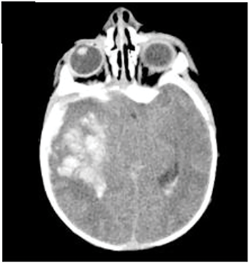
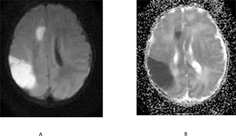
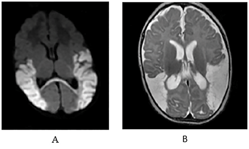
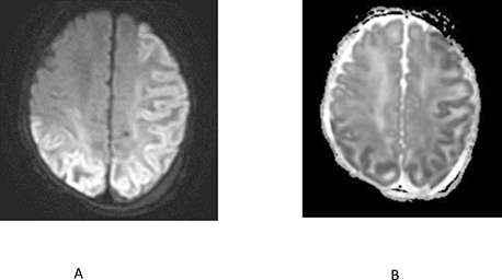
Over the past decade there has been much debate [Reference Dammann, Ferriero and Gressens3,Reference Chalak, Ferriero, Gressens, Molloy and Bearer4,Reference Molloy, Branagan, Hurley, Quirke, Devane and Taneri5] as to what constitutes HIE, indeed whether it can be diagnosed given the limited data present around the time of birth, particularly as it relates to cerebral blood flow (CBF) and oxygen delivery [Reference Molloy and Bearer6]. The ability to diagnose an infant at high risk for evolving hypoxic–ischemic brain injury is highly relevant because therapeutic hypothermia (TH), the only treatment known to improve neurologic outcome in term and near-term infants after HIE, has become the standard of care, and should be administered within 6 h of birth to achieve neuroprotection [Reference Shevell7]. However, as noted, many argue that identification of a high-risk infant is fraught with problems. Several points need clarification. First, infants at high risk for evolving HIE can be identified by a constellation of clinical and biochemical findings starting during labor and delivery and extending to the initial early hours following birth. However, other clinical conditions during labor and delivery may modulate the early clinical examination and need to be considered during the evaluation. These include the presence of clinical chorioamnionitis (CA) with maternal fever as the leading sign, a difficult delivery (usually instrumented) with the potential for intracranial hemorrhage (ICH), intrapartum acquired sepsis, and maternal medications. During the first 24 h, additional causes of NE to be considered include hypoglycemia, neonatal arterial ischemic stroke (NAIS), seizures in the absence of overt signs of depressed level of consciousness, ICH, and evolution of early onset sepsis (Box 1.1).
Postnatal Day One
Postnatal Day Two and Beyond
Hyperbilirubinemia
Inborn errors of metabolism
Neonatal onset epileptic encephalopathy
Transient encephalopathies
Brain malformations
*May initially present on the second postnatal day
Beyond 24–72 h, NAIS, ICH, malignant epileptic syndromes (epileptic encephalopathies), hyperbilirubinemia (kernicterus), inborn errors of metabolism (urea cycle disorders, nonketotic hyperglycinemia, mitochondrial disorders, organic acidemias, among others), bacterial and viral infections may become apparent. The approach to the diagnosis and management of NE has been well described in two fairly recent reports [Reference Glass8,Reference Hanna, Rosenbaum and Bartlett9]. In this chapter, we will initially focus on HIE as representing a distinct cause of NE able to be identified within 6 h and as such allow infants to receive TH, as well as describe the other conditions in the differential diagnosis and management of NE as it presents at the time of birth, in the first 24 h, 24–48 h, and beyond (Box 1.1). Clearly these time frames are arbitrary, and presentation of NE may vary in time of onset, depending on the severity of the underlying condition.
For many years there has been considerable debate in the literature as to the relationship and/or contribution of HIE (previously used synonymously with the term birth asphyxia [BA]) to the subsequent development of cerebral palsy (CP). This is briefly discussed next.
Perinatal HIE/NE and the Contribution to Subsequent CP
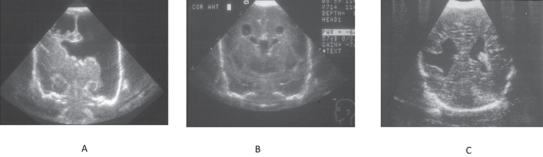
Hypoxic–ischemic brain injury that occurs around the time of birth is the most recognized cause of the severe, long-term neurologic deficits in infants and children termed CP. This is often defined as a nonprogressive motor disorder of early onset affecting approximately 2–3 per 1,000 school-aged children. Brain injury resulting in CP may occur during the antepartum, peripartum, or postnatal period. However, overwhelming evidence suggests that the genesis of CP [Reference Aravamuthan, Fehlings and Novak10,Reference te Velde, Morgan, Novak, Tantsis and Badawi11,Reference McIntyre, Goldsmith and Webb12] in approximately 60–70% of cases is antenatal in onset [Reference Nelson and Ellenberg13,Reference Nelson and Leviton14,Reference Nelson15]. The timing of the antepartum insult is critical to the evolution of a specific lesion. Thus, cerebral ischemia prior to the 20th week of gestation results in a neuromigrational defect, for example, schizencephaly. Ischemia occurring between the 28th and the 34th weeks of gestation usually results in periventricular leukomalacia (PVL). When ischemia occurs between the 34th and 40th weeks of gestation, focal/multifactorial cerebral injury usually develops (Figure 1.1) [Reference Aravamuthan, Fehlings and Novak10]. By contrast, the proportion of CP attributed to perinatal events is small and has been estimated to represent approximately 15–20% of cases [Reference Aravamuthan, Fehlings and Novak10,Reference Hollung, Bakken and Vik16,Reference Novak, Hines, Goldsmith and Barclay17]. We will briefly review the evidence supporting the substantial antenatal contribution to CP.
Antenatal ischemic injury to the brain is dependent upon the timing of the insult. A, schizencephaly; B, periventricular leukomalacia; C, focal cerebral infarction.
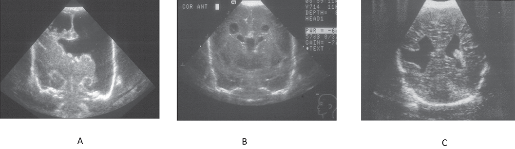
Figure 1.1 Long description
Panel A shows a large, irregular anechoic cavity extending from the cortical surface toward the ventricular region. Panel B shows multiple small, rounded anechoic areas clustered near the central region. Panel C shows two asymmetric anechoic regions within the cerebral parenchyma.
Radiologic Evidence
Magnetic resonance imaging was used to retrospectively analyze the brains of 29 patients born at term suffering from CP. Three major patterns were noted. They included (1) gyral anomalies, suggestive of polymicrogyria, consistent with mid second trimester injury; (2) isolated PVL reflecting late second- or early third-trimester injury; and (3) watershed cortical or deep gray nuclear damage, consistent with late third-trimester, perinatal, or postnatal injury. Overall, the contribution of these abnormalities was noted in 55% of patients [Reference Påhlman, Gillberg and Himmelmann18]. In a second study, CT brain scans obtained from 83 children born at term were used to evaluate the most likely etiology of hemiplegic CP. The most common finding was periventricular atrophy considered to represent PVL (37%), often as a complication of preterm birth. As this lesion was found in children born at term it was considered to reflect an in-utero etiology of CP. Cerebral maldevelopment, due to a very early intrauterine lesion, was detected in 17%, whereas cortical/subcortical lesions were seen in 16% of the children. Altogether the CT findings indicated a prenatal origin of hemiplegia in approximately 70% of cases [Reference Whitney, Warschausky, Ng, Hurvitz, Kamdar and Peterson19].
Clinical Evidence
A study of children with spastic CP (n = 183) were compared to a matched group of control children (n = 549). There was a 2.8-fold increased risk for spastic CP in those children with clinical signs of BA as compared to controls. It was estimated that the possible cause of subsequent brain damage related to intrapartum “asphyxia” was approximately 8% of children [Reference Blair and Stanley20].
Contribution of Intrapartum versus Antenatal Factors in the Genesis of NE/HIE
A study involving 405 infants ≥35 weeks’ gestation with early-onset encephalopathy, meeting criteria for perinatal asphyxia, and with neuroimaging findings consistent with acute HIE, were compared with 239 neurologically normal infants. Case infants were fivefold more often delivered by emergency cesarean section (CS). On logistic regression analysis, only one antepartum factor (gestation ≥41 weeks) and several intrapartum factors – prolonged rupture of membranes (PROM), abnormal cardiotocography, thick meconium, and sentinel event – remained independently associated with HIE (area under the curve [AUC] 0.88; p < 0.001). Notably 3.7% of cases and 19.7% of controls had no identifiable risk factors for HIE [Reference Fahey, Maclennan, Kretzschmar, Gecz and Kruer21]. These findings strongly point to the intrapartum period as important in the evolution of HIE.
In another study of infants with HIE, 87% had intrapartum complications that increased risk, a sentinel event (36%, relative risk [RR] = 16.1), clinical CA (40%, RR = 5.2), or both (RR = 11). Urinary tract infection during pregnancy (RR = 2.6) was a potential antenatal risk factor. It was concluded that a significant proportion of HIE is preceded by intrapartum events [Reference Jin, Lewis and Bakhtiari22]. In another study involving 351 term infants >36 weeks who presented with NE (n = 262) or seizures (n = 90) without other signs of global encephalopathy, MRI images showed evidence of an acute insult in 80%, whereas evidence of established injury was noted in 2 infants (<1%). In the seizure group, acute focal damage was noted in the majority of infants. Two infants also had evidence of antenatal injury. These findings point to the immediate perinatal period as the most important precursor to NE [Reference Cooper, Fahey and Mackay23].
In another study of infants with moderate or severe NE, the incidence was found to be 1.64 per 1,000 term live births. The main cause was perinatal hypoxic ischemia (52%). The timing was intrapartum in 56%, antepartum in 13%, and ante-intrapartum in 10% of cases. In 19% of cases, no underlying cause for NE was identified [Reference Jöud, Sehlstedt, Källén, Westbom and Rylander24]. Another large study comprised singleton neonates of gestational age (GA) ≥36 weeks, who met criteria for NE within 3 days of birth [Reference Novak, Jackman and Finch-Edmondson25]. Clinically recognized seizures (60%) were the most prevalent condition; 49% had a 5-min Apgar score of ≤3 and 18% had a reduced level of consciousness. An abnormal maternal or fetal condition predated labor in 46%; maternal hypertension (16%) or small for gestational age (SGA) (16%) were the most frequent risk factors. The most prevalent perinatal event was elevated maternal temperature ≥37.5°C (27% of cases). Acute asphyxial indicators were reported in 35% and inflammatory indicators in 24%. Much of NE remained unexplained. This group had very heterogenous inclusion criteria and only 18% had evidence for reduced level of conscious, contributing to the unclear etiology of NE in many cases.
Summary
1. Most children with CP (50–80%) have their genesis during the antepartum period.
2. The processes that lead to HIE occur around birth in approximately 80–85% of cases.
3. The etiology of perinatal HIE cannot be identified in approximately 19% of cases.
4. In two studies the prevalence of HIE was between 1 and 1.64/1,000 term infants, respectively.
Perinatal Conditions Associated with NE on the First Postnatal Day
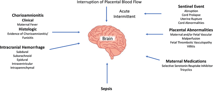
Neonatal encephalopathy that occurs around the time of birth may be related to interruption of placental blood flow (PBF), which may be acute (a sentinel event) or intermittent (fetal heart rate [FHR] abnormality), ICH, CA/maternal fever, intrapartum-acquired sepsis, placental abnormalities, and maternal medications (Figure 1.2). Factors that may affect the brain around delivery and manifest as NE during the first postnatal day. The most distinctive of these states is HIE secondary to interruption of PBF, reflective of asphyxia (see section on Perinatal Hypoxic–Ischemic Encephalopathy). Over the years it has become apparent that not all cases of “acute” brain injury present immediately at birth and are triaged directly to the neonatal intensive care unit (NICU). Rather, some infants are initially triaged to the well-baby nursery (WBN) and subsequently present with clinical signs of encephalopathy including seizures during the first 24 h. Two case series presented next highlight a group of infants with delayed clinical presentation of perinatal injury.
Factors that may affect the brain around delivery and manifest as neonatal encephalopathy during the first postnatal day.
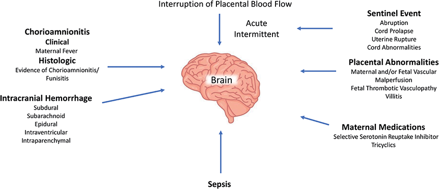
Figure 1.2 Long description
The diagram shows a brain at the centre surrounded by the following factors: Interruption of Placental Blood Flow includes Acute and Intermittent. Sentinel Event includes Abruption, Cord prolapse, Uterine Rupture, and Cord Abnormalities. Placental Abnormalities include Maternal and/or Foetal Vascular Malperfusion, Foetal Thrombotic Vasculopathy, and Villitis. Maternal Medications include Selective Serotonin Uptake Inhibitor and Tricyclics. Sepsis is listed as a separate factor. Intracranial Haemorrhage includes Subdural, Subarachnoid, Epidural, Intraventricular, and Intraparenchymal. Chorioamnionitis includes Clinical with Maternal Fever, and Histologic with Evidence of Chorioamnionitis or Funisitis.
First Case Series (January–December 1993)
Between January and December 1993, 12 of 14,518 term infants were delivered at Parkland Memorial Hospital Dallas and presented clinically with seizures during the first postnatal day [Reference Blair, Langdon, McIntyre, Lawrence and Watson26]. Five infants (0.34 per 1,000 live births) were triaged directly to the NICU and seven (0.48 per 1,000 live births) to the WBN. Of the latter, complications during labor included meconium-stained amniotic fluid (MSAF), FHR abnormalities, and CA. Six received no resuscitation, whereas one needed brief positive pressure ventilation (PPV). The Apgar scores were ≥8 in all cases. The cord umbilical arterial pH was <7.00 in one case. All seven developed seizures in the first 24 h (mean 18 h). The presumed cause of the seizures was HI (n = 4) based on timing of seizures, associated systemic signs (oliguria), cranial ultrasound findings (CUS), findings of ventricular effacement and increased periventricular echogenicity, and a negative workup for other causes of seizures. Additional causes included NAIS (n = 2) and subdural hemorrhage (SDH) (n = 1). Infants admitted to the NICU all exhibited respiratory depression and were intubated in the delivery room (DR). The cord arterial pH was <7.00 (mean 6.75 ± 0.12) and the base excess (BE) was ≤–14 mEq/l in all cases (mean –19 ± 5). Onset of seizures was within 16 h (mean 11 h). The CUS was abnormal in all cases. One infant died of severe brain injury.
Second Case Series (2004–2009)
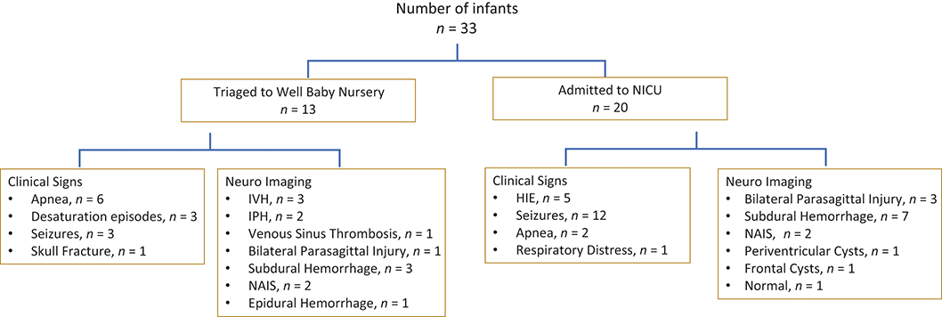
Between January 2004 and December 2009, 33 infants of 29,597 term infants delivered at New York Presbyterian Hospital, New York presented clinically with early signs consistent with intrapartum brain injury [Reference Whitney, Kamdar, Ng, Hurvitz and Peterson27]. The underlying conditions included HIE (n = 8, 0.27/1,000), or after neuroimaging, SDH (n = 10, 0.34/1,000), intraventricular hemorrhage (IVH) (n = 3), or intraparenchymal hemorrhage (IPH) (n = 2, 0.17/1,000), and NAIS (n = 4, 0.14/1,000 live term births), reflecting an overall rate of 1.12/1,000 term live births. Of the 33 infants, 13 (39%) were asymptomatic in the DR and triaged to the WBN. Delayed presentation included apnea (n = 6) and, upon neuroimaging, IVH (n = 3), IPH (n = 2), and sinus venous thrombosis (SVT) (n = 1) (Figure 1.2). Three infants presented with desaturation episodes and were noted to have SDH (n = 1) and NAIS (n = 2), and one infant with a visible fracture had an epidural hemorrhage on neuroimaging (Figure 1.3). Twenty (61%) infants were triaged directly to the NICU due to perinatal depression requiring respiratory support. Diagnoses included HIE with seizures (n = 4) presenting from 6 to 48 h post-delivery; one infant who was comatose from birth died within 6 h. One infant required prolonged respiratory support but had a normal neurologic course. In the remaining 14 infants, clinical signs included seizures (n = 12) and apnea (n = 2) with resolution within 24 h. On MRI the associated changes included SDH (n = 7), basal ganglia and bilateral parasagittal injury (n = 3), NAIS (n = 2), periventricular cysts (n = 1), and normal (n = 1). Electroencephalogram (EEG) seizures were noted in nine cases. Placental pathology revealed changes in 16/18 cases including CA (n = 7) and fetal thrombotic vasculopathy (FTV) (n = 5); the latter was associated with NAIS in 3/4 cases.
Schema of infants who present with neurologic signs in the first 24 h and triaged initially to the well-baby nursery (n = 13) or to the neonatal intensive care unit (n = 33).
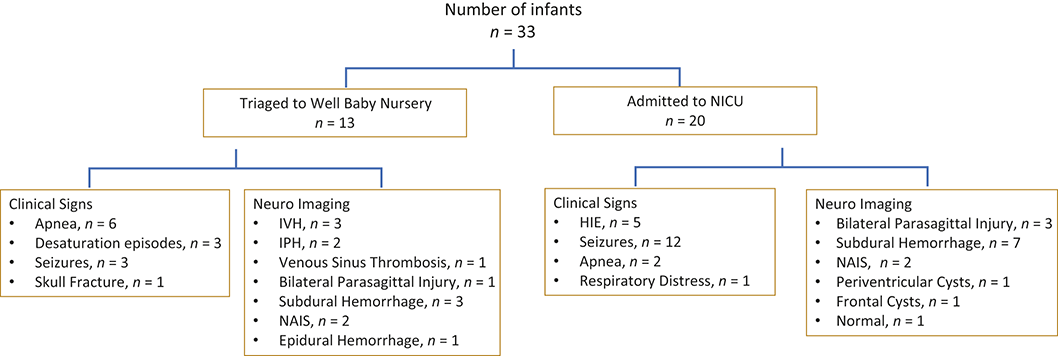
Figure 1.3 Long description
The flowchart begins with the heading number of infants n equals 33. The infants are divided into two groups. One group is labelled triaged to well baby nursery, n equals 13. Under this group, clinical signs include apnoea, n equals 6; desaturation episodes, n equals 3; seizures, n equals 3; and skull fracture, n equals 1. Also under this group, neuroimaging includes IVH, n equals 3; IPH, n equals 2; venous sinus thrombosis, n equals 1; bilateral parasagittal injury, n equals 1; subdural haemorrhage, n equals 3; NAIS, n equals 2; and epidural haemorrhage, n equals 1. The second group is labelled admitted to NICU, n equals 20. Under this group, clinical signs include HIE, n equals 5; seizures, n equals 12; apnoea, n equals 2; and respiratory distress, n equals 1. Also under this group, neuroimaging includes bilateral parasagittal injury, n equals 3; subdural haemorrhage, n equals 7; NAIS, n equals 2; periventricular cysts, n equals 1; frontal cysts, n equals 1; and normal, n equals 1.
Summary
1. Clinical signs of NE including HIE may manifest in the DR or be delayed with onset of clinical signs in the first 24 h of life.
2. Hypoxic–ischemic encephalopathy appears to be less frequent in occurrence (range 0.27–0.34/1,000 births), whereas ICH and SDH were relatively more common.
3. With the exception of infants who progressed to HIE, no perinatal marker was able to prospectively identify the remaining infants destined for brain injury; such infants were only identified at the time of clinical signs/symptoms.
4. Histologic CA was associated with both SDH and FTV with cerebral infarction (CI), consistent with prior studies [Reference Bourke-Taylor, Joyce, Morgan, Reddihough and Tirlea28,Reference De Felice, Toti, Laurini, Stumpo, Picciolini and Todros29].
Perinatal Hypoxic–Ischemic Encephalopathy
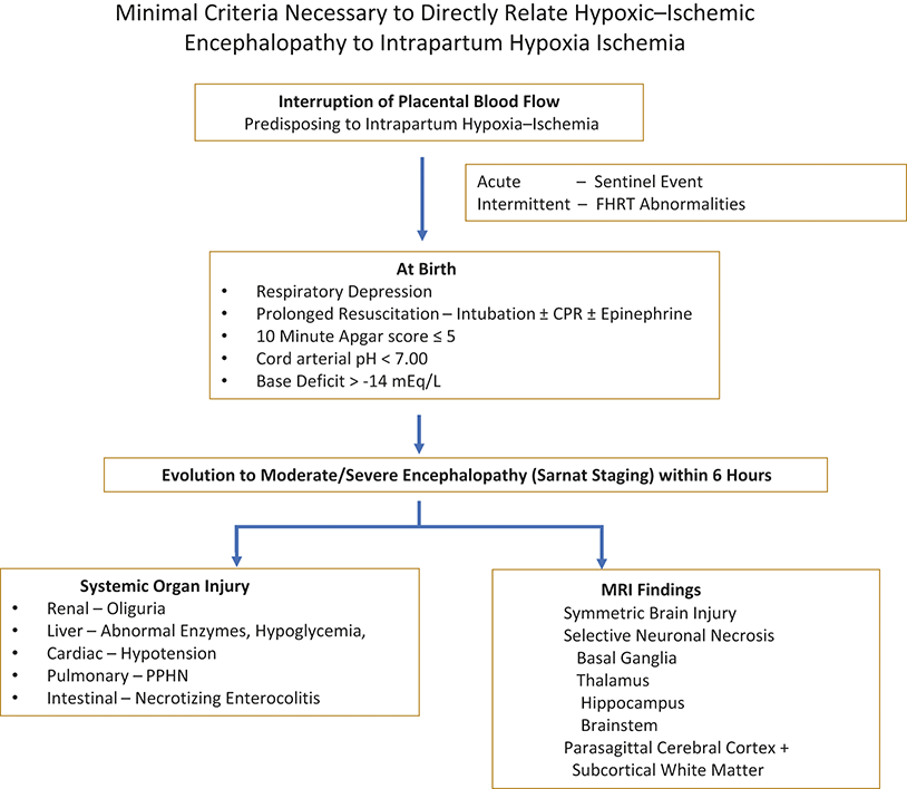
Perinatal HIE remains the most prominent recognized cause of NE with an incidence of approximately 1–1.5 per 1,000 live term births [Reference Kurinczuk, White-Koning and Badawi2]. The diagnosis of HIE is based on a constellation of clinical and biochemical findings noted during labor and around the time of delivery, with postnatal evolution of moderate/severe encephalopathy, presence of systemic organ dysfunction, and evidence on MRI of symmetric brain injury located to the deep nuclear gray matter or watershed cortical and white matter injury (Figure 1.4) [Reference Volpe1,Reference Hollung, Bakken and Vik16]. The constellation of findings described above will be discussed in greater detail in the subsequent sections.
Minimal criteria necessary to implicate intrapartum hypoxia ischemia in the genesis of hypoxic–ischemic encephalopathy.
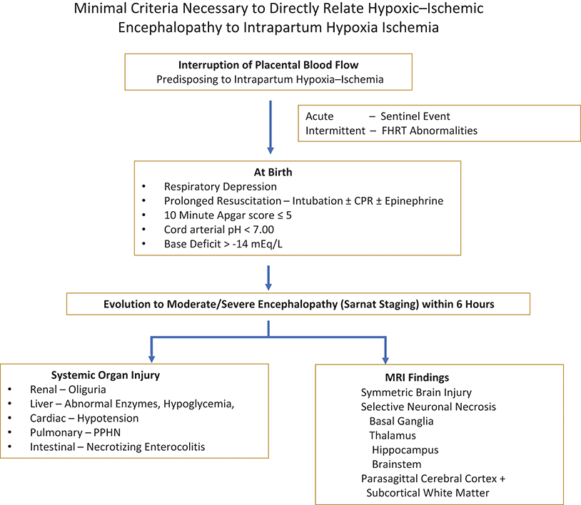
Figure 1.4 Long description
The flowchart begins with Interruption of Placental Blood Flow predisposing to Intrapartum Hypoxia-Ischemia. An additional pathway shows Acute Sentinel Event. Another pathway shows Intermittent - F H R T Abnormalities. The sequence continues to At Birth, followed by listed findings. These include Respiratory Depression; Prolonged Resuscitation - Intubation plus or minus C P R plus or minus Epinephrine; 10 Minute Apgar score less than or equal to 5; Cord arterial p H less than 7.00; and Base Deficit greater than minus 14 milliequivalent per litter. The next step shows Evolution to Moderate or Severe Encephalopathy (Sarnat Staging) within 6 Hours. Two outcome categories are shown next. One category shows Systemic Organ Injury, including Renal - Oliguria; Liver - Abnormal Enzymes, Hypoglycaemia; Cardiac - Hypotension; Pulmonary - PPHN; and Intestinal - Necrotizing Enterocolitis. The other category shows M R I Findings, including Symmetric Brain Injury; Selective Neuronal Necrosis; Basal Ganglia; Thalamus; Hippocampus; Brainstem; and Parasagittal Cerebral Cortex plus Subcortical White Matter.
Pathway to Brain Injury
Studies with term fetal or neonatal monkeys provide important insights into the relation of FHR changes to hypoxia ischemia (HI) and subsequent neuropathology [Reference McNamara, Morgan and Novak30,Reference Novak, Morgan and Adde31]. With severe “total” asphyxia the resultant neuropathology involved principally the thalamus and brainstem. Partial interference in respiratory gas exchange (partial asphyxia) resulted in slower evolving hypoxia and acidosis, followed by late FHR decelerations, a reduction in cardiac output, hypotension, and eventual evidence for cerebral ischemia. Depending on the duration and severity, brain injury became apparent from one to several hours after the insult. The subsequent injury was located within a watershed distribution, that is, parasagittal cerebral cortex and white matter as well as basal ganglia (BG) [Reference McNamara, Morgan and Novak30,Reference Novak, Morgan and Adde31,Reference Kinney, Volpe and Volpe32]. The topography of the lesions is similar to that described in humans [Reference Kinney, Volpe and Volpe32].
Definition of Asphyxia
Birth asphyxia develops during the first and second stages of labor secondary to interruption of PBF, with impaired gas exchange, which leads, if persistent, to hypoxemia and hypercapnia. The process is identified by fetal acidosis (measured in umbilical arterial blood), which broadly reflects the degree of anaerobic metabolism that evolves during periods of hypoxia or increased oxygen demand [Reference Rainaldi and Perlman33]. The umbilical arterial pH that best defines asphyxia remains unclear. Fetal acidemia had originally been defined as an umbilical arterial pH < 7.2. With this definition the rate of ‘‘asphyxia’’ ranged from 5% to 20% and the risk of neonatal neurologic sequelae was low [Reference Einspieler, Prayer and Marschik34]. Subsequently it was noted that severe fetal acidemia, that is, umbilical arterial pH < 7.00, reflects a degree of acidemia where the risk of adverse neurologic sequelae is increased [Reference Olsen, Brown and Eeles35,Reference Einspieler, Bos, Libertus and Marschik36,Reference Goodwin, Belai, Hernandez, Durand and Paul37]. A cord pH of <7.0 is present in from 0.3 to 3.9 per 1,000 of term newborns [Reference Prechtl, Einspieler, Cioni, Bos, Ferrari and Sontheimer38,Reference Kraus, Saucedo, Byrne, Chalak, Pruszynski and Spong39]. A systematic review evaluated the association of umbilical arterial pH <7.0 with the incidence of HIE and the proportion of cases of CP in term infants. The incidence of an arterial pH <7.0 at birth was 3.7 of 1,000 term newborns of which 17.2% survived with neonatal neurologic morbidity, 16.3% had seizures, and 5.9% died during the neonatal period. The incidence of HIE was 2.5 of 1,000 live births. The proportion of CP associated with intrapartum HI was 14.5%. This adds further support for the relatively small contribution of intrapartum HIE to CP [Reference Alexander, Amery and Salt40].
Circulatory Changes Accompanying Interruption of Placental Blood Flow (Asphyxia) to Preserve Cerebral Perfusion
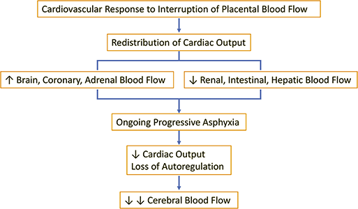
When PBF is compromised, cardiac output redistributes to protect more vital organs (e.g., brain, myocardium, and adrenal glands) (Figure 1.5). This chemoreflex-mediated alteration of blood flow [Reference Kwong, Eeles, Olsen, Cheong, Doyle and Spittle41] is at the expense of decreased flow to “less vital” organs such as the kidney or intestine, as discussed in detail in Chapter 5 [Reference Cohn, Sacks, Heymann and Rudolph42,Reference Peeters, Sheldon, Jones, Makowski and Meschia43,Reference Jensen, Garnier and Berger44]. With time there is loss of cerebral vascular autoregulation resulting in a pressure passive circulation, eventual diminution in cardiac output with resultant hypotension, and ultimately a critical decrease in cerebral perfusion and oxygen delivery (Figure 1.4) [Reference Volpe45,Reference Dawes, Jacobson, Mott, Shelley and Stafford46,Reference Himmelmann, Horber and De La Cruz47]. The mechanisms involved in the blood flow redistribution include peripheral vasoconstriction, triggered by a carotid chemoreflex, and endocrine factors and then maintained or subsequently modified by endocrine and local components [Reference Himmelmann, Horber and De La Cruz47,Reference Morrison, Gardner, Fletcher, Bloomfield and Giussani48,Reference Jones, Sheldon, Peeters, Makowski and Meschia49]. The critical relationship between blood pressure and CBF has been categorized in the experimental model. Thus, with initial arterial hypoxemia, fetal vascular resistance can decrease by at least 50% to maintain CBF with no or a minimal decrease in cerebral oxygen delivery [Reference Koehler, Jones and Traystman50,Reference Haataja, Mercuri and Guzzetta51,Reference Johnson, Palahniwk and Tween52]. Critical to this state is a normal or elevated mean arterial blood pressure. However, persistent, severe hypoxemia leads to hypotension, and cerebral vascular resistance cannot decrease further, resulting in a marked reduction in CBF and a high likelihood of ischemic cellular injury [Reference Volpe45,Reference Romeo, Ricci and van Haastert53].
Fetal circulatory response to interruption of placental blood flow.
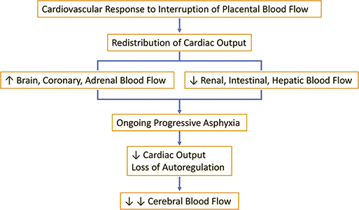
Figure 1.5 Long description
The flowchart begins with Cardiovascular Response to Interruption of Placental Blood Flow. This leads to Redistribution of Cardiac Output. Two outcomes are shown next. One path shows Increased Brain, Coronary, Adrenal Blood Flow. The other path shows Decreased Renal, Intestinal, Hepatic Blood Flow. These paths lead to Ongoing Progressive Asphyxia. The next step shows Decreased Cardiac Output and Loss of Autoregulation. The final step shows Decreased Cerebral Blood Flow.
Respiratory Breathing Patterns in Response to Asphyxia
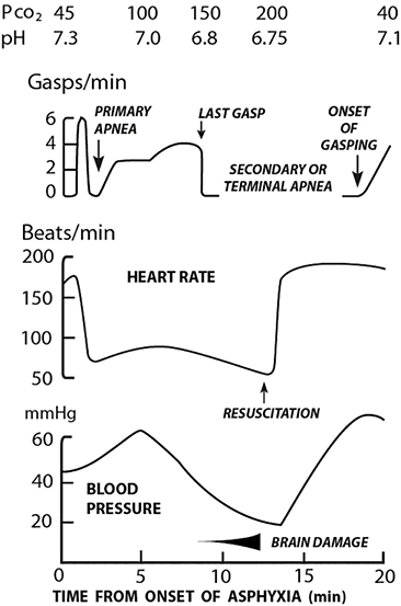
In addition to the cardiovascular changes that occur with asphyxia, characteristic changes in breathing patterns occur. Critical to understanding the relationship between respiratory and circulatory changes is the work of Dawes et al. (Figure 1.6) [Reference Dawes, Jacobson, Mott, Shelley and Stafford46,Reference Dawes54]. In rhesus monkeys, asphyxia was induced by umbilical cord ligation and covering the head with a small bag of warm saline with a characteristic series of changes seen. Within 30 s of total asphyxia, a brief period of rapid rhythmic respiratory effort occurred. This culminated in apnea (primary) and bradycardia, which lasted for approximately 30–60 s (Figure 1.6). The animal then began to have gasping respirations, but spontaneous regular respiration could be induced via prompt physical stimulation and PPV after reoxygenation. If no intervention was performed, the gasping lasted for approximately 4 min. It gradually became weaker until a terminal “last gasp” occurred. This was deemed to be secondary apnea with a heart rate (HR) invariably <60 bpm and pH < 7.00. Unless resuscitation was initiated, death followed. These changes appeared to be pH related, that is, when the arterial pH was >7.17, asphyxiated monkeys were initially able to maintain rhythmic respirations followed by apnea and gasping respirations. By contrast, when the arterial pH was <7.14 there was no onset of rhythmic respiration; rather, the monkey progressed directly to gasping. Notably for every 1-min delay in initiating resuscitation at this stage, it took approximately 2 min before gasping was observed and 4 min until respirations became apparent [Reference Dawes54]. In this regard a nonbreathing newborn observed immediately following delivery is likely to be in primary apnea because in most cases they respond to stimulation and PPV with a rapid increase in HR. For those in secondary apnea with pH < 7.0, the likelihood of cardiopulmonary resuscitation (CPR) is markedly increased.
Respiratory and circulatory responses to asphyxia. Note with primary apnea the arterial pH is >7.0 and recovery in most cases will respond to stimulation and rarely positive pressure ventilation, whereas at the point of secondary apnea the heart rate is often <60 bpm and the arterial pH < 7.00. In this situation resuscitation is more intensive and often will require intubation ± CPR.
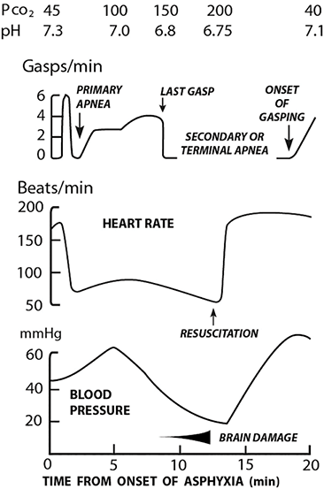
Figure 1.6 Long description
The three graphs have a shared horizontal axis labelled Time From Onset of Asphyxia in minutes, scaled from 0 to 20 in increments of 5. At the top of the figure, text lists P subscript C O 2 values of 45, 100, 150, 200, and 40, with corresponding pH values of 7.3, 7.0, 6.8, 6.75, and 7.1. The upper graph is labelled Gasps per minute. The vertical axis is scaled from 0 to 6 in increments of 2. The tracing begins with a sharp decrease labelled Primary Apnoea. This is followed by a moderate increase in gasping activity labelled Last Gasp. The tracing then reaches zero during a phase labelled Secondary or Terminal Apnoea. The tracing increases again at a point labelled Onset of Gasping. The middle graph is labelled Beats per minute and Heart Rate. The vertical axis is scaled from 50 to 200 in increments of 50. The tracing begins near 200, decreases, and continues at lower values. A point along the tracing is labelled Resuscitation, after which the tracing rises toward higher values. The lower graph is labelled Blood Pressure, with units shown as m m H g. The vertical axis is scaled from 20 to 60 in increments of 20. The tracing initially increases, then decreases to lower values, and later increases again following resuscitation. Along the horizontal axis beneath this graph, a segment is labelled Brain Damage, extending approximately from 10 to 15 minutes. Values are estimated.
Noncirculatory Responses to Asphyxia
Several biologic factors aid in preserving critical organ viability during and following asphyxia. Cerebral metabolic rate is lower in the fetus than in the term infant or adult, creating a more favorable ratio of energy supply and demand. Additionally, the neonatal brain has the capacity to use alternate energy sources when needed [Reference Romeo, Cioni, Scoto, Pizzardi, Romeo and Guzzetta55]. During relative oxygen and glucose depletion, energy substrates such as lactate and ketones become critical for cerebral metabolism [Reference Yager, Heitjan, Towfighi and Vannucci56,Reference Dawes, Mott and Shelley57]. The fetal and neonatal myocardium is more resistant to HI than the adult myocardium [Reference Thompson58]. In addition to the brain and heart, fetal hemoglobin may also allow for a greater tolerance to a hypoxic environment [Reference Wimberley59], because at low oxygen tensions (i.e., below a “crossover pO2”), a left-shifted fetal hemoglobin–oxygen dissociation curve may be advantageous in delivering more oxygen to tissues [Reference Aberman60,Reference Romeo, Cowan and Haataja61].
Summary
1. The adaptive mechanisms briefly described above allow the fetus to maintain CBF and oxygen under most adverse clinical states. These adaptations likely account for the relatively low occurrence of perinatal HIE, that is, approximately 1–1.5/1,000 term deliveries despite many more infants being at risk secondary to intrapartum complications.
Pathophysiology of Acute/Subacute Hypoxic–Ischemic Brain Injury
When the compensatory mechanisms described above are overwhelmed and CBF can no longer meet demand, a cascade of biochemical events ensues. These events are complex, interrelated, and will ultimately lead to cell death without intervention. With compromised CBF, oxygen delivery is reduced, there is a switch to anaerobic glycolysis with a reduction in high-energy phosphate compounds [i.e., adenosine triphosphate (ATP) and phosphocreatinine]. Lactic acid accumulates and membrane ion pumps fail (Na+/K+ ATPase and Na+/Ca2+ exchanger). With membrane pump failure, sodium and water influx into cells, leading to cell swelling. Calcium also flows into cells, which initiates the release of excitatory amino acids such as glutamate into the extracellular space. This overexcitation leads to more calcium influx, fostering an excitotoxic cycle [Reference Romeo, Cowan and Haataja62,Reference Maitre, Burton and Duncan63]. Further consequences include formation of free radicals, production of nitric oxide, and lipid peroxidation of cell membranes. The endpoint of cell death is classically described to occur via necrosis or apoptosis. Necrosis represents a rapid and severe breakdown of cellular function that occurs with the primary HI insult [Reference Romeo, Cowan and Haataja62,Reference Maitre, Burton and Duncan63,Reference te Velde, Tantsis and Novak64]. Following resuscitation, cerebral perfusion and oxygenation is restored, along with partial restoration of energy sources over 30 min, followed by a latent phase in which the patient has near-normal oxidative metabolism. However, there is a subsequent progressive decrease in high-energy phosphates from 24 to 48 h later, that is, secondary energy failure [Reference Romeo, Cowan and Haataja62,Reference Vannucci, Towfighi and Vannucci65,Reference McNamara, Scott, Boyd, Webb, Taifalos and Novak66]. During this state, reperfusion injury occurs due to extended reactions from the primary insult, characterized by inflammation, generation of reactive oxygen species and free radicals, and eventual apoptotic cell death. Apoptosis may be induced through caspase-dependent or gene transcription (caspase-independent) processes [Reference te Velde, Tantsis and Novak64]. Because of its delayed nature, preventing apoptosis has become an enticing target for therapeutic hypothermia (TH) and other therapies [Reference Romeo, Cowan and Haataja62,Reference Maitre, Byrne and Duncan67].
Pathologic Brain Injury following Perinatal Asphyxia in Term Infants
Brain injury following interruption of PBF (asphyxia) is HI in nature and is seen in characteristic locations at autopsy or upon MRI. The major neuropathologic types of injury in the term infant include selective neuronal necrosis (SNN) and parasagittal cerebral injury. Multi-/focal injury is a rare form attributed to HI. Three basic patterns of SNN are described. First, diffuse neuronal injury occurs with very severe and prolonged insults with involvement of hippocampus, perirolandic cortex, deep nuclear structures (caudate, putamen, globus pallidus, and thalamus), and brainstem (cranial nerve nuclei, pons, and inferior olivary nuclei). Second, a cerebral cortical – deep nuclear neuronal – predominance occurs with moderate to severe insults. Deep nuclear involvement includes BG, especially the putamen and thalamus. Third, deep nuclei–brainstem neuronal predominance occurs with severe, relatively abrupt insults (e.g., abruption placentae). Parasagittal cerebral injury refers to a lesion of cerebral cortex and subcortical white matter that occurs involving bilateral parasagittal and superomedial aspects of the cerebral convexities. In the more severe injury this extends to the lateral cerebral convexity, particularly in the parieto-occipital regions [Reference Rainaldi and Perlman33,Reference Romeo, Cowan and Haataja62].
MRI
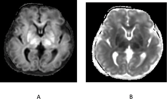
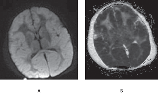
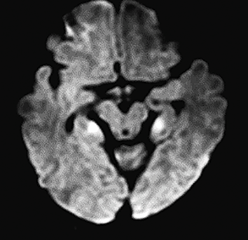
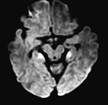
MRI imaging has proved invaluable in diagnosing brain injury in infants with HIE following intrapartum HI [Reference Morgan, Romeo and Chorna68,Reference Ryll, Wagenaar, Verhage, Blennow, de Vries and Eliasson69,Reference Ouwehand, Smidt and Dudink70]. The major types of brain injury attributed to HI are illustrated in Figures 1.7–1.9. Heterogeneous patterns are common [Reference Steinman, Gorno-Tempini and Glidden71], as elements of more than one of these patterns are frequently appreciated. Partial lesions may also be found, as described in a recent study where 10 infants that underwent TH had isolated bilateral hippocampal injury [Reference Guttmann, Flibotte and DeMauro72]. MRI findings associated with poor outcome include involvement of the BG and thalamus, posterior limb of the internal capsule, and loss of gray–white matter differentiation [Reference Morgan, Romeo and Chorna68,Reference Ryll, Wagenaar, Verhage, Blennow, de Vries and Eliasson69,Reference Ouwehand, Smidt and Dudink70].
There is extensive confluent restricted water diffusion on the diffusion-weighted imaging (DWI) sequences involving the basal ganglia with corresponding hypointensity on the apparent diffusion coefficient (ADC) maps.
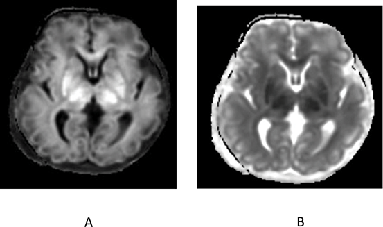
Figure 1.7 Long description
Panel A shows an axial brain scan with increased signal intensity in the bilateral basal ganglia regions relative to surrounding brain tissue. The ventricles are visible centrally, and cortical structures are visible peripherally. The basal ganglia appear as symmetric areas of higher signal. Panel B shows an axial brain scan at the same level. The bilateral basal ganglia regions appear darker relative to the surrounding brain tissue. The ventricular system and cortical outlines are visible, with symmetric low-signal areas in the basal ganglia compared with panel A.
Diffuse cerebral edema involving the entire bilateral cerebral cortex and deep gray matter that demonstrate profound confluent diffusion hyperintensity and corresponding ADC hypointensity. Mass effect on the brain is noted with swelling of the cortical gyri and diffuse sulcal effacement.
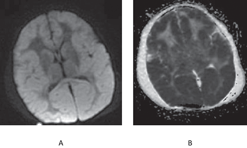
There is restricted diffusion in the bilateral medial temporal lobes/hippocampus.
Clinical and Biomarkers Used to Identify Infants at High Risk for Evolving HIE
Cord Arterial pH < 7.00 Only
A study of 35 term infants born with an umbilical arterial pH < 7.00 who exhibited no difficulties in the DR and triaged to the WBN revealed the following. All infants exhibited an uncomplicated neonatal course and were discharged home within 36–48 h [Reference King, Jackson and Burks73]. These findings indicate that a cord arterial pH < 7.00 in and of itself does not portray an adverse outcome in most cases.
CPR in the DR
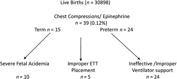
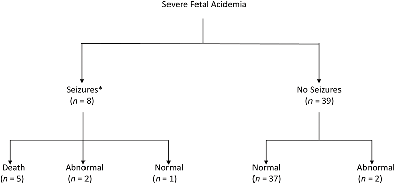
A study of 39 infants who received CPR in the DR demonstrated the importance of an associated cord arterial pH < 7.00 (Figure 1.10). All term infants who received CPR with a cord pH > 7.0 had a normal neurologic course, whereas infants who received CPR with a cord pH < 7.0 all had an abnormal neurological examination including HIE and seizures [Reference Perlman and Risser74]. In another study, 47 infants with a cord arterial pH < 7.00 were admitted to intensive care of whom 37 (95%) exhibited a benign neurologic course and two had mild hypotonia [Reference Toldo, Varishthananda and Einspieler75]. Eight infants progressed to severe brain injury with signs of HIE and seizures (Figure 1.11). These infants were 234-fold more likely to have received CPR in the DR as compared to infants with a benign neonatal course (Figure 1.11) [Reference Toldo, Varishthananda and Einspieler75].
Intensive resuscitation in the delivery room. Note that 39 infants were administered cardiopulmonary resuscitation [26].
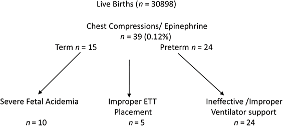
Figure 1.10 Long description
The chart begins with Live Births (n equals 30898). This leads to Chest Compressions or Epinephrine, with n equals 39 (0.12 percent). From this point, three paths are shown. One path is labelled Term n equals 15 and ends at Severe Foetal Acidemia with n equals 10. A second path ends at Improper E T T Placement with n equals 5. A third path ends at Preterm n equals 24 and leads to Ineffective or Improper Ventilator support with n equals 24.
Infants born in the presence of fetal acidence (cord arterial pH < 7.00) and admiitted to intensive care. Note that 8 presented with seizures. Note that 6/8 of these infants required CPR. The odds ratio estimate for developing seizures with CPR was 234.
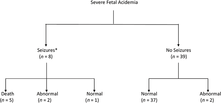
Figure 1.11 Long description
The classification chart begins with Severe Foetal Acidemia at the top. Two paths are shown below. One path is labelled Seizures asterisk with n equals 8. From this group, three outcome categories are shown: Death with n equals 5, Abnormal with n equals 2, and Normal with n equals 1. The second path is labelled No Seizures with n equals 39. From this group, two outcome categories are shown: Normal with n equals 37 and Abnormal with n equals 2. At the bottom of the figure, a line of text reads 6 of 8 infants required C P R in the D R. The O R estimate for developing seizures following.
Combination of High-Risk Markers
In another study, 96 term infants considered to be at high risk for HIE/seizures were delivered in the presence of high-risk markers including MSAF, FHR abnormalities, sentinel event, PPV in the DR, intubation, a 5-min Apgar score of ≤5, umbilical cord arterial pH < 7.00, and or a BD of >–14 mEq/L. Seizures developed in 5 (5.2%) infants. A combination of markers i.e., low pH, DR intubation and a low 5-minute Apgar score showed a strong relationship with seizures with an odds ratio (OR) of 340 (95% confidence limits (CI) 17.3 and 6480); sensitivity 80%, specificity 98.8% and positive predictive value 80% (Table 1.1) [Reference Blair, Langdon, McIntyre, Lawrence and Watson26].
| Predictor | Odds ratio | 95% CI Lower–Upper | Sensitivity | Specificity | PPV |
|---|---|---|---|---|---|
| 5-min Apgar ≤ 5 | 20.9 | 2.2–200 | 80 | 85 | 22.2 |
| pH < 7.00 | 47 | 2.7–892 | 100 | 81 | 23.8 |
| pH ≤ 7.00 DR intubation | 163 | 7.9–3343 | 100 | 94 | 50 |
| pH ≤ 7.00 DR intubation 5-min Apgar ≤ 5 | 340 | 17.8–6480 | 80 | 98.8 | 80 |
Summary
1. CPR with a normal pH was associated with an uncomplicated neonatal course.
2. CPR with a cord arterial pH < 7 increased the likelihood of abnormal outcome.
3. The presence of severe fetal acidemia coupled with intubation or CPR provides initial objective evidence of a severe acute intrapartum insult of sufficient duration or severity to increase the risk of subsequent brain injury.
Prolonged Resuscitation
Several studies have also demonstrated an increased risk of adverse outcome with a longer need for resuscitation. In a study of 178 infants with postasphyxial HIE, there was an increased risk of severe adverse outcome, that is, death or major neurosensory impairment, with delayed onset of breathing. The risk was 42% for infants with first breath at 1–9 min, 56% at 10–19 min, and 88% at >20 min. In addition, the need for CPR was one of the three most important variables predicting adverse outcome [Reference Juneja, Einspieler and Khosla76]. In another study of 375 infants with postasphyxial HIE, significant predictors of outcome were need for CPR >1 min, onset of respiration >30 min, and birth depression (BD) >16 mEq/L [Reference Shah, Beyene, To, Ohlsson and Perlman77]. The rate for severe adverse outcome was 93% for infants who had all three predictors.
Postnatal Acid–Base Status
There is increasing evidence that early postnatal metabolic acidosis is associated with an increased risk for neurodevelopmental (ND) deficits. In a retrospective review of 35 term infants with HIE, an initial BD ≥20 mmol/L obtained within 30 min of life had a predictive value of 94% for death or severe disability at 18 months [Reference Gorter, Ketelaar, Rosenbaum, Helders and Palisano78]. In a secondary analysis of data from the multicenter whole-body cooling study, an important predictor for death/disability was BD > 22mmol/L in the first postnatal gas [Reference Ryll, Krumlinde-Sundholm and Verhage79]. Finally, infants treated with selective head cooling, those infants with more severe deficits at follow up could be distinguished from infants with a normal outcome by the presence of a severe acidosis, that is, pH < 7.00 noted within the first hour following delivery [Reference Takenouchi, Cuaycong, Ross, Engel and Perlman80].
Subacute HI Injury Presenting at Birth
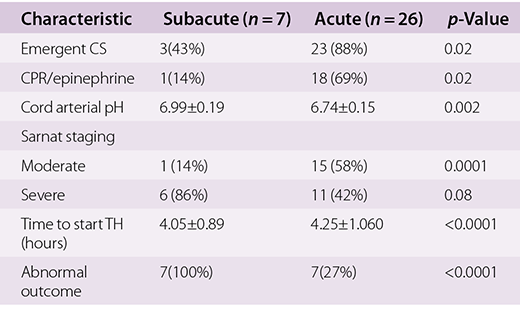
From the preceding section infants at high risk for acute HIE can be identified based on evidence of a sentinel event and/or FHR abnormalities during labor plus a combination of postnatal markers, that is, cord arterial pH < 7.00, prolonged ± CPR, and a low Apgar score. However, a recent study a group of infants delivered with severe encephalopathy were considered to have suffered a subacute insult based on the following characteristics: absence of an acute sentinel event, often with decreased fetal movements for more than 6 h before delivery and with severe encephalopathy immediately following birth without the need for CPR and often with a cord arterial pH > 7.00 [Reference Kasdorf, Grunebaum and Perlman81]. Compared to infants who presented with acute interruption of PBF (placental abruption), infants with subacute injury were significantly less likely to deliver via emergent CS, required less CPR in the DR, and exhibited a higher cord arterial pH and BD compared with the acute group. Infants with subacute injury were more likely to present with severe rather than moderate encephalopathy (Table 1.2). An abnormal outcome was universal with subacute injury as opposed to 27% in those infants with an acute insult (p = 0.0001).

Table 1.2 Long description
The table has four columns: Characteristic, Subacute (n equals 7), Acute (n equals 26), and p-Value. It reads as follows. Row 1. Emergent C S: 3 (43 percent); 23 (88 percent); 0.02. Row 2. C P R or epinephrine: 1 (14 percent); 18 (69 percent); 0.02. Row 3. Cord arterial p H: 6.99 plus minus 0.19; 6.74 plus minus 0.15; 0.002. Under Sarnat staging: Row 4. Moderate: 1 (14 percent); 15 (58 percent); 0.0001. Row 5. Severe: 6 (86 percent); 11 (42 percent); 0.08. Row 6. Time to start T H (hours): 4.05 plus minus 0.89; 4.25 plus minus 1.060; less than 0.0001. Row 7. Abnormal outcome: 7 (100 percent); 7 (27 percent); less than 0.0001.
CS, cesarean section; TH, therapeutic hypothermia.
Case Presentation
This was a 3,415 g 39 5/7-week female with an uncomplicated pregnancy. The mother presented with decreased fetal movements for approximately 30 h. Upon arrival to labor the FHR tracing was flat with no variability and a biophysical profile of 2. The infant was delivered via emergent CS and delivered with a weak cry and soon stopped breathing. She required PPV for a HR < 100 bpm with a good response, and was intubated at 4.5 min of life for continued poor respiratory effort. Apgar scores were 2, 5, 7 at 1, 5, and 10 min, respectively. Her cord arterial pH was 7.12, pCO2 63, and BD 9 mEq/L. Initial postnatal blood gas showed pH 7.33, pCO2 33, BD 13, and a lactate of 17. Initial blood glucose was 36 mg/dL, which was treated. The initial hematocrit was 13% and the infant was transfused. A maternal Kleihauer Betke test was 10.5%, suggesting a significant fetomaternal hemorrhage. She developed eye deviation with lip smacking at approximately 2.5 h of life. An EEG was concerning for seizure activity. The infant was treated with anticonvulsants and underwent TH for 72 h. The EEG progressed to a burst suppression pattern. An MRI was obtained, which demonstrated diffuse cerebral edema involving the entire bilateral cerebral cortex and deep gray matter with profound confluent diffusion hyperintensity and corresponding hypointensity of ADC (Figure 1.8). The parents requested comfort care.
This case is representative of infants presenting with subacute injury; all received TH within 6 h, with a uniformly unfavorable outcome. This is consistent with a presumed “subacute” insult, remote from delivery, with further compromise during delivery. Bonifacio and colleagues similarly showed that all infants with decreased fetal movements had some degree of injury with persistence of a watershed predominant pattern, despite TH. The authors suggested that TH may be less beneficial when there has been HI remote from the time of birth [Reference Bonifacio, Glass and Vanderpluym82]. In this regard, Wyatt et al. found that encephalopathy grade at time of randomization was the single most predictive factor for adverse outcome in the TH population, with moderate encephalopathy associated with better outcome at 18 months of age [Reference Pagnozzi, Shen and Doecke83]. Experimental observations in the fetal lamb highlight the importance of time to initiation of TH on the extent of subsequent brain injury [Reference Gunn, Gunn, de Haan, Williams and Gluckman84]. Thus, cooling initiated within 30 min of ischemia significantly reduced cytotoxic edema and the extent of neuronal loss [Reference Gunn, Gunn, de Haan, Williams and Gluckman84]. When TH was delayed until 5.5 h after the primary insult, and before the onset of seizures, partial neuroprotection was still demonstrated [Reference Gunn, Gunn, Gunning, Williams and Gluckman85]. However, a less uniform effect was seen, particularly within the hippocampus. Cytotoxic edema as measured by cortical impedance was also decreased, yet there was no prevention of delayed seizures [Reference Gunn, Bennet, Gunning, Gluckman and Gunn86]. Finally, when cooling was initiated at 8.5 h after birth, after the onset of postasphyxial seizures, no neuroprotection was observed [Reference Gunn, Bennet, Gunning, Gluckman and Gunn86].
Systemic Organ Injury in Infants with Asphyxia
Given the redistribution of cardiac output with interruption of PBF there is a high likelihood of systemic organ injury (see Figure 1.5). This has been evaluated in three studies. Findings vary in part based on the entry criteria and on the methodology used to determine organ injury. The first study prospectively evaluated term infants with a 5-min Apgar score <5 and an umbilical arterial cord pH <7.20 [Reference Perlman, Tack, Martin, Shackelford and Amon87]. No evidence of organ injury was noted in 35%, whereas 37% had CNS injury (with moderate to severe encephalopathy), 40% had renal injury (oliguria), 29% had cardiac injury as shown by abnormal electrocardiogram (ECG) and/or echocardiogram, and 34% had pulmonary injury shown by need for mechanical ventilation (MV) for >48 h. Renal injury was the best systemic marker of potential brain injury with likelihood of poor neurologic outcome increased 2.8-fold with transient and 5.1-fold with persistent oliguria [Reference Perlman and Tack88]. A second study evaluated term infants with a 5-min Apgar score < 7, scalp pH < 7.20, and PPV > 1 min. Abnormal outcome included CNS, defined as moderate to severe encephalopathy (72%); renal, oliguria (42%); pulmonary, FIO2 > 40% and/or need for MV (26%); cardiac, ECG abnormalities suggestive of ischemia (18%); and gastrointestinal, persistent bloody aspirates or frank necrotizing enterocolitis (29%). No organ involvement was noted in 18% [Reference Martin-Ancel, Garcia-Alix, Gaya, Cabanas, Burgueros and Quero89]. A third study enrolled term infants with a 5-min Apgar score < 5, metabolic acidosis (i.e., BD >16 mmol/L), the need for MV, and evidence of moderate to severe encephalopathy [Reference Shah, Riphagen and Beyene90]. Eighty infants had HIE. All infants with HIE had evidence of multiple organ dysfunction (at least one organ dysfunction). No relation between individual or combinations of organ involvements and long-term outcomes was observed.
Summary
1. Systemic organ injury is common in infants with moderate to severe (MS HIE.)
2. Associated renal involvement was observed in approximately 40% of cases, cardiac abnormalities in 30%, and pulmonary in approximately 30–35% of cases.
Clinical Presentation of Infants at High Risk for Evolving Encephalopathy
Infants at high risk for evolving moderate to severe HIE can be identified by a constellation of clinical and biomarkers (Figure 1.4). During labor the presence of a sentinel event and/or FHR abnormalities is the first indicator that the fetus has been stressed. In the DR, if the infant is depressed, that is, absent breathing and hypotonia, the resuscitative steps become important. If bradycardia is unresponsive to intubation and PPV, necessitating CPR ± epinephrine, with a concurrent cord arterial pH < 7.0, this suggests a state of secondary apnea. The duration of the resuscitation efforts will be reflected as a low 10-min Apgar score. The combination of these criteria places the infant at very high risk for evolving MS encephalopathy. The infant should be transferred to intensive care and evaluated for TH. During this period of evaluation every effort should be made to avoid hyperthermia and hypoglycemia.
Evaluation for Moderate to Severe Encephalopathy
Clinical Examination
The Sarnat staging is frequently used for assessing the extent of encephalopathy (Table 1.3) [Reference Sarnat and Sarnat91]. In the original description the maximum staging of encephalopathy was determined in the first 2–3 days of life. This has been compressed to within the first 6 h for the purposes of administering TH. Thus, this represents a sicker group of infants than originally described. We consider the level of consciousness critical to the assessment of encephalopathy. The presence of lethargy is consistent with moderate encephalopathy and is invariably accompanied by other signs, for instance, decreased tone or weak suck. The infant becomes a strong candidate for TH if the other criteria mentioned previously are present. A stuporous/comatose infant should be cooled expeditiously in the absence of any contraindication. Infants with mild encephalopathy should be monitored repeatedly within the 6-h time frame for evolution to moderate to severe encephalopathy.
| Stage 1 | Stage 2 | Stage 3 | |
|---|---|---|---|
| Level of consciousness | Hyperalert | Lethargic | Stuporous/comatose |
| Muscle tone | Normal Mild distal flexion | Mild hypotonia (usually central) Strong distal flexion | Flaccid Intermittent decerebration |
| Stretch reflexes | Overactive | Overactive | Decreased/absent |
| Suck | Weak | Weak or absent | Absent |
| Gag | Weak | Weak or absent | Absent |
| Seizures | None | Common | Uncommon |
Amplitude Electroencephalograph
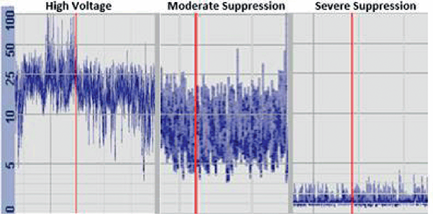
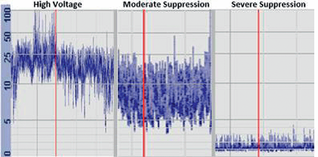
An amplitude electroencephalograph (aEEG) was used as an entry criterion for infants who received selective head cooling. aEEG recordings are characterized as high voltage (normal) (15–25 µV), moderate suppression (upper > 10 and lower ~5 µV), severe suppression (entire band width <5 µV) (Figure 1.12) [Reference al Naqeeb, Edwards, Cowan and Azzopardi91].
Representative aEEG tracings that are characterized as high voltage (normal) (15–25 µV), moderate suppression (upper > 10 and lower ~5 µV), severe suppression (entire band width <5 µV) [Reference Shankaran, Laptook and Ehrenkranz94].
TH as a Neuroprotective Strategy to Reduce Evolving HI Injury
TH has been shown to be effective therapy for NE secondary to HI. Multiple randomized trials have demonstrated that reductions in core temperature either alone or in combination with reduced head temperature [Reference Gluckman, Wyatt and Azzopardi93] consistently reduce death or disability at 18 months [Reference Shankaran, Laptook and Ehrenkranz94,Reference Azzopardi, Strohm and Edwards95,Reference Jacobs, Morley and Inder96]. A Cochrane meta-analysis indicated that TH reduced death or major neurodevelopmental disability in survivors from 61% in noncooled infants versus 46% in infants treated with hypothermia, yielding a risk ratio of 0.75 (95% CI 0.68 to 0.83),with number needed to treat (NNT) for an additional beneficial outcome of 7 (95% CI 5 to 10) [Reference Shevell7]. A second similar meta-analysis showed a beneficial effect in infants that extended to those with moderate encephalopathy (RR, 0.67; 95% CI 0.56 to 0.81) and those with severe encephalopathy (RR 0.83; 95% CI 0.74 to 0.92). The NNT to prevent 1 is 6 for infants with moderate encephalopathy and 7 for those with severe encephalopathy [Reference Tagin, Woolcott, Vincer, Whyte and Stinson97]. The neuroprotective effects of TH persisted at 6–7 years (RR 0.77; 95% CI 0.63 to 0.94) [Reference Shankaran, Pappas and McDonald98,Reference Azzopardi, Strohm and Marlow99]. Given the beneficial effects of hypothermia, the Committee on the Fetus and Newborn of the American Academy of Pediatrics provided an overview of the available data and expectations for centers that provide this therapy [Reference Papile, Baley and Benitz100].
Supportive Care
Ventilation
Changes in carbon dioxide (CO2) are important to monitor carefully as hypercarbia increases and hypocarbia decreases CBF [Reference Kasdorf, Perlman, Perlman and Inter101,Reference Rosenberg, Jones, Traystman, Simmons and Molteni102]. In term infants, several studies have demonstrated an association of hypocarbia (typically <35 mmHg) and adverse ND outcome especially in the setting of HIE [Reference Klinger, Beyene, Shah and Perlman103,Reference Pappas, Shankaran and Laptook104,Reference Lingappan, Kaiser, Srinivasan and Gunn105]. A retrospective cohort study of 198 term infants with moderate to severe HIE treated with TH showed an association of lowest PaCO2 averaged over days 1–4 with identification of brain injury on MRI [OR (95% CI) 1.07 (1.00 to 1.14); p = 0.04] [Reference Lopez Laporte, Wang and Sanon106]. It is recommended that CO2 be maintained in the normal range in mechanically ventilated infants at risk for HIE. This goal may be clinically difficult to achieve as infants with HIE often demonstrate hypocapnia, which may reflect the underlying CNS disease[Reference Nadeem, Murray, Boylan, Dempsey and Ryan107].
Fluid Status
Infants who have suffered HI often progress to a fluid overload state. This may reflect renal failure secondary to acute tubular necrosis or to the syndrome of inappropriate antidiuretic hormone release (Figure 1.5). This often presents with increase in weight, low urine output, and hyponatremia. Although fluid restriction is common practice, a randomized controlled trial of neonates with HIE treated with TH showed no difference in death or ND disability at 6 months between infants randomized to normal fluid versus restricted fluid intake (defined as 2/3 of normal intake) in the first 4 days of life [Reference Tanigasalam, Plakkal, Vishnu Bhat and Chinnakali108]. Thus, fluid and electrolyte administration should be individualized.
Maintenance of Adequate Perfusion
Given the high likelihood of a pressure-passive cerebral circulation, management should aim to maintain the arterial blood pressure within a normal range for age and gestation. Hypotension is not uncommonly related to myocardial dysfunction, endothelial cell damage, or rarely to volume loss. Treatment should be directed toward the cause, that is to say: inotropic support for myocardial dysfunction, and volume replacement for intravascular depletion [Reference Wyckoff, Perlman and Laptook109].
Renal Function
Acute renal injury is common in infants in the setting of HIE (see above) and appears to be modulated by TH. A 2021 meta-analysis showed a significant difference between the incidence of acute renal injury in infants treated with TH versus controls (RR = 0.81, p = 0.03) [Reference van Wincoop, de Bijl-Marcus, Lilien, van den Hoogen and Groenendaal110]. In a randomized controlled trial of infants treated with TH versus standard treatment, the incidence of acute renal injury was approximately 50% less with TH versus standard treatment (p < 0.05) [Reference Tanigasalam, Bhat, Adhisivam and Sridhar111]. The mechanism of this potential protective effect is not entirely understood.
Control of Blood Glucose Concentration
Experimental studies suggest that both hyperglycemia and hypoglycemia may exacerbate brain damage. In adult experimental models of HI, hyperglycemia accentuates brain damage, whereas in immature rodents significant hyperglycemia (blood glucose 600 mg/dL) entirely prevented HI brain damage [Reference Vannucci and Mujsce112]. The effects of hypoglycemia in experimental neonatal models vary, as do the mechanisms of inducing hypoglycemia. Thus, insulin-induced hypoglycemia is detrimental to the immature rat brain subjected to HI. However, with fasting-induced hypoglycemia, a high degree of protection was noted, and may be secondary to increased concentrations of ketone bodies, presumably serving as alternative substrate to the immature brain [Reference Yager113]. Clinically, hypoglycemia when associated with HI appears detrimental to brain. Thus, term infants delivered in the presence of severe fetal acidemia (umbilical arterial pH < 7.0) who presented with an initial blood glucose concentration <40 mg/dL versus >40 mg/dL were 18 times more likely to progress to moderate to severe encephalopathy [Reference Salhab, Wyckoff, Laptook and Perlman114]. Post-hoc analysis of the Cool Cap Study noted an unfavorable outcome at 18 months more often in infants with hypoglycemia (≤40 mg/dL) and hyperglycemia (≥150 mg/dL) within the first 12 h following randomization [Reference Basu, Kaiser and Guffey115]. After adjusting for Sarnat stage and 5-min Apgar score, hyperglycemic infants randomized to TH had a significant reduced risk of death and/or severe disability at 18 months (adjusted risk ratio 0.8; 95% CI 0.66 to 0.99); hypoglycemia or normoglycemia infants did not show any benefit. Derangements in glucose levels have also been associated with varying patterns of brain injury on MRI including when treated with TH. Thus, infants with hypoglycemia had higher adjusted odds of watershed or focal–multifocal injury, whereas infants with labile glucose levels had predominantly basal ganglia or global injury on MRI compared to infants with normal glucose values [Reference Basu, Ottolini and Govindan116].
Seizure Management
HI cerebral injury is a common cause of early-onset neonatal seizures. Experimental evidence strongly suggests that repetitive seizures disturb brain growth and development as well as increase the risk for subsequent epilepsy [Reference Dzhala, Ben-Ari and Khazipov117,Reference Holmes, Gairsa, Chevassus-Au-Louis and Ben-Ari118]. Human studies show conflicting evidence. Glass et al. found that clinical seizures in the setting of HIE pre-TH were associated with worse cognitive and motor outcome at age 4 years [Reference Glass, Glidden, Jeremy, Barkovich, Ferriero and Miller119]. The Cool Cap study also demonstrated that aEEG seizures at time of enrollment were independently associated with an unfavorable outcome [Reference Gluckman, Wyatt and Azzopardi120]. By contrast, the presence of clinical seizures at any time during hospitalization was not associated with death or moderate to severe disability at 18 months in a secondary analysis of the Eunice Kennedy Shriver National Institute of Child Health and Human Development (NICHD) whole-body cooling trial [Reference Kwon, Guillet and Shankaran121]. The optimal management of neonatal seizures in the setting of HIE treated with TH remains unclear [Reference van Rooij, Hellstrom-Westas and de Vries122,Reference DeLaGarza-Pineda, Mailo and Boylan123]. Clinical and/or EEG seizures are invariably treated with an anticonvulsant, commonly phenobarbital as a first-line agent, and treatment is continued if seizures persist until the anticonvulsant therapy has been optimized
Adjuvant Neuroprotective Agents
Multiple alternative and adjuvant agents that target various mechanisms in the cascade of events that lead to neuronal necrosis and apoptosis have been evaluated without much success. Melatonin has been evaluated in experimental and human studies [Reference Robertson, Faulkner and Fleiss124]. A recent meta-analysis concluded that clinical data supporting the neuroprotective effects in neonates are limited [Reference Ahmed, Pullattayil, Robertson and More125]. Erythropoietin has been shown in experimental rodent models to reduce the extent of brain injury [Reference Juul, McPherson and Bammler126]. The drug was subsequently evaluated in a large multicenter, double-blind, randomized, placebo-controlled trial in conjunction with TH versus a placebo group where the incidence of death or neurodevelopmental impairment was comparable between treatment groups. Moreover, the number of serious adverse events per child was significantly higher in the erythropoietin versus the placebo group. Currently there are no adjunctive therapies that enhance the neuroprotection of TH.
Summary
1. HIE is a distinct form of NE and is readily diagnosed when criteria as outlined in the text are met.
2. If criteria are met, TH should be implemented within 6 h.
3. TH is associated with improved outcomes.
Additional Causes of NE Presenting During the First Postnatal Day
Chorioamnionitis
Background
Chorioamnionitis (CA) is defined as inflammation of the amniochorionic membranes of the placenta and is further categorized as (1) clinical CA based on maternal signs including fever, uterine tenderness/abdominal pain, maternal or fetal tachycardia, foul-smelling vaginal discharge, and/or (2) histologic CA (HCA) based on microscopic evidence of an inflammatory response [Reference Seong, Lee and Kang128]. Clinical CA has been diagnosed in approximately 1–3% of pregnancies with intact membranes and 6–10% of those with PROM [Reference Seaward, Hannah and Myhr129]. Most infants (>90%) born to mothers with clinical CA are asymptomatic and discharged without issues [Reference Shalak, Laptook, Jafri, Ramilo and Perlman130]. It has been questioned whether infants presenting with evolving HIE can be distinguished from encephalopathy secondary to CA. In this brief review we will compare and contrast these two conditions with regard to clinical presentation.
Case Presentation
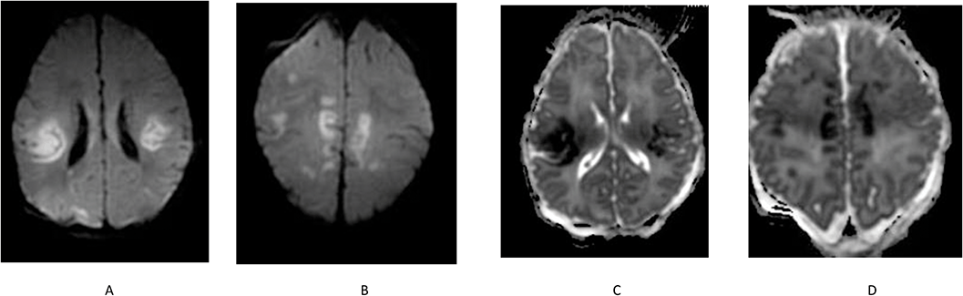
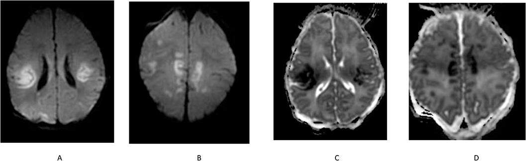
The infant was a 3,155 g 40 1/7-week female delivered via CS secondary to arrest of descent. There was PROM of 36 h duration. The infant emerged meconium stained, depressed, hypotonic with a HR at 1 min < 100 bpm. PPV was initiated with improvement in HR >100 bpm. The infant was intubated for lack of respiratory effort. Apgar scores were 1 and 8 at 1 and 5 min, respectively. Initial temperature was 38.2°C. No cord blood gas was available. Initial blood gas revealed a pCO2 40, pH 7.23, BD –10 mEq/L. She was extubated within 12 h. No blood pressure issues were noted. She was started on antibiotics. Seizures developed at approximately 36 h of life. An EEG revealed seizures emanating from the right frontocentral region with continuous diffuse slowing. She was treated with anticonvulsants. An EEG 3 days later showed improved continuity. A spinal tap was unremarkable. An MRI showed multiple infarcts (Figure 1.13). Magnetic resonance venography (MRV) was normal. Placental pathology was consistent with acute CA and funisitis. She was discharged home on phenobarbital on day of life 13.
Multifocal areas of cortical/subcortical T2 hyperintensity (A and B) with corresponding ADC-restricted diffusion (C and D) are identified throughout the supratentorial region in multiple vascular distributions most prominent within the posterior perisylvian frontoparietal junctions, right greater than left. These are most compatible with multiple acute infarctions.
Summary
1. Several important issues are raised. The infant had an elevated temperature at birth, and exhibited BD, was hypotonic, developed seizures at 36 h of life, and had bilateral symmetric lesions on MRI in a distribution distinct from that noted with HI injury. There was placental evidence of CA and funisitis.
In the sections below, the relationship between clinical CA and maternal fever and neonatal neurologic signs including BD, NE, and seizures will be explored, and the potential link of maternal fever and cytokine release to subsequent NE will be reviewed.
Hyperthermia and Neonatal Brain Injury
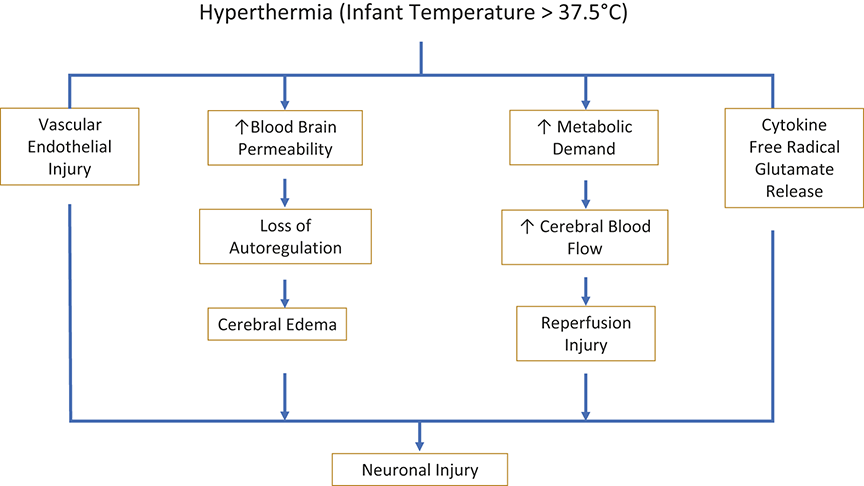
Hyperthermia in an infant is defined as a rectal or axillary temperature >37.5°C, usually secondary to an elevated maternal temperature. Importantly the fetus must maintain a temperature gradient of approximately 0.5°C higher than the mother to dissipate heat across the placenta [Reference Kasdorf and Perlman131]. Hyperthermia may induce a relative hypermetabolic state [Reference Kasdorf and Perlman131,Reference Perlman132] that can be deleterious to neonatal brain [Reference Baumgart133,Reference Sedin, Martin and Walsh MC134,Reference Slinko, Caspersen and Ratner135]. The increase in metabolic demand is associated with a simultaneous increase in CBF, which may exacerbate the extent of reperfusion injury following prior hypoxia–ischemia [Reference Busija, Leffler and Pourcyrous136,Reference Inder, Volpe and Volpe137]. Moreover, an elevated temperature may alter blood–brain barrier permeability, promoting cerebral edema [Reference Natah, Srinivasan, Pittman, Zhao and Dunn138]. The association of hyperthermia and brain injury may be related to several mechanisms including free radical production, glutamate and glycine release, and increased metabolic demands with depletion and subsequent slow recovery of phosphocreatine and adenosine triphosphate levels (Figure 1.14) [Reference Kasdorf and Perlman131]. In addition, there is a clear link between hyperthermia and cytokine release as well as hemostatic changes including thrombosis, which may lead to adverse neurologic outcome [Reference Bouchama, Roberts and Al Mohanna139]. In a baboon experimental model subjected to heatstroke, a significant increase in prothrombin time, activated partial thromboplastin time, and decreases in D-dimer, fibrinogen, and platelets was noted [Reference Leon, Blaha and DuBose140]. Additionally cytokine release with a marked elevation of interleukin (IL)-6 was demonstrated compared to controls [Reference Leon, Blaha and DuBose140], and in mice exposed to heat strain significant elevations in IL-6, IL-10, and IL-1b were noted [Reference Leon and Helwig141].
Potential pathways to neuronal injury with hyperthermia (temperature > 37.5°C).
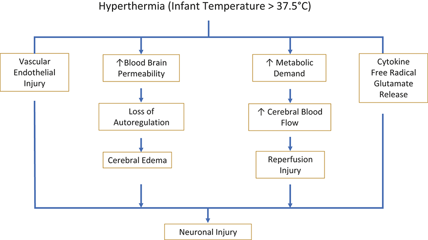
Figure 1.14 Long description
The flowchart begins with Hyperthermia (Infant Temperature greater than 37.5 degrees Celsius). From this, multiple parallel pathways are shown. One pathway shows Vascular Endothelial Injury. A second pathway shows Increased Blood Brain Permeability, which proceeds to Loss of Autoregulation and then to Cerebral Edema. A third pathway shows Increased Metabolic Demand, which proceeds to Increased Cerebral Blood Flow and then to Reperfusion Injury. A fourth pathway shows Cytokine Free Radical Glutamate Release. All pathways converge at the bottom of the diagram at Neuronal Injury.
The Association of Maternal Fever and BD, NE, and Seizures
BD
A clinical study demonstrated a significant relationship between maternal fever (temperature > 38°C), BD (5-min Apgar < 5) and need for assisted ventilation ± chest compressions [Reference Perlman142]. In a second study term infants born to mothers with clinical CA whose infants had a higher rectal temperature at 30 min, were 3.9-fold more likely to have BD (requiring PPV for >2 min, and/or intubation) and were 2.8-fold more likely to be admitted to the NICU [Reference Shalak, Perlman, Jackson and Laptook143].
Maternal fever and NE
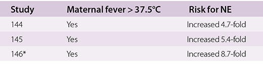
Numerous studies have assessed maternal fever and subsequent NE. The risk for NE ranged from 3.1- to 8-fold among studies. The risk was highest with associated acidosis (see Table 1.4) [Reference Impey, Greenwood, MacQuillan, Reynolds and Sheil144,Reference Blume, Li, Loch and Koepsell145,Reference Impey, Greenwood, Black, Yeh, Sheil and Doyle146].

Table 1.4 Long description
The table has three main columns: Study, Maternal Fever greater than 37.5 degrees Celsius, and Risk for N E. It reads as follows. Row 1. Study 144: yes; increased 4.7-fold. Row 2. Study 145: yes; increased 5.4-fold. Row 3. Study 146 asterisk: yes; increased 8.7-fold.
*Associated acidosis.
Maternal Fever and Neonatal Seizures
Several studies have described seizures in association with intrapartum maternal fever [Reference Lieberman, Lang, Richardson, Frigoletto, Heffner and Cohen147,Reference Lieberman, Eichenwald, Mathur, Richardson, Heffner and Cohen148,Reference Glass, Pham, Danielsen, Towner, Glidden and Wu149]. In one study, infants with seizures were threefold more likely when born to mothers with fever >100.4°C during labor even after controlling for numerous factors, such as epidural use [Reference Lieberman, Eichenwald, Mathur, Richardson, Heffner and Cohen148]. In another study, maternal fever was associated with a significant 2.9-fold adjusted increased risk for seizures [Reference Glass, Pham, Danielsen, Towner, Glidden and Wu149].
Cytokine Responses in Infants with Clinical CA and Associated HIE/NE
The association between the inflammatory cytokine response [IL-6, IL-8, and regulated on activation, normal T-cell expressed and secreted (RANTES) in cord blood, at 6 and 30 h] in term infants born to mothers with clinical CA and BD, abnormal neurologic examination, and HIE was assessed [Reference Shalak, Laptook, Jafri, Ramilo and Perlman130]. Cord cytokine concentrations were higher in infants with CA than in controls. IL-6 increased at 6 h, followed by a fivefold decline by 30 h. In contrast, IL-8 and RANTES progressively decreased over time. There was no relationship between cytokines and BD, but the neurologic examination correlated significantly with IL-6 at 6 h (r < 0.5). All cytokine concentrations were significantly higher in infants with HIE/seizures versus none at 6 h of age. There were no differences in cord or postnatal concentrations for any of the cytokines evaluated between infants with and without HCA. In a second study, serum neuronal glial fibrillary acidic protein (GFAP), ubiquitin carboxyl-terminal hydrolase L1(UBCH), and inflammatory biomarkers (cytokines measured in serum from umbilical artery at 6–24, 48, 72 h) in term newborns with mild, moderate, or severe HIE who received TH was undertaken to determine whether measurements could stratify severity of HIE. At birth, serum GFAP and UBCH increased significantly with the severity of HIE, and serial GFAP remained elevated in neonates with MS HIE. IL-6, IL-8, and vascular endothelial growth factor (VEGF) were significantly greater at 6–24 h with MS HIE. Serial values were unaffected by TH or rewarming. Elevated GFAP, IL-1, IL-6, IL-8, tumor necrosis factor (TNF), interferon (IFN), and VGEF at 6–24 h were associated with abnormal neurological outcomes at 15–18 months [Reference Chalak, Sánchez, Adams-Huet, Laptook, Heyne and Rosenfeld150]. It was concluded that both neuronal and inflammatory biomarkers were elevated in infants with MS encephalopathy. A third prospective study evaluated measures of 10 pro- and anti-inflammatory cytokines including IL-1α, IL-1β, IL-6, IL-8, IL-10, TNF-α, IFN-γ, VEGF, and granulocyte macrophage/colony-stimulating factor (GM-CSF) from term infants requiring resuscitation at birth. Infants with NE and abnormal neuroimaging had significantly elevated GM-CSF at 0–24 h and IL-8, IL-6, and IL-10 at 24–48 h. Elevated IL-10 levels were associated with mortality [Reference Hare, Watson and O’Neill151]. In another study from the same investigator, infants with NE who received TH had significantly higher levels of erythropoietin (Epo), IL-6, and IL-1ra versus controls at baseline. Following lipopolysaccharide (LPS) stimulation an excessive Epo and IL-1ra response was noted. By contrast, IL-8, IL-2, IL-6, TNF-α, GM-CSF, VEGF, and IFN-γ were significantly lower than controls following LPS stimulation. GM-CSF, IFN-γ, IL-1β, IL-1ra, and VEGF were higher on days 1–2 in NE infants with abnormal neuroimaging [Reference O’Dea, Kelly and McKenna152].
Summary
1. A host of cytokines are released to varying degree in infants born to mothers with clinical CA and play an important role in the evolution of MS encephalopathy.
Cytokine Storm
The above observations raise the possibility of whether the increased release of cytokines in infants born to mothers with CA and subsequent NE may reflect a “cytokine storm release,” akin to that described in adults [Reference Fajgenbaum and June153]. It is an umbrella term characterized by constitutional symptoms, systemic inflammation, and multiorgan dysfunction that can lead to multiorgan failure if inadequately treated. Laboratory findings include nonspecific markers of inflammation such as C-reactive protein and prominent elevations in serum inflammatory cytokine levels, as described above [Reference Fajgenbaum and June153].
Effects of the Inflammatory Response on CBF
Experimental Studies
Preterm fetal sheep 107–109 days of gestation were given an intravenous infusion of GM-CSF and an intra-amniotic infusion of endotoxin, which induced necrotizing funisitis and CA in all cases [Reference Saito, Matsuda and Okuyama154]. Cerebral hemodynamics were assessed with near-infrared spectroscopy. A significant decrease in blood oxygen content and increase in brain total hemoglobin levels was observed after endotoxin infusion. Fetuses subjected to a hemodynamic insult – that is, acute hemorrhage and/or exchange transfusion – showed an abrupt decrease in total brain hemoglobin levels. Most fetuses in each treatment group progressed to PVL versus no control fetuses.
Cerebral Blood Flow Velocity
In a neonatal study, cerebral blood flow velocity (CBFV) measurements determined by Doppler ultrasound in the anterior and middle cerebral arteries (ACA, MCA) were obtained between 24 and 72 h after birth in CA-exposed and control infants [Reference Koch, Wagner and Jenkins155]. CA infants had decreased resistance in most major cerebral vessels versus controls. Male HCA infants had lower resistance and demonstrated a loss of correlation of vascular resistance between vessels in all cases. These were tightly correlated in control infants. Male fetuses also had significantly greater release of proinflammatory cytokines versus females after ex-vivo LPS stimulation. Abnormal CBFV correlated negatively with neurodevelopmental outcome at 12 months [Reference Koch, Wagner and Jenkins155]. A second prospective observational study measured resistance index (RI), pulsatility index (PI), and peak systolic flow velocity from internal carotid, vertebral, and middle cerebral arteries utilizing Doppler within 48 h in premature infants (<34 weeks) exposed to clinical CA in the absence of early-onset neonatal sepsis. Significantly higher cord blood IL-6 concentrations, lower resistance (RI and PI), and higher blood flow velocities were recorded in all cerebral arteries of the CA group. A significant correlation was observed between the increase in CBFV and elevated IL-6 concentrations [Reference Basu, Dewangan, Barman, Anupurba, Shukl and Kumar156].
Proinflammatory Cytokines and Myocardial Status in Premature Infants
The relationship between CA cord blood inflammatory cytokine concentrations IL-6, IL-1β, and TNF-α, blood pressure (BP), HR, cardiac output, and middle CBFV were measured shortly after birth in a cohort of premature infants. CA was defined by placental histology and was significantly associated with increased IL-6, IL-1β, and HR; and with decreased mean and diastolic BP [Reference Yanowitz, Jordan and Gilmour157]. IL-6 concentration correlated inversely with systolic, mean, and diastolic BP. Right ventricular cardiac output was significantly elevated in infants with fetal vessel inflammation.
Summary
1. Infants born to mothers with CA, elevations of cytokines and in particular IL-6 are associated with abnormalities in CBF and myocardial function which increases the vulnerability of brain injury.
Placental Evidence of for HCA and its Relationship to NE
This relationship has been evaluated in several studies (Table 1.5). The first case-control study showed no difference in HCA in infants with HIE and three control groups [Reference Bingham, Gundogan, Rand, Farrar, Tucker and Laptook158]. Importantly, other changes observed included fetal thrombotic vasculopathy (FTV), chronic villitis with obliterative fetal vasculopathy and maternal vascular malperfusion (MVM) [Reference Bingham, Gundogan, Rand, Farrar, Tucker and Laptook158]. In a second study, placental abnormalities were identified in 85% of cases described as acute (20%), which included CA and meconium staining, and chronic (21%), including MVM, villitis of unknown etiology, and fetal vascular malperfusion (FVM) in infants with moderate or severe HIE treated with TH. There was no specific relationship identified between CA and NE [Reference Chalak, Redline, Goodman, Juul and Chang159]. In a third study, placentas of infants with NE indicated that vascular malperfusion of subacute or chronic origin on the fetal side of the placenta was associated with increased risk of NE [Reference Vik, Redline and Nelson160]. A fourth study of infants with moderate–severe HIE found that acute inflammation, including fetal inflammatory reaction (FIR), was 6.2-fold higher than in controls [Reference Espinoza, Brundler and Hasan161]. In a fifth study of neonates with HIE requiring TH, CA with or without fetal response and patchy/diffuse chronic villitis (CV) were independently and significantly associated with severity of NE. Importantly, diffuse CV was ninefold more likely to predict abnormal neurodevelopmental outcome [Reference Mir, Johnson-Welch, Nelson, Brown, Rosenfeld and Chalak162]. In a sixth study the placental features of FTV, funisitis, and accelerated villous maturation were independently associated with NE. The acute and chronic features suggest that NE may result from acute stress in an already compromised infant [Reference McDonald, Kelehan and McMenamin163]. A seventh study noted a significant association between CA with fetal vasculitis and chorionic plate meconium and MRI brain injury [Reference Wintermark, Boyd, Gregas, Labrecque and Hansen164]. In another study of term infants with NE, CA and those without injury, the basal ganglia–thalamic (BGT) injury pattern was found to be 12.7-fold more likely to be associated with chronic villitis [Reference Harteman, Nikkels, Benders, Kwee, Groenendaal and de Vries165]. The importance of placental assessment providing insight into mechanisms contributing to and timing of neonatal HIE has been the subject of recent review [Reference Volpe166].
| Reference | Patient population | Placental findings |
|---|---|---|
| Reference Bingham, Gundogan, Rand, Farrar, Tucker and Laptook158 | Singletons ≥36 weeks (n = 240) | Acute CA in infants with HIE was comparable to SGA and infants receiving PPV without HIE. Other histologic findings included FTV, chronic villitis, obstructive fetal vasculopathy and MVM |
| Reference Chalak, Redline, Goodman, Juul and Chang159 | Singletons ≥36 weeks (n = 321) | Acute (CA and meconium staining) and chronic changes (MVM, villitis, FVM) were noted in 20% each. There was no relationship between CA and NE |
| Reference Vik, Redline and Nelson160 | Singletons ≥35 weeks (n = 326) | FVM was associated with NE. No effect of CA |
| Reference Espinoza, Brundler and Hasan161 | Singletons ≥35 weeks (n = 73) | Acute CH, funisitis was associated with 6.2-fold increased risk for moderate/severe NE |
| Reference Mir, Johnson-Welch, Nelson, Brown, Rosenfeld and Chalak162 | Singletons ≥36 weeks (n = 120) | CH and diffuse villitis were independently associated with NE. By logistic regression diffuse chronic villitis predicted neurodevelopmental outcome by 9,2-fold |
| Reference McDonald, Kelehan and McMenamin163 | Singletons ≥37 weeks NE (n = 93). Controls (n = 387) | Funisitis, FTV, and accelerated villous maturation were independently associated with NE |
| Reference Wintermark, Boyd, Gregas, Labrecque and Hansen164 | Singletons ≥36 weeks (n = 23) | CA and fetal vasculitis and chorionic plate meconium were associated with NE |
| Reference Harteman, Nikkels, Benders, Kwee, Groenendaal and de Vries165 | Singletons ≥36 weeks (n = 375) | CA was a common finding in infant with and without BG and WMI injury. Chronic villitis was associated with BG injury |
BG, basal ganglia; CA, chorioamnionitis; FTV, fetal thrombotic vasculopathy; FVM, fetal vascular malperfusion; HIE, hypoxic–ischemic encephalopathy; MVM, maternal vascular malperfusion; NE, neonatal encephalopathy; PPV, positive pressure ventilation; SGA, small for gestational age; WMI, white matter injury.
Summary
1. Most infants born to mothers with CA are asymptomatic.
2. Maternal fever is associated with BD, NE, and seizures.
3. The cumulative data suggest that the link between clinical CA and NE may be mediated through cytokine release (“cytokine storm”).
4. CA is associated with changes in CBFV and depressed myocardial function. A prominent role for IL-6 is suggested.
5. There is no consistent relationship between HCA and NE. The associated placental findings of MVP, FVM, and chronic villitis when present may play an important independent or additive role in the genesis of NE.
6. If the criteria for TH are met and there is evidence of CH, the infant should receive TH unless there are known contraindications.
Intracranial Hemorrhage
Intracranial hemorrhage (ICH) remains an important problem in the term infant [Reference Inder, Perlman, Volpe and Volpe167]. The hemorrhage may be confined to the subdural (SDH), subarachnoid (SAH), and epidural spaces (EPH) or evolve within the brain parenchyma and/or ventricular system. Although trauma has often been invoked with causing most hemorrhagic lesions in term infants, it is likely that other factors are involved in many cases. The incidence of ICH attributed to “trauma” appears to be declining, which may reflect improved perinatal care and increasing recognition that some lesions once attributed to trauma may be unrelated to the delivery process. Indeed, the occurrence of in utero SDH has been documented more often following wider use of prenatal ultrasonography [Reference Gunn, Mok and Becroft168,Reference Hanigan, Ali, Cusack, Miller and Shah169]. Moreover, SDH has also been documented after spontaneous vaginal delivery [Reference Chamnanvanakij, Rollins and Perlman170].
SDH
A recent report indicates that SDH has become the most common lesion identified in newborn infants undergoing MRI with an occurrence of 0.34/1,000 term infants [Reference Whitney, Kamdar, Ng, Hurvitz and Peterson27]. The clinical expression varies depending on the size and location of the blood. The commonest presentation is unexplained respiratory depression in the DR, apnea, or seizures. Other signs include lethargy, hypotonia, or rarely a full fontanel [Reference Inder, Perlman, Volpe and Volpe167,Reference Chamnanvanakij, Rollins and Perlman170]. However, depending on location, in particular within the posterior fossa, even small amounts of blood sometimes result in obstructive hydrocephalus or striking neurological deficits [Reference Inder, Perlman, Volpe and Volpe167]. Chronic SDH over the convexity usually presents with macrocephaly, seizures, obstructive hydrocephalus, or striking neurological deficit.
SAH
SAH can be categorized as either primary or secondary in origin. Primary SAH refers to hemorrhage in the subarachnoid space most frequently from rupture of small veins bridging the leptomeninges [Reference Inder, Perlman, Volpe and Volpe167,Reference Perlman171]. Secondary SAH is due to extension of subdural, intraventricular, or intraparenchymal hemorrhage and occurs less often. Coagulation disorders and rupture of an intracranial aneurysm or arteriovenous malformation are occasionally responsible [Reference Inder, Perlman, Volpe and Volpe167]. The most common clinical presentation is seizures, with onset usually on the second postnatal day. In the interictal period, the infants appear remarkably well. A second presentation is that of recurrent apnea, with onset may be in the first 24 h. Rarely, a third presentation is that of more extensive bleeding into the CSF causing seizures, a bulging anterior fontanel or diminished response, or intraparenchymal hemorrhages and occurs less often [Reference Inder, Perlman, Volpe and Volpe167].
EPH
EPH is a rare lesion accounting for approximately 2% of all cases of neonatal ICH observed at autopsy [Reference Perlman171]. Bleeding is derived from branches of the middle meningeal artery or from major veins or venous sinuses. Often, there is overlying fracture. The importance of an EPH is that it can rapidly expand, resulting in acute progression of encephalopathy in the hours following delivery. Such infants need emergent surgical exploration to relieve pressure and stop bleeding.
ICH
ICH in the term infant is uncommon. Blood may be found within the germinal matrix, ventricles, or brain parenchyma (Figure 1.15). The thalamus appears to be a common site of hemorrhage in the term infant and frequently also includes IVH [Reference Trounce, Dodd, Fawer, Fielder, Punt and Levene172]. Typically the hemorrhage develops later, that is, during the second week of life, but it may occur earlier. Predisposing factors include prior HI cerebral injury, sepsis, coagulopathy [Reference Roland, Flodmark and Hill173,Reference Bray and Luban174], and arteriovenous malformations [Reference Heck, Gailloud and Cohen175], or may be observed in association with subarachnoid or SDH [Reference Perlman171]. Recent data point to an important role of SVT in the pathway to ICH. Thus, in a study by Wu et al. one-third of infants with IVH had evidence of SVT [Reference Wu, Hamrick and Miller176]. Primary IVH is rare in the term infant. The clinical presentation of ICH is characterized by sudden onset of marked neurologic abnormalities, including seizures, evidence of increased intracranial pressure, and downward eye deviation. A bulging fontanel or blood in the CSF may be present. Hemiparesis or other focal neurological deficits are uncommon. The prognosis is surprisingly good with primary thalamic hemorrhage [Reference Inder, Perlman, Volpe and Volpe167].
CT scan that demonstrates both intraventricular hemorrhage and intraparenchymal hemorrhage. The infant was initially admitted to the WBN and presented at 10 h with seizures.
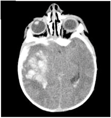
NAIS
NAIS or perinatal stroke has been defined as “a group of heterogeneous conditions” in which there is focal disruption of CBF secondary to arterial or SVT or embolization between 20 weeks of fetal life through the 28th postnatal day and confirmed by neuroimaging or neuropathological studies [Reference Lee, Croen and Backstrand177, 178, 179]. NAIS is relatively common with an estimate of 1 in 4,000 births, albeit two recent reports suggest the incidence is closer to 1 in 7,000 births [Reference Whitney, Kamdar, Ng, Hurvitz and Peterson27,Reference Leon, Kalachakra, Andrews, Thomas, Mir and Chalak178].
Case Presentation
AA is a 3,340 g 38 6/7-week female infant. Delivery was via CS. The baby emerged with a spontaneous cry and was given oxygen and CPAP for 6 min. Apgar scores were 7 and 8 at 1 and 5 min, respectively. The cord arterial blood gas revealed a pH 7.32 pCO2 35 and BD 8 mEq/L. The infant was transferred to the WBN. On DOL 2 the mother noted that the infant’s cry appeared different, with poor feeding. Over a 2-h period, the infant was observed to have four episodes of generalized tonic–clonic movements of legs and arms each lasting approximately 1–2 min. She was transferred to the NICU and treated with anticonvulsants. A video EEG showed slowing but no EEG seizures. MRI revealed extensive restriction diffusion within the right parasagittal frontal lobe and right parietotemporal lobes, with correlating decreased signal on the ADC maps compatible with an acute infarction (Figure 1.16). Magnetic resonance angiography (MRA) and MRV were both negative. A thrombophilia work up was negative. No placental pathology was available.
Infant with a stroke with presentation on DOL 2 with seizures. The MRI revealed extensive diffusion restriction within the right parasagittal frontal lobe and right parietotemporal lobes, with correlating decreased signal on the ADC maps compatible with an acute infarction.
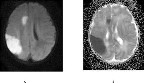
Pathogenesis
The pathogenesis of NAIS is not completely understood; however, several risk factors have been identified including maternal CA and PROM (threefold increased risk for both) [Reference Lee, Croen and Backstrand179], maternal hypertension (twofold increased risk, 180), intrapartum fever (threefold increased risk, early-onset sepsis/meningitis OR 5.8) [Reference Harteman, Groenendaal, Kwee, Welsing, Benders and de Vries181], emergency CS, male sex [Reference Srivastava, Dunbar and Shevell182], and SGA (threefold increase) [Reference Sorg, von Kries and Klemme183]. A meta-analysis of 11 studies found additional factors including preeclampsia, oligohydramnios, FHR abnormalities, and resuscitation at birth [Reference Li, Miao and Xu184]. A recent review noted that perinatal inflammation often induced by infection was the only consistent independent risk factor for NAIS [Reference Giraud, Guiraut, Chevin, Chabrier and Sébire185]. The mechanism(s) of injury remain unclear and may include elevated temperature, cytokine release associated with infection, alterations in coagulation, and a subsequent prothrombotic state associated with hyperthermia as discussed earlier [Reference Bouchama, Roberts and Al Mohanna186,Reference Leon and Helwig187]. Additional possible causes include inherited or acquired coagulopathies, trauma, and hypoxia–ischemia. However, a specific cause often cannot be identified.
Clinical Studies Evaluating Infants with NAIS
The clinical course of eight term infants with NAIS was evaluated using Doppler assessment of CBFV from the MCA and ACA during the acute phase of the disease [Reference Perlman, Rollins and Evans188]. Seven of eight patients required no DR resuscitation; one infant required brief PPV. All infants were initially admitted to the WBN. Infants were identified on the basis of abnormal clinical findings, that is, seizures (n = 6), hypotonia and apnea (n = 2) observed during the first 48 h. Neuroimaging revealed nonhemorrhagic left focal (n = 6) and right focal MCA infarction (n = 2). Transient ipsilateral decreases in CBFV as compared to the contralateral unaffected side was noted upon clinical presentation in four infants. These differences were not present at subsequent CBFV measurements. In a second study risk factors for HIE were compared to those infants with NAIS. All infants with NAIS had a negative thrombophilia work up. Infants with NAIS had a significantly higher incidence of seizures whereas infants with HIE were notable for a high occurrence of CS delivery, often secondary to a nonreassuring FHR tracing, suggesting that NAIS was not associated with perinatal risk factors that allow for early identification [Reference Adami, Grundy, Poretti, Felling, Lemmon and Graham189].
Placental Findings with NAIS
Placental examination provides a plausible argument that many cases of NAIS have associated placental changes. In a study of 46 children with NAIS, FVM occurred 4.8-fold and amniotic fluid inflammation 2.6-fold more often in NAIS versus controls. In addition, evidence of a “stress response” (meconium plus elevated nucleated red blood cells) was 31-fold more common in the NAIS compared to a control group. These placental findings reflect subacute-to-chronic intrauterine stressors and were associated with both arterial and venous stroke [Reference Bernson-Leung, Boyd and Meserve190]. A second report in 12 infants with symptomatic NAIS (n = 5) or SVT (n = 7) noted the following placental changes: thrombo-inflammatory processes (n = 6), sudden catastrophic events (n = 5), decreased placental reserve (n = 3), and a stressful intrauterine environment (n = 2) [Reference Elbers, Viero, MacGregor, DeVeber and Moore191].
Summary
1. Current perinatal markers are insensitive in identifying infants at risk for NAIS.
2. Initial clinical signs often include focal motor seizures and less frequently apnea.
3. Presentation is usually toward the end of the first postnatal day.
4. NAIS should be readily differentiated from HIE.
5. There is a predominant left-sided involvement of unclear etiology.
6. Cerebral embolism is likely the most common cause of stroke in this setting, although the site of origin for the embolism is rarely apparent.
7. Evaluations including thrombophilia workup, MRA, and MRV are invariably negative.
8. When placental pathology is available histologic thrombo-inflammatory findings are common as well as findings representing subacute-to-chronic intrauterine stressors
9. Multiple risk factors may predispose to NAIS, limiting efforts at prevention.
Cerebral Venous Sinus Thrombosis
SVT is a relatively rare complication with a varying incidence that has been typically estimated in neonates to be between 1 and 3 per 100,000 [Reference Berfelo, Kersbergen and van Ommen192,Reference Moharir, Shroff and Pontigon193]. The clinical features of SVT typically include seizures, depressed level of consciousness, or diffuse jitteriness, and less commonly focal deficits. Symptoms developed at a median postnatal age of 1.5 days [Reference Berfelo, Kersbergen and van Ommen192]. All SVT cases were confirmed with MRI/MRV. Mortality was 19% [Reference Berfelo, Kersbergen and van Ommen192]. Risk factors for SVT include PROM, CA, neonatal sepsis, meningoencephalitis, dehydration, prothrombotic disorders, and congenital heart disease, among others [Reference Ramenghi, Cardiello and Rossi194]. In the study by Moharir et al., lack of anticoagulation predicted propagation; complete thrombus recanalization occurred in 90% by 3 months [Reference Moharir, Shroff and Pontigon193]. In a study of infants who underwent TH, SVT was detected in 27% of infants [Reference Radicioni, Bini and Chiarini195]. Blanket temperature was independently associated with cerebral SVT [Reference Radicioni, Bini and Chiarini195]. By contrast, in another study of 209 infants who received TH, SVT was uncommon [Reference Munster, El-Shibiny and Szakmar196]. Brain MRI with MRV is the imaging modality of choice for SVT [Reference Teksam, Moharir, Deveber and Shroff197]. IVH in a term neonate should alert the clinician to the possibility of deep CVST [Reference Wu, Hamrick and Miller198].
Hypoglycemia
Background
Glucose is an essential metabolic fuel for the brain. Central nervous function is rapidly perturbed by hypoglycemia [Reference Perlman, Volpe and Volpe199]. Under conditions where glucose is limited, alternative substates such as lactate, pyruvate free fatty acids, and a variety of keto acids, such as ketone bodies, may help spare brain function and structure [Reference Hawdon, Ward Platt and Ainsley-Green200,Reference Harris, Weston and Harding201].
Case Presentation
GG is a 2,460 g 39-week gestation SGA male infant born via emergent CS secondary to fetal decelerations. The infant emerged limp with a weak cry with HR > 100 bpm. Initial resuscitation included CPAP for about 1 min for weak respiratory effort. Apgar scores were 7 and 8 at 1 and 5 min, respectively. Cord arterial pH was 6.95, pCO2 77, and BD 15.2 mEq/L. He was admitted to the NICU due to mild respiratory distress requiring nasal cannula and room air. Initial lactate was 11, which normalized over 24 h. He soon developed hypoglycemia with a blood glucose < 20 mg/dL, which required four glucose boluses for correction. Within 6 h of birth, leg movements concerning for seizures were noted. He was placed on a vEEG within 12 h, which detected seizure activity, without a clinical correlate and was indicative of severe diffuse cerebral dysfunction. Anticonvulsants were initiated. The vEEG improved with eventual resolution of seizures. A spinal tap was unremarkable. He was maintained and discharged on phenobarbital. The placental pathology revealed maternal and fetal vascular malperfusion, intervillous thrombus, mild chronic multifocal villitis, and fetal membranes filled with meconium-filled macrophages. MRI brain revealed bilateral cortical restricted diffusion involving the temporal, parietal, and occipital lobes (Figure 1.17). Follow-up MRI at 3 months demonstrated evolution of cystic encephalomalacia in the temporal, parietal, and occipital lobes.
MRI brain of an infant with hypoglycemia showing bilateral cortical restricted diffusion involving the temporal, parietal, and occipital lobes (A). Follow-up evolution of cystic encephalomalacia in the temporal, parietal, and occipital lobes was noted by 3 months (B).
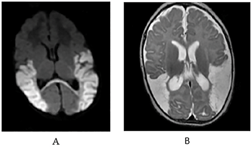
Summary
1. Although there were risk factors – that is, decelerations and severe fetal acidemia – there was no overt concerns for intrapartum HI and the infant was never considered a candidate for TH.
2. Hypoglycemia was severe and early but responded to glucose infusions and had stabilized by 10 h.
3. Placental pathology revealed both maternal and fetal vascular malperfusion that likely was a precursor to the bilateral occipital injury noted on the MRI.
4. Seizures developed early, that is within 6 h, and likely reflect an in-utero injury to the brain.
5. MRI revealed bilateral parieto-occipital brain injury likely secondary to alterations in CBF during the intrapartum period.
Relationship between Hypo-/Hyperglycemia and Brain Injury in Infants with HIE/NE
A retrospective review was undertaken to determine the potential contribution of initial hypoglycemia (defined as blood glucose ≤ 40 mg/dL) to the development of neonatal brain injury in 185 term infants with severe fetal acidemia (umbilical arterial pH < 7.00) [Reference Salhab, Wyckoff, Laptook and Perlman114]. Infants with a blood sugar ≤40 mg/dL versus >40 mg/dL were 6.3-fold more likely to have an abnormal neurologic outcome. By multivariate logistic analysis, four variables significantly associated with abnormal outcome, including: initial blood glucose (OR 18.5), cord arterial pH ≤6.90 versus >6.90 (OR 9.8), a 5-min Apgar score ≤5 versus >5 (OR 6.4), and requirement for intubation ± CPR versus neither (OR 4.7).
A second prospective cohort of term neonates evaluated the impact of hypoglycemia (glucose < 46 mg/dL) in the first 24 h on MRI changes and early neurodevelopmental outcome at one year [Reference Tam, Haeusslein and Bonifacio202]. Neonatal hypoglycemia was associated with a 3.72-fold increased odds of corticospinal tract injury and 4.82-fold increased odds of a one-point worsened neuromotor score and a 15-point lower cognitive and language score on the Bayley Scales of Infant Development. A third study investigated the association of neonatal hypoglycemia (defined as ≤40 mg/dL) and hyperglycemia (defined as >150 mg/dL) with neurocognitive outcomes in 234 infants with moderate–severe HIE who received TH. By univariate analysis unfavorable outcome was more common with hypoglycemia (81%, p = 0.004), hyperglycemia (67%, p = 0.01), and any glucose derangement within the first 12 h (67%, p = 0.002) versus normoglycemic infants [Reference Basu, Kaiser and Guffey115]. A study from the same group of investigators determined whether the early glycemic profiles in infants with HIE [defined as follows: normoglycemia (glucose concentrations of >47 to ≤150 mg/dL); hypoglycemia (≥1 glucose measurement ≤47 mg/dL); hyperglycemia (≥1 concentration >150 mg/dL)] and labile glucose (both hypoglycemia and hyperglycemia) were associated with distinct patterns of brain injury on MRI. Infants with normal versus abnormal blood glucose values had a normal brain MRI in 60% versus 32%, respectively (p = 0.02) [Reference Basu, Ottolini and Govindan116]. In particular, the odds of predominant watershed or focal–multifocal injury was sixfold higher in association with hypoglycemia and labile glucose, whereas the odds of predominant BG or global injury was 5.6-fold higher with labile glucose levels versus infants with normal blood glucose values [Reference Basu, Ottolini and Govindan116].
Further, a prospective cohort of neonates ≥36 weeks’ gestational age with NE underwent continuous glucose monitoring for 72 h, with MRI at day 4 of life, and follow-up at 18 months. Maximum glucose during the first 48 h best predicted BG AUC = 0.81) and watershed injury (AUC = 0.85) [Reference Kamino, Widjaja and Brant203]. Minimum glucose was not predictive of brain injury. At ND follow-up, a glucose threshold of >180 mg/dL during the first 48 h was associated with significantly higher child behavior problem checklist, worse neuromotor score, and higher odds for CP; this association was lost after adjusting for brain injury severity [Reference Kamino, Widjaja and Brant203]. A retrospective analysis of newborn infants with HIE found that hyperglycemia on the first day was associated with poor outcome in newborn infants undergoing TH [Reference Chouthai, Sobczak, Khan, Subramanian, Raman and Raghavendra204]. A systematic analysis evaluated the association between neonatal hypoglycemia and hyperglycemia with adverse outcome and found that children exposed to neonatal hypoglycemia had twofold higher odds of ND impairment or death and exposure to hyperglycemia was associated with a threefold higher risk of death or neurodisability at 18 months [Reference Puzone, Diplomatico and Caredda205].
Summary
1. Both hypoglycemia and hyperglycemia are associated with worse brain injury in infants with HIE ± TH.
2. Hypoglycemia is likely related to an already anaerobic state with depleted high-energy stores. Hyperglycemia likely reflects underlying brain injury with an inability to utilize glucose.
3. In infants who have HIE and undergo TH, close attention to glucose management may potentially reduce the risk of evolving brain injury.
Neonatal Sepsis
Neonatal infection can sensitize the newborn brain to injury. In this regard neonatal early-onset sepsis remains a significant cause of morbidity and mortality [Reference Schrag, Farley and Petit206]. Group B streptococcus (GBS) is the most common pathogen among term infants followed by Escherichia coli [Reference Stoll, Puopolo and Hansen207,Reference Wortham, Hansen and Schrag208]. The risk of sepsis in the setting of maternal fever and NE remains unclear but important to delineate [Reference Nelson and Penn209].
Case Presentation
IB was a 2,800 g 37-week male infant born to a mother via CS secondary to maternal fever and persistent fetal tachycardia with a nonreassuring FHR. The mother was GBS negative. Rupture of membranes occurred <5 h prior to delivery. The infant emerged limp, apneic, and cyanotic with poor color and low HR. He was intubated and administered CPR for 3 min. The Apgar scores were 2, 6, and 8 at 1, 5, and 10 min, respectively, with a cord arterial pH of 7.30. In the NICU, he soon became jittery with posturing. He had no gag or suck but was breathing spontaneously. Initial postnatal pH was 7.21, with pCO2 35 and a BD –13 mEq/L. A complete blood count revealed a white blood count of 10,000 with 39 bands and immature to mature ratio of 0.81. The platelets were 239,000. C-reactive protein was 8.4. Antibiotics were started. He was transferred to a tertiary NICU. Examination there revealed a spontaneously breathing intubated infant, lethargic but responsive to stimulation, a weak suck and absent gag, central hypotonia with intermittent sensory-induced clonus. The assessment was moderate encephalopathy. Selective head cooling was initiated close to 6 h. Within 24 h the blood cultures were positive for Listeria monocytogenes. The placental pathology was significant for acute CA with focal acute funisitis and acute villitis with associated necrosis. A spinal tap was unremarkable. A vEEG was abnormal due to the presence of left-sided rhythmic runs of predominantly sharp activity consistent with seizures persisting over 2 days. MRI revealed multifocal areas of cortical/subcortical T2 hyperintensity with corresponding ADC-restricted diffusion identified throughout the supratentorial region in multiple vascular distributions, and most prominent within the posterior perisylvian frontoparietal junctions, right greater than left, strongly suggestive of multiple acute infarctions (Figure 1.18). MRA and MRV were normal.
MRI from an infant with acute CA who underwent TH. Multifocal areas of cortical/subcortical T2 hyperintensity (A) with corresponding ADC-restricted diffusion (B) are identified throughout the supratentorial region in multiple vascular distributions most prominent within the posterior perisylvian frontoparietal junctions, left greater than right. These are most compatible with multiple acute infarctions.
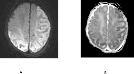
Summary
1. This infant met criteria for TH and was treated even though sepsis was high on the differential diagnosis. Antibiotics were initiated early.
2. The principal diagnosis was NE secondary to Listeria sepsis.
3. The MRI findings are not consistent with those typically seen with classic intrapartum hypoxia–ischemia.
Neonatal Encephalopathy on the Second Postnatal Day and Beyond
Hyperbilirubinemia
Physiologic jaundice is noted in every newborn with a serum bilirubin peak of approximately 6 mg/dL by 48–72 h of life with subsequent decline [Reference Perlman, Volpe and Volpe210,Reference Gartner, Lee, Vaisman, Lane and Zarafu211]. The rate of rise of bilirubin is more rapid with hemolysis (ABO incompatibility) [Reference Bahr, Christensen, Agarwal, George and Bhutani212,Reference Bhutani, Zipursky and Blencowe213]. Neurons are selectively susceptible to bilirubin toxicity. The most commonly affected regions include the BG (particularly the globus pallidus and subthalamic nucleus), the hippocampus, various cranial nerve nuclei (particularly the oculomotor, vestibular, and auditory), various other brainstem nuclei, and certain cerebellar nuclei (particularly the dentate) [Reference Perlman, Volpe and Volpe210]. The clinical presentation is usually on the second day of life and beyond. Most infants exhibit definite neurologic findings with initial features of lethargy, sleepiness, a paucity of movements, hypotonia, poor suck, and a slightly high-pitched cry. During an intermediate phase the infant appears more stuporosed coupled with irritability and increased extensor tone with arching of the neck (retrocollis) and back (opisthotonos). Without intervention the encephalopathy becomes more pronounced with deep stupor and coma, absent suck, and a shrill cry [Reference Perlman, Volpe and Volpe210]. Although the acute neurologic syndrome has become increasingly rare, acute bilirubin encephalopathy is still estimated to occur in 1.6 per 100,000 live births [Reference Bahr, Christensen, Agarwal, George and Bhutani212,Reference Bhutani, Zipursky and Blencowe213]. Importantly, early neurologic signs are nonspecific for hyperbilirubinemia and may be seen with the other causes of NE discussed in this review.
Metabolic Causes of Neonatal encephalopathy
Nonketotic Hyperglycinemia (NKH)
NKH is an inborn error of metabolism in which large amounts of glycine accumulate in body fluids. Glycine functions as a neurotransmitter, and during early development the glycine receptor is excitatory while the “classic” glycine receptor is inhibitory [Reference Perlman, Volpe and Volpe214]. Clinical features typically present in the first 2 days of life with ineffective suck, impaired ventilatory effort (apnea), hypotonia, seizures, myoclonus, and frequent hiccups. Metabolic features include hyperglycinemia.
Hyperammonemia
Hyperammonemia results from enzyme defects of the first four of five steps of the Krebs–Henseleit cycle, which include carbamoyl phosphate synthetase, ornithine transcarbamylase, argininosuccinic acid synthetase, and argininosuccinase [Reference Perlman, Volpe and Volpe214,Reference Enns215]. Ammonia and the conversion product glutamine may have a variety of toxic effects on the brain. The clinical syndrome invariably has its onset between 24 and 72 h with features that include poor feeding, disturbed level of consciousness (stupor, coma), and hyperventilation with concomitant respiratory alkalosis presumably reflecting a central effect of hyperammonemia.
Organic Acid Disorders
The most common of the organic acid disorders are propionic and methylmalonic acidurias, which result in serious neurological disturbances. Propionate and methylmalonate are vital intermediates in the catabolism of protein and fats. The accumulation of these acids results in a disturbance of myelin and involvement of the BG [Reference Perlman, Volpe and Volpe214,Reference Enns215]. Onset of symptoms is in the first days of life with an abrupt onset of vomiting, tachypnea, stupor, and seizures. Metabolic features include profound acidosis, propionic ± methylmalonic acidemia, hyperglycinemia, and hyperammonemia.
Disorders of Pyruvate and Mitochondrial Metabolism
Pyruvate dehydrogenase deficiency is the most common defect in oxidative metabolism [Reference Robinson, MacKay, Chun and Ling216]. The neonatal form is characterized by lactic acidosis with ketosis often present, with occasional hyperammonemia and severe neurologic compromise with predominant hypotonia [Reference Byrd, Krohn and Winkler217].
Mitochondrial Disease
Mitochondrial disorders can cause dysfunction of any organ system, either alone or in any combination. Neuromuscular signs and symptoms occur most commonly in the first days and include hypotonia, seizures, lethargy, feeding and respiratory difficulties, deafness, dystonia, or seizures [Reference Perlman, Volpe and Volpe214,Reference Sue, Hirano, DiMauro and De Vivo218].
Sulfite Oxidase Deficiency/Molybdenum Cofactor Deficiency
Sulfite oxidase deficiency may occur in isolation or combined with xanthine oxidase deficiency (molybdenum cofactor deficiency). Neonates typically present with feeding difficulties, vomiting, and drug-resistant seizures in the first few days after birth. A diagnosis may be made from a sulfite strip test on a fresh urine sample [Reference Sass, Gunduz and Araujo Rodrigues Funayama219].
Isolated Seizures
Metabolic disorders that may present with isolated neonatal seizures, without obvious clinical or laboratory markers of an underlying inborn error of metabolism, include NKH, sulfite oxidase deficiency, pyridoxine-responsive seizures, and folinic acid-responsive seizures. Seizures may be a prominent component in peroxisomal disorders (e.g., Zellweger syndrome), mitochondrial disorders (Leigh’s syndrome), organic acidemias, and urea cycle defects. These disorders are characterized by routine laboratory abnormalities (e.g., prominent hyperammonemia, lactic acidemia, or metabolic acidosis) that should establish an underlying diagnosis [Reference Perlman, Volpe and Volpe214,Reference Enns215].
Medication-Related Encephalopathy
Several maternal medications administered during the perinatal period may affect the neonate either transplacentally or via breastfeeding, resulting in encephalopathy marked by irritability, feeding difficulties, altered crying, jitteriness, apnea, or convulsions [Reference Rodriguez-Kovacs, Tien and Plunkett220,Reference Sanz, De-las-Cuevas, Kiuru, Bate and Edwards221,Reference Tobon, Habecker and Forray222]. Poor neonatal adaptation or withdrawal syndrome is common among neonates exposed to selective serotonin reuptake inhibitors in the third trimester of pregnancy. Such infants are 3.5-fold more likely to have a 1-min Apgar score of <5, and fivefold more likely to be admitted to the NICU and require respiratory support [Reference Hogue, Temple-Cooper and Lagzdins223,Reference Nörby, Forsberg, Wide, Sjörs, Winbladh and Källén224,Reference Marchand, Meassick and Wolf225].
Genetics and HIE/Neonatal Seizures
Several studies have evaluated the relationship between genetic mutations and HIE/seizures. Thus, term newborns with NE including seizures were evaluated using whole-exome sequencing (WES) and a genetic diagnosis was established in 36% of cases [Reference Bruun, DesRoches and Wilson226]. These included autosomal-dominant de-novo variants SCN2A (p.Met1545Val) (associated with benign familial neonatal–infantile seizures), KCNQ2 (p.Asp212Tyr) (representing a continuum of overlapping neonatal epileptic phenotypes including self-limited familial neonatal epilepsy and neonatal-onset developmental and epileptic encephalopathy), and GNAO1 (p.Gly40Arg) (a disease-causing gene for the autosomal-dominant early infantile epileptic encephalopathy). In a second study utilizing WES, pathogenic or likely pathogenic variants displayed a significantly higher incidence of seizure with a concomitant lower frequency of abnormal MRI findings [Reference Yang, Chen and Liu227]. Epileptic encephalopathy-related genes accounted for 46% of all genetic defects with seizures. A third study evaluated infants treated with TH using microarray and WES and genetic mutations were noted in 35% of cases, including CDKL5 (associated with seizures, developmental delay, and severe intellectual disability) and KIF1A (associated with a neurodegenerative disorder) [Reference Woodward, Murthy, Mineyko, Mohammad and Esser228]. Treatment was changed in two patients because of confirmed genetic etiology.
Conclusions
NE is an important clinical problem with an overall prevalence of 3 per 1,000 live births.
HIE is the most distinctive cause noted in approximately 1–1.5 per 1,000 live term births.
Neurologic signs in infants with HI brain injury may be immediate or delayed for hours following delivery, despite the presence of significant brain injury on neuroimaging.
HIE remains the most distinctive cause of NE presenting in the DR and can be readily identified early, based on a combination of clinical and biochemic markers with evolution to MS encephalopathy within 6 h.
Clinical CA with maternal fever as the leading sign can present in the DR with BD, subsequent NE, and/or seizures.
ICH can present at birth but more commonly during the first or second postnatal day.
Rapidly progressive NE particularly in association with a difficult delivery should raise concern for an epidural or a large SDH.
NAIS tends to present late on the first or on the second postnatal day as an unanticipated event with focal clonic seizures as the leading sign.
Hypoglycemia can result in brain injury as a primary event but increases the risk of exacerbating brain injury during the course of HIE.
On the second postnatal day and beyond there are a host of important but relatively rare conditions including extreme hyperbilirubinemia, hyperammonia, inborn errors of metabolism, and epileptic encephalopathies that manifest with a variety of clinical signs including progressive encephalopathy. Work up should include where relevant measures of bilirubin, lactate, and organic acids, and genetic work up for mutation in genes known to be associated with seizures.
Numerous opportunities for research are available and critically necessary. Examples include what predisposes and results in NAIS (associations with HCA and fetal thrombotic vasculopathy are noted; however, the clinical presentation is invariably delayed for up to 24 h and beyond), and what are the mechanisms that link clinical CA and specifically maternal fever to neonatal neurologic signs and pathology.