Introduction
The class Pentastomida, belonging to the subphylum Crustacea, comprises endoparasitic organisms with a vermiform morphology (Brusca et al. Reference Brusca, Giribet and Moore2023; Christoffersen and De Assis Reference Christoffersen and De Assis2013). These parasites possess a single mouth associated with two pairs of specialised chitinous hooks, which are used for attachment to the tissues of their host’s respiratory tract (Ali and Riley Reference Ali and Riley1983). This structure is directly related to their haematophagous habit, enabling them to feed on the host’s body fluids (Christoffersen and De Assis Reference Christoffersen and De Assis2013).
Among pentastomids, the family Raillietiellidae comprises a single genus, Raillietiella Sambon, Vaney & Sambon (1910), which includes approximately 45 species (Christoffersen and De Assis Reference Christoffersen and De Assis2013). In Brazil, the following species are known: Raillietiella freitasi Motta & Gomes, 1968, R. furcocerca Diesing, 1863, R. mottae Almeida, Freire & Lopes, 2008, and R. gigliolii Hett, Reference Hett1924 (Christoffersen and De Assis Reference Christoffersen and De Assis2013). The latter is a small-sized species (less than 26 mm), characterised by a proportionally large cephalothorax, a slender abdomen (less than 1 mm wide), and two small but distinct papillae located above the oral opening – morphological traits that tend to be less conspicuous in larger species of the genus Raillietiella (Hett Reference Hett1924).
Initially, it was described as R. furcocerca by Diesing (Reference Diesing1836), whose sampling included pentastomids from both snakes and amphisbaenians. Later, Hett revised the material collected by Giglioli (Reference Giglioli1923) and formally described the species based on female specimens. The first description of a male R. gigliolii was provided by Ali et al. (Reference Ali, Riley and Self1984), during his revision of the genus Raillietiella, based on specimens collected from Amphisbaena alba Linnaeus, 1758, in Trinidad (Caribe). In this analysis, a distinctive feature of the species was identified: a markedly curved copulatory spicule, with a trumpet-like shape, which allowed the long-standing taxonomic confusion between R. furcocerca and R. gigliolii to be resolved.
The first record of R. gigliolii in Brazil was made by Rego (Reference Rego1983), who provided a detailed morphological description of specimens parasitising A. alba, significantly contributing to the understanding of the species morphology and systematics in the country. Subsequently, in the Northeast region, Almeida et al. (Reference Almeida, Ferreira, Brito and Christoffersen2006) identified three male specimens of R. gigliolii in the respiratory tract of A. alba, representing the first record of the species in that region. In later studies, the species was again recorded in A. alba and, for the first time, in Amphisbaena vermicularis Wagler, 1824 (Almeida et al. Reference Almeida, Sales, Santana, Vieira, Ribeiro, Alves and Nóbrega2009). To date, R. gigliolii appears to be restricted to the genus Amphisbaena, possibly due to the marked morphological and ecological differences these reptiles exhibit in comparison to other members of the order Squamata (Vitt and Caldwell Reference Vitt and Caldwell2013). In this context, it is plausible to assume that the pentastomids associated with these hosts also possess particular biological traits, as previously suggested by Ali et al. (Reference Ali, Riley and Self1984; Reference Ali, Riley and Self1985).
From this perspective, identification based solely on morphological characteristics may lead to taxonomic errors in the description of parasites (Kelehear et al. Reference Kelehear, Spratt, Dubey, Brown and Shine2011). A recent example is provided by Silva et al. (Reference Silva, Oliveira, Brandão, Figueirêdo Chaves, Monteiro, Koroiva, Guedes, Vasconcellos and Oliveira Almeida2024), who observed different female morphotypes of R. mottae parasitising distinct hosts. Initially, based only on traditional taxonomy, these morphological variations suggested the presence of different species. However, molecular analyses revealed that all specimens shared exactly the same nucleotide sequence, confirming that they belong to a single species.
Thus, the present study aims to characterise specimens of R. gigliolii through morphological and molecular analyses. Comparison with available molecular sequences from other members of the group will contribute to a more accurate phylogenetic understanding of the raillietiellids.
Materials and methods
Morphometry
The hosts whose pulmonary parasites were subsequently deposited in a scientific collection were collected from two localities in the Northeast of Brazil: a remnant area of Atlantic Forest (07°10’ S, 35°05’ W; datum WGS84), in the municipality of Cruz do Espírito Santo, state of Paraíba; and a humid forest area (07°16’ S, 39°26’ W; datum WGS84), located on the slopes of the Chapada do Araripe, in the municipality of Crato, state of Ceará. In turn, the hosts used in the molecular analyses were collected in the district of Zé Gomes, municipality of Exu, state of Pernambuco (07°29’20.7" S, 39°34’10.0" W; datum WGS84), also in the northeast region of Brazil.
For the morphometry, all specimens of R. gigliolii deposited in the collection of the Zoology Laboratory at the Universidade Regional do Cariri (LZ-URCA), in the state of Ceará, Brazil, were analysed. This collection, built over 20 years of scientific research, stands out for housing one of the largest pentastomid collections in Latin America.
The pentastomids were mounted on temporary slides using Hoyer’s medium, and the following morphological variables were recorded: body length and width, as well as measurements of the buccal structure, cephalothorax, and tail. The dimensions of the anterior and posterior hooks were also measured (AB, length of the barb; and BC, total hook length). For each morphological characteristic, means and standard deviations were calculated. The hook measurements followed the protocol described by Ali et al. (Reference Ali, Riley and Self1984, Reference Ali, Riley and Self1985) and Kelehear et al. (Reference Kelehear, Spratt, Dubey, Brown and Shine2011), in which AB corresponds to the distance from the tip of the hook to the anterior inner surface of the hook’s arm, while BC refers to the distance from the anterior portion of the hook’s arm to the outermost point of the posterior base. The shape of the hooks (rhomboid or pointed) and the measurements of the copulatory spicules in male specimens were also assessed.
The identification of the specimens as belonging to the species R. gigliolii was based on a direct comparison of their morphological features with the descriptions provided by Rego (Reference Rego1983) and Ali et al. (Reference Ali, Riley and Self1984, Reference Ali, Riley and Self1985). In traditional descriptions, the male spicules are long, strongly curved, and exhibit a distinctive trumpet-shaped distal end – a diagnostic feature of the species. Also notable are two small but clearly visible papillae above the oral opening, which are typically less apparent in larger Raillietiella species. The pronounced size difference between the hooks, with the posterior hooks being larger than the anterior, further supports the identification, as this is another characteristic trait of the species (Ali et al. Reference Ali, Riley and Self1984, Reference Ali, Riley and Self1985; Rego Reference Rego1983).
All measurements, expressed in micrometres (μm), were taken using a Zeiss Axio Imager M2 microscope coupled with an image analysis system (Carl Zeiss Microscopy GmbH, Königsallee 9–21).
Sample preparation for molecular procedures
With the aim of finding suitable pentastomids for molecular characterisation, all amphisbaenians from Caatinga areas in the Northeast of Brazil, provided by the collection of the Laboratory of Biology and Ecology of Wild Animals at the Universidade Federal do Cariri – UFCA, were used.
The individuals were dissected; the respiratory tracts were completely removed and carefully examined under a stereoscopic microscope. All pentastomids found were collected and stored in glass vials containing 95% ethanol, with the aim of subsequent DNA extraction, amplification, and sequencing.
All molecular procedures were carried out at the Termitology Laboratory of the Universidade Federal da Paraíba, under controlled room temperature ranging between 20°C and 22°C. To prevent contamination of the genetic material with host blood, the cephalothorax of the parasites was carefully separated prior to extraction. The specimens, previously preserved in absolute ethanol, were left to rest for 15 minutes to allow complete evaporation of residual ethanol before the commencement of laboratory procedures.
DNA extraction, PCR, and sequencing
The whole-genome DNA of three specimens was extracted using Zymo Research Quick-DNA/RNA viral MagBead. PCR amplifications were performed for the cytochrome c oxidase subunit 1 mitochondrial gene (COI) using BF2 (5′-GCH CCHGAYATRGCHTTYCC-3′) and BR2 (5′-TCDGGRTGNCCRAARAAYCA-3′) described by Elbrecht and Leese (Elbrecht and Leese Reference Elbrecht and Leese2017). All PCRs consisted of 25 μl reagent mixture containing: 10×reaction buffer (Promega); 25 mM MgCl₂; 2 mM dNTPs; 2 mM primers and GoTaq® G2 Hot Start Poly- merase (Promega); 2 μl template DNA. The PCR cycling program was run as follows: initial denaturation step with 3 min at 95°C, 35 cycles of denaturation for 30 s at 95°C, annealing for 45 s at 50°C and extension for 1 min at 72°C, and final extension for 5 min at 72°C. PCR products were bidirectionally sequenced in ABI 3130 Genetic Analyzer (Applied Biosystems).
We used GENEIOUS v. 9.1.3 (Kearse et al. Reference Kearse, Moir, Wilson, Stones-Havas, Cheung, Sturrock, Buxton, Cooper, Markowitz and Duran2012) to check the sequence quality of the strands by comparison to their respective chromatograms and to assemble and edit if necessary. Sequences of Raillietiella present in GenBank were incorporated into analyses for genetic regions. In the phylogenetic analysis, we aligned sequences using MAFFT v. 7.017 (Katoh et al. Reference Katoh, Misawa, Ki and Miyata2002), a module implemented in GENEIOUS v. 9.1.3. We constructed maximum likelihood (ML) trees with RAxML v. 8.2.12 (Stamatakis Reference Stamatakis2014) using GTR GAMMA I model and 10,000 bootstrap (BP) replicates. The GTR+I+G evolutionary model was used for the phylogenetic analyses because it is the most comprehensive evolutionary model and includes all other evolutionary models (Abadi et al. Reference Abadi, Azouri, Pupko and Mayrose2019). Our sequences were deposited in GenBank under accession PQ635383, PQ635384 and PQ635385.
Results
We examined 31 specimens of R. gigliolii (Figure 1) deposited in the LZ-URCA Collection, of which 17 were recorded from the host A. alba and 14 from A. vermicularis. Detailed morphometric measurements of males, adult females, and juvenile females, along with the corresponding values reported by Rego (Reference Rego1983) and Ali et al. (Reference Ali, Riley and Self1984), are presented in the comparative table (Table 1). The eggs (Figure 2) had an average size of 85 μm in length and 52.5 μm in width. The copulatory spicule of the male, which is trumpet-shaped and highlighted in Figure 3, had a length of 472 μm.
Cephalothorax of a female Raillietiella gigliolii from Amphisbaena alba, housed in the LZ-URCA Collection.

Morphometric comparison between Raillietiella gigliolii from this study and data from Rego (Reference Rego1983) and Ali et al. (Reference Ali, Riley and Self1984)

All measurements are expressed in micrometres (μm). Values presented as ± correspond to the mean followed by one standard deviation (±1 SD). Abbreviations: n = number of individuals; L = length; W = width; AB and BC = hook segments (see methodology).
Detail of the maximum width of the abdomen showing the uterus full of eggs of the adult female of Raillietiella gigliolii.

Details of the ventral region of a male Raillietiella gigliolii. (A) Anterior region, cephalothorax; (B) copulatory spicules with a ‘trumpet-shaped’ appearance, a diagnostic feature of the species.

In addition to these measurable characteristics, the R. gigliolii specimens exhibited a similar morphotype, evidenced by the apparent absence of body rings, a trapezoidal cephalothorax wider than the abdomen, and well-developed, pointed anterior and posterior hooks. Well-defined apical and dorsal papillae were also observed. In males, the presence of strongly curved copulatory spicules, shaped similarly to a trumpet, stood out, reinforcing the consistent morphological pattern of the species.
The specimens exhibited similar morphotypes regarding the shape of the hooks (Figure 4), which remained consistent both between male and female individuals and among specimens parasitising different hosts, specifically A. alba and A. vermicularis.
Anterior hook (A) and posterior hook (B) of Raillietiella gigliolii, from the same individual, showing their respective AB and BC measurements and pointed shape.

For the molecular analysis, specimens from eleven amphisbaenians belonging to different species were dissected. Among these, eight individuals were of the species Leposternon polystegum (Duméril, 1851), two of A. alba and one of A. vermicularis. The presence of pentastomids was recorded in only one A. alba specimen. Eight pentastomid specimens were extracted from the parasitised host and were previously analysed morphologically following the criteria established by Ali et al. (Reference Ali, Riley and Self1985), which confirmed their identification as R. gigliolii. However, only three of these specimens were suitable for molecular analysis. The exclusion of the remaining samples was primarily due to DNA degradation, possibly caused by prolonged preservation in ethanol or insufficient quality of the biological material for efficient extraction. The genetic sequences obtained from these three individuals were duly deposited in GenBank under the accession numbers PQ635383, PQ635384 and PQ635385.
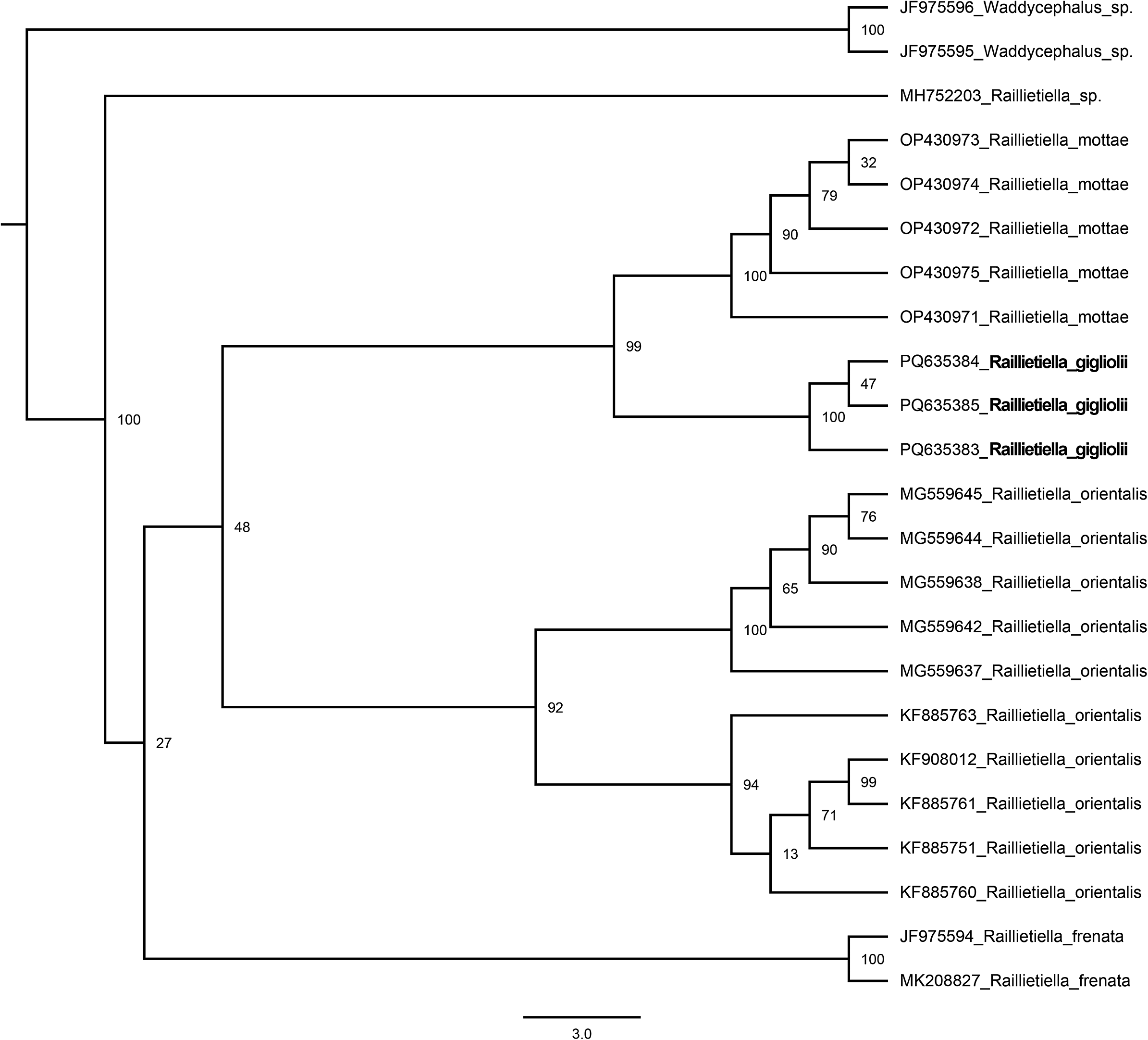
The Maximum Likelihood (ML) phylogenetic analysis of mitochondrial COI gene sequences revealed distinct groupings among species of the genus Raillietiella, with strong statistical support for several clades (bootstrap values indicated at the nodes) (Figure 5). Samples identified as R. gigliolii formed a monophyletic clade (support of 100), clearly separated from other analysed species such as R. orientalis, R. mottae, and R. frenata. The sequences assigned to R. gigliolii showed complete nucleotide identity, with no variations among them, indicating an absence of genetic divergence within the sampled group.
Maximum likelihood phylogenetic tree based on mitochondrial COI gene sequences for representatives of the genus Raillietiella.

Discussion
According to Ali et al. (Reference Ali, Riley and Self1984), R. gigliolii was initially grouped with pentastomids parasitising snakes. However, following a thorough taxonomic revision based on morphological and ecological data, this species was reclassified into group IV, which consists exclusively of pentastomids parasitising amphisbaenians (Ali et al. Reference Ali, Riley and Self1985). This new classification more accurately reflects the evolutionary and ecological peculiarities of R. gigliolii, which proves to be highly specialised, with stable morphology and low phenotypic plasticity, as evidenced by the specimens analysed in this study (Ali et al. Reference Ali, Riley and Self1985; Christoffersen and Assis Reference Christoffersen and De Assis2013).
Unlike pentastomids that parasitise lizards, many of which exhibit marked morphological variability, including differences in hook shape (Kelehear et al. Reference Kelehear, Spratt, Dubey, Brown and Shine2011; Silva et al. Reference Silva, Oliveira, Brandão, Figueirêdo Chaves, Monteiro, Koroiva, Guedes, Vasconcellos and Oliveira Almeida2024), R. gigliolii is, to date, represented by a single known morphotype. In all specimens analysed, both anterior and posterior hooks display a sharp, well-defined shape, with no perceptible structural variation.
Nevertheless, the differences in hook measurements observed between this study and those reported by Rego (Reference Rego1983) and Ali et al. (Reference Ali, Riley and Self1984) are understandable (see Table 1). Morphometric variation in these appendages has already been documented in pentastomids and is influenced by factors such as host species and host body size (Kelehear et al. Reference Kelehear, Spratt, Dubey, Brown and Shine2011; Silva et al. Reference Silva, Oliveira, Brandão, Figueirêdo Chaves, Monteiro, Koroiva, Guedes, Vasconcellos and Oliveira Almeida2024). For example, Kelehear et al. (Reference Kelehear, Spratt, Dubey, Brown and Shine2011) examined 34 specimens of R. frenatus from Rhinella marina and Hemidactylus frenatus, reporting substantial variation in hook size. Similarly, Silva et al. (Reference Silva, Oliveira, Brandão, Figueirêdo Chaves, Monteiro, Koroiva, Guedes, Vasconcellos and Oliveira Almeida2024) showed, based on approximately 50 individuals of R. mottae collected from different lizard species, that about 36% of the variation in hook size was linearly related to host size.
The results obtained also reinforce the systematic framework proposed by Christoffersen and Assis (Reference Christoffersen and De Assis2013), demonstrating that R. gigliolii exhibits a conserved and homogeneous morphology consistent with the morphological criteria they established. According to these authors, characteristics such as the morphology of the hooks, the structure of the cephalothorax, the pattern of body segmentation, and the shape of the copulatory spicules are fundamental for species delimitation within the genus Raillietiella. In the present study, the analysed specimens of R. gigliolii displayed hooks with uniform shape and size, a well-defined and structurally similar cephalothorax, regular body segmentation, and copulatory spicules with characteristic shape, aligning fully with the parameters set out by Christoffersen and Assis (Reference Christoffersen and De Assis2013).
Several studies over the years have reported misidentifications of species within the genus Raillietiella, primarily due to the plasticity of certain morphological characteristics. For example, in Kelehear et al. (Reference Kelehear, Spratt, Dubey, Brown and Shine2011), R. indica Gedoelst, 1921 and R. frenata Ali, Riley & Self, 1981, both recorded in the lungs of the cane toad (Rhinella marina Linnaeus, 1758), were initially classified as distinct species based on morphological differences. However, subsequent molecular analyses demonstrated that they were a single species, confirmed as R. frenata. Similarly, Silva (2024) observed variations in the morphotype of female R. mottae from different hosts, initially raising the hypothesis of two distinct species. Through PCR amplification of the mitochondrial COI gene (barcode), combined with a detailed morphological reassessment, the authors confirmed that they were the same species, leading to the redescriptions of R. mottae.
To date, R. gigliolii appears to be specific to A. alba and A. vermicularis (Almeida et al. Reference Almeida, Ferreira, Brito and Christoffersen2006; Almeida et al. Reference Almeida, Sales, Santana, Vieira, Ribeiro, Alves and Nóbrega2009), as specimens of R. gigliolii analysed in this study were found only in these two hosts, showing no morphological divergence and demonstrating that it is a valid species. This structural uniformity reinforces the cohesion of the group and supports the taxonomic delimitation of R. gigliolii as a well-defined morphospecies (Vitt and Caldwell Reference Vitt and Caldwell2013).
The complete nucleotide identity among the three sequences analysed in this study reinforces the effectiveness of the COI gene as a reliable tool for the taxonomy of pentastomids (Elbrecht and Leese Reference Elbrecht and Leese2017). This delimitation corresponds to the process of biodiversity identification at the species level (Carstens et al. Reference Carstens, Pelletier, Reid and Satler2013), which, besides being fundamental for the compilation of an inventory with species names, also provides valuable information for various fields of biology, such as evolutionary biology, conservation, and biogeography (Dayrat Reference Dayrat2005). Such a finding supports a more precise taxonomic identification based on consistent morphological characters and reinforces the importance of the integrative approach in systematics, demonstrating its usefulness both for species with high morphological variability and for those with conserved morphology, as is the case with R. gigliolii (Elbrecht and Leese Reference Elbrecht and Leese2017). Moreover, it corroborates other studies that employed the same marker and found its high resolution in the specific identification of parasitic helminths (Hebert et al. Reference Hebert, Cywinska, Ball and DeWaard2003).
The maximum likelihood phylogenetic analysis, based on mitochondrial COI gene sequences, demonstrated complete genetic identity among the R. gigliolii specimens, which formed a monophyletic clade with maximum statistical support. The three sequences analysed showed 100% nucleotide similarity, with no detectable genetic divergence, indicating high genetic stability and further supporting the clear taxonomic delimitation of the species. In this context, it is worth noting that DNA barcoding adopts a threshold of approximately 2% genetic divergence as the standard criterion for species distinction (Hebert et al. Reference Hebert, Cywinska, Ball and DeWaard2003). To ensure the effectiveness of this method, it is essential to establish and maintain reference databases that compile genetically validated and accurately identified sequences, enabling precise comparisons between organisms (Ekrem et al. Reference Ekrem, Willassen and Stur2007; Monchamp et al. Reference Monchamp, Taranu, Garner, Rehill, Morissette, Iversen, Fugere, Littlefair, da Costa and Desforges2023).
The results obtained contribute to the understanding of the evolution and systematics of the group by indicating that R. gigliolii does not exhibit morphotype variability, unlike what is observed in other species of the genus (Kelehear et al. Reference Kelehear, Spratt, Dubey, Brown and Shine2011; Silva et al. Reference Silva, Oliveira, Brandão, Figueirêdo Chaves, Monteiro, Koroiva, Guedes, Vasconcellos and Oliveira Almeida2024). This absence of variation is significant, considering that morphological plasticity can obscure true taxonomic boundaries when only morphological traits are used, as discussed by Almeida (2009) regarding morphological variations in Raillietiella from different geographic regions.
Despite the strong foundation provided by the molecular analyses, it was not possible to include genetic data from pentastomid samples originating from A. vermicularis. Although the R. gigliolii specimens found in A. alba and A. vermicularis exhibit similar morphology, no parasites were recovered from A. vermicularis individuals for molecular analysis. This absence may be related to the difficulty in sampling these hosts, which have relict fossorial habits and predominantly live underground, making their collection challenging and contributing to the scarcity of records (Colli and Zamboni Reference Colli and Zamboni1999). Therefore, future studies are necessary to confirm, based on molecular evidence, whether the parasites infecting both hosts indeed belong to the same species.
Acknowledgements
RGS, AAMT, and SCR thank the Fundação Cearense de Apoio ao Desenvolvimento Científico e Tecnológico (FUNCAP) and Secretaria de Ciência, Tecnologia e Educação Superior do Ceará (Secitece) for research fellowship grants (respective processes: BMD-0008-02422.01.18/24; PVS-0215-00125.01.00/23 and BP5-0197-00161.01.00/22). WOA and AV are both recipients of research productivity fellowships awarded by the Conselho Nacional de Desenvolvimento Científico e Tecnológico (CNPq, Brazil), under grants 308534/2021–2 and 309820/2020–0, respectively. Finally, RECRFC (88887.929233/2023-00) and RRF (88887.950422/2024-00) received scholarships from Coordenação de Aperfeiçoamento de Pessoal de Nível Superior – Brasil (CAPES).
Competing interests
None.
Ethical standard
All applicable international, national, and/or institutional guidelines for the care and use of animals were followed. Research was permitted by Biodiversity Authorization and Information System of the Brazilian Institute of Environment and Renewable Natural Resources (No. 92112-1).