Introduction
Setaria cervi (Williams, Reference Williams1955) is a parasite belonging to the phylum Nemathelminthes and superfamily Filarioidea. It is a species that occurs in the abdominal cavity of cattle, buffalo, bison, yak and various species of deer, but rarely in sheep. Another closely related species is S. digitata, which also inhabits the peritoneal cavity of cattle and other ungulates. Setaria species are known to cause cerebrospinal nematodiasis and lumbar paralysis as well as ocular infection in cattle (Shin et al., Reference Shin, Cho and Wee2002; Tung et al., Reference Tung, Lai, Ooi, Yang and Wang2003). The parasite is generally considered to be non-pathogenic in its natural hosts, but the transmission of the infective larvae through mosquito vectors to its abnormal hosts (goats, sheep or horses) can result in a serious and often fatal cerebrospinal nematodiasis (Wijesundera et al., Reference Wijesundera, Chandrasekharan, Karunanayake and Rajapakse1997, Reference Williams1999). Initially S. digitata was considered synonymous to S. labiatopapillosa and S. cervi (Purvis, Reference Purvis1931; Baylis, Reference Baylis1936a, Reference Baylisb). Later on, other workers (Skrjabin & Shikhobalova, 1945; Sarwar, Reference Sarwar1946) considered S. digitata as an independent species. The resemblance of S. cervi to various filarial species has been reported earlier (Singhal et al., Reference Singhal, Saxena and Johri1973). Setaria leichungwingi (Shoho, Reference Shoho1976), a parasite of water buffaloes (Bubalus bubalis Linn.) in Asia, is closely related to S. labiatopapillosa (Gidel & Brengues, Reference Gidel and Brengues1972; Shoho & Sachs, Reference Shoho and Sachs1975). Setaria pujoli, S. scalprum and S. saegeri are other closely related species that occur in the smaller antelopes of Africa (Durette-Desset, Reference Durette-Desset1973; Watermeyer et al., Reference Watermeyer, Boomker and Putteril2003).
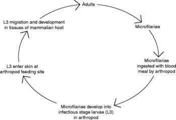
The life cycle of filarial parasites encompasses five major stages, delineated by four complete moults of the cuticle (Scott, Reference Scott, Nutman and Pasuol2000). Two of these moults occur in the arthropod vector, which takes up blood microfilariae (L1; mf) and permits rapid development of parasites to the L3 or infective larval stage. Once L3 s gain access to the mammalian host, they continue developing over many weeks and through two further moults into dioecious adult worms. After mating, the females release large numbers of mf which continue the cycle when ingested in the blood meal of the arthropod (fig. 1). Each of the major lifecycle stages (mf, L3 and adults) has unique immunological characteristics. In the adult stage of its life cycle, S. cervi inhabits the peritoneal cavity of cattle and releases thousands of microfilariae (mf) every day. The adults survive for several years, causing considerable damage to various organs and being responsible for the disease setariasis; they therefore represent a major target in development of novel veterinary medicines (Bush, Reference Bush1951; Ansari, Reference Ansari1963).
Generalized life cycle of filarial worms.

The peritoneal transplantation, periodicity and morphology of mf of S. cervi have been studied in white rats (Ansari, Reference Ansari1963, Reference Ansari1964). Suggestions have also been put forward that many neurological syndromes of unknown aetiology in humans are due to infection with Setaria species (Innes, Reference Innes1951; Innes & Shoho, Reference Innes and Shoho1952; Nelson, Reference Nelson1965; Kadenatsii et al., Reference Kadenatsii, Sokolov and Mamaev1974). Several common antigens between the bovine (S. cervi) and human (Brugia malayi) filarial parasites have been demonstrated (Kaushal et al., Reference Kaushal, Kaushal, Ghosh and Talwar1994). Sertaria cervi has been widely used as a test organism for the in vitro screening of prospective antifilarial compounds (Singhal et al., Reference Singhal, Chandra, Chawla, Gupta and Saxena1969, Reference Singhal, Chandra and Saxena1972a, Reference Singhal, Chandra and Saxenab, Singhal et al., Reference Singhal, Saxena and Johri1973; Anwar et al., Reference Anwar, Srivastava and Ghatak1978; Srivastava & Ghatak, Reference Srivastava and Ghatak1983a). The S. digitata–Mastomys coucha model has also been found to be amenable to chemotherapeutic and immunobiological investigations in experimental filariasis (Srivastava et al., Reference Srivastava, Sethi and Ghatak1983, 1984; Srivastava & Ghatak, Reference Srivastava and Ghatak1983b; Mukhopadhyay et al., Reference Mukhopadhyay, Dash and Ravindran1996).
Some Setaria species, such as S. digitata, S. marshalli, S. marshalli pandei, S. equina and S. labiatopapillosa, have been studied by scanning electron microscopy at critical points in the anterior and posterior parts of the adult worms (Srivastava et al., Reference Srivastava, Maitra, Shipstone and Ghatak1985). These studies have contributed towards the identification of Setaria species, in addition to the characteristics which have been demonstrated by common light microscopy (Shoho & Uni, Reference Shoho and Uni1977; Zdarska & Scholl, Reference Zdarska and Scholl1978; Almeida et al., Reference Almeida, Deobhankar, Bhopale, Zaman and Renapurkar1991; Rhee et al., Reference Rhee, Choi, Park and Jang1994). The morphology of the larval stages of S. labiatopapillosa, S. leichungwingi, S. equina and S. digitata has also been studied (Li & Yu, Reference Li and Yu1990; Das & Das, Reference Das and Das1995).
The protein profiles of adult S. digitata and S. marshalli (Tung et al., Reference Tung, Lai, Ooi, Yang and Wang2003) have been compared using SDS-PAGE (Subhachalat & Adachi, Reference Subhachalat and Adachi1997). The results obtained have shown that one band with a molecular size of 69 kDa is present only in S. marshalli, while this band has not been detected in S. digitata. There are no differences of the major bands between males and females of the respective worm species. Further investigation has involved two-dimensional gel electrophoresis, in which one spot having molecular weight ranging from 64 to 73 kDa has been confirmed in S. marshalli but not in S. digitata.
Biochemical composition
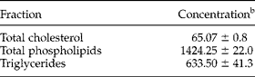
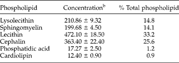
Adults and mf of S. cervi have been analysed for various biochemical constituents (Rathaur et al., Reference Rathaur, Anwar and Ghatak1980; Malhotra et al., Reference Malhotra, Kaushal and Ghatak1986; Kumar, Reference Kumar1987a, Reference Kumarb). Both stages of the parasite are rich in protein, carbohydrate and lipids, while nucleic acids are detectable in only small amounts. Microfilariae and adults contain 10 and 25% dry matter, 57.4 and 63.0% protein, 8.9 and 14.8% carbohydrate and 12.8 and 9.2% lipid, respectively. Reducing sugars account for 52 and 67%, respectively, of the total carbohydrate content in mf and adult worms. Glycogen accounts for 95% of total carbohydrate in the cold trichloroacetic acid-soluble fraction of adult worms, while its concentration in mf is very low. Glucose is the main reducing sugar, while concentrations of fructose have been found to be very low. Microfilariae contain more lipids, nucleic acids, mucopolysaccharides and total phosphorus than adults. All the major classes of lipids have been demonstrated in S. cervi adults (Ansari et al., Reference Ansari, Yusufi and Siddiqi1973) as well as mf (Rathaur et al., Reference Rathaur, Anwar and Ghatak1980). The phospholipid percentage is higher in mf than in adults. The nucleic acid content of adults and mf revealed 0.3 and 1.44% of DNA and 0.6 and 1.29% RNA of the dry weight respectively (Rathaur et al., Reference Rathaur, Anwar and Ghatak1980). The lipid composition of S. cervi is depicted in tables 1 and 2.
Lipid fractions isolated from S. cervi a; reproduced according to Ansari et al. (1973).
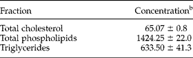
a The results are expressed as the mean ± SD of the mean, based on four separate determinations.
b Values given are in terms of mg% of wet weight of S. cervi.
Properties of individual phospholipids in S. cervi a; reproduced and modified according to Ansari et al. (1973).
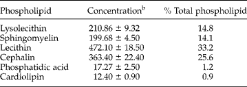
a The results are expressed as the mean ± SD of the mean, based on four separate determinations.
b Values given are in terms of mg% of wet weight of S. cervi.
The amino acids that have been identified include cysteine, lysine, arginine, histidine, aspartic acid, serine, glycine, threonine, glutamic acid, tryptophan, tyrosine, proline, valine, leucine, isoleucine and phenylalanine (Rathaur et al., Reference Rathaur, Anwar and Ghatak1980). The amino acid pattern of both mf and adult worms is very similar (Rathaur et al., Reference Rathaur, Anwar and Ghatak1980). SDS-PAGE analysis has shown the presence of 35–40 protein bands in adults and 25–29 protein bands in mf, in the molecular weight range of 10,000–200,000. These results demonstrate the complex nature of somatic extracts of the adult stage as compared to mf, and also reveal some qualitative and quantitative differences between these stages (Ansari et al., Reference Ansari, Yusufi and Siddiqi1973).
Carbohydrate metabolism
The pathways of carbohydrate metabolism have been investigated in detail in both mf and adult stages of S. cervi (Pandya, Reference Pandya1961).
Glycolysis
Glycolysis proceeds in a manner similar to that found in vertebrate muscles. Nearly all the enzymes involved in the glycolytic pathway have been partially purified and characterized (Anwar et al., Reference Anwar, Ansari, Ghatak and Murti1977; Saxena et al., Reference Saxena, Bharadwaj, Rathaur and Ghatak1986). The complete sequence of glycolytic enzymes has been demonstrated with high activity levels in S. cervi (Saz & Dunbar, Reference Saz and Dunbar1975; Anwar et al., 1977). Several key enzymes related to carbohydrate metabolism have been assayed in S. digitata (Banu et al., Reference Banu, Dhandayuthapani and Nellaiappan1991). In the cytosolic fraction; pyruvate kinase (PK), phosphoenolpyruvate carboxykinase (PEPCK), malate dehydrogenase (MDH) and malic enzyme (ME) have been found (Banu et al., Reference Banu, Dhandayuthapani and Nellaiappan1991).
The ratios of PK : PEPCK indicate the metabolic group to which the parasite belongs. It has been reported earlier that parasites having a low PK : PEPCK ratio (0.05–0.10) are carbon dioxide fixers, and those possessing a high ratio (2–10) are lactate fermenters. The ratio in S. digitata is 0.6, which lies between the two extremes, suggesting that it is likely to utilize both pathways for energy generation. Such intermediate ratios have been observed in Litomosoides carinii (1.9), Hymenolepis diminuta (0.18), Schistocephalus solidus (1.7) and F. hepatica (Bueding & Saz, Reference Bueding and Saz1968; Brazier & Jaffe, Reference Brazier and Jaffe1973; Prichard, Reference Prichard1976; Korting & Barrett, Reference Korting and Barrett1977).
It has been found that PK of S. digitata shows increased activity in the presence of fructose-1,6-bisphosphate (FBP), as in the case of Fasciola hepatica and Moniezia expansa (Behm & Bryant, Reference Behm and Bryant1975; Prichard, Reference Prichard1976). This indicates the possibility of FBP playing a regulatory role.
Histochemical localization of aldolase has been demonstrated in the mf of S. cervi (Khatoon et al., Reference Khatoon, Ullah, Baqui and Ansari1984). Transcuticular uptake of methyl glucose by S. cervi has also been demonstrated (Roy et al., Reference Roy, Masood and Srivastava1983).
Besides glucose, other monosaccharides such as fructose and galactose are also accepted as substrates, although less preferentially. β-d-Galactosidase activity has been found to be localized in the digestive tract of S. digitata. The enzyme shows a K m of 3.636 mM for the substrate 6-bromo-2-naphthyl β-d-galactoside. PAGE has revealed that the enzyme exists as a single entity. Some medicinal plants and the drug filarin have been found to effectively inhibit the enzyme. β-Galactosidase activity may serve as a potential target for the development of novel antifilarial drugs (Banu et al., Reference Banu, Kalyani and Nellaiappan1990).
The comparative efficacy of hetrazan, levamisole and tetramisole on the enzymes of the glycolytic pathway of adult S. cervi worms has been studied (Khatoon et al., Reference Khatoon, Baqui, Ullah and Ansari1983). Biochemical assays revealed the decreased activities of glucose-6-phosphatase, fructose-1,6-bisphosphatase, lactate dehydrogenase and adenosine triphosphatase in the drug-treated worms. Hetrazan was relatively more effective than tetramisole and levamisole. Some anthelmintic studies on S. cervi based on histochemical alterations in glucose-6-phosphatase, adenosine triphosphatase and malic dehydrogenase have also been carried out (Khatoon et al., Reference Khatoon, Baqui and Ansari1982) and the effect of glycosides of Streblus asper on motility, glucose uptake and enzymes of carbohydrate metabolism of S. cervi females has also been studied (Singh et al., Reference Singh, Chatterjee and Srivastava1994).
Fermentative pathway (conversion of pyruvate to lactate)
The outstanding feature of glucose degradation is the production of organic end products such as lactic and pyruvic acids. These worms generally survive in hypoxia and it could be assumed that glycolysis is the main source of energy production.
Lactate dehydrogenase (LDH) of S. digitata has been studied in detail (Banu et al., Reference Banu, Nellaiappan and Dhandayuthapani1989). The enzyme exists as isozymes (four in the female and three in the male) and their separation depends on the percentage of gel and pH. Parasitic LDH has been found to exhibit an optimum pH of 7.0–8.0 and shows resistance to high temperature. Filarin and diethylcarbamazine (DEC) have been found to inhibit LDH from S. digitata.
The evidence that S. digitata is a facultative anaerobe has been derived from the fact that the closely related S. cervi is a confirmed lactate excretor (Barrett, Reference Barrett1981) and S. digitata itself has a very active LDH (Banu et al., Reference Banu, Nellaiappan and Dhandayuthapani1989). Therefore, the pyruvate formed from oxaloacetate (OA) after dismutation, as well as that formed from malate via malate decarboxylation, may cross over to the cytosol where it forms a substrate for LDH, as has been demonstrated in case of Echinococcus granulosus, E. multilocularis and Dicrocoelium dendriticum (Köhler & Hanselmann, Reference Köhler and Hanselmann1973; McManus & Smyth, Reference McManus and Smyth1982).
Further evidence that lactate formation is favoured has been obtained from the fact that the PK : PEPCK ratio (1.37) in the presence of FBP is 1.0. It seems that the presence or absence of FBP may play an important role in deciding the fate of phosphoenolpyruvate (PEP) in S. digitata. In the absence of FBP, carbon dioxide fixation of PEP to OA may be predominant, once the FBP is formed, PK may be favoured resulting in the formation of lactate.
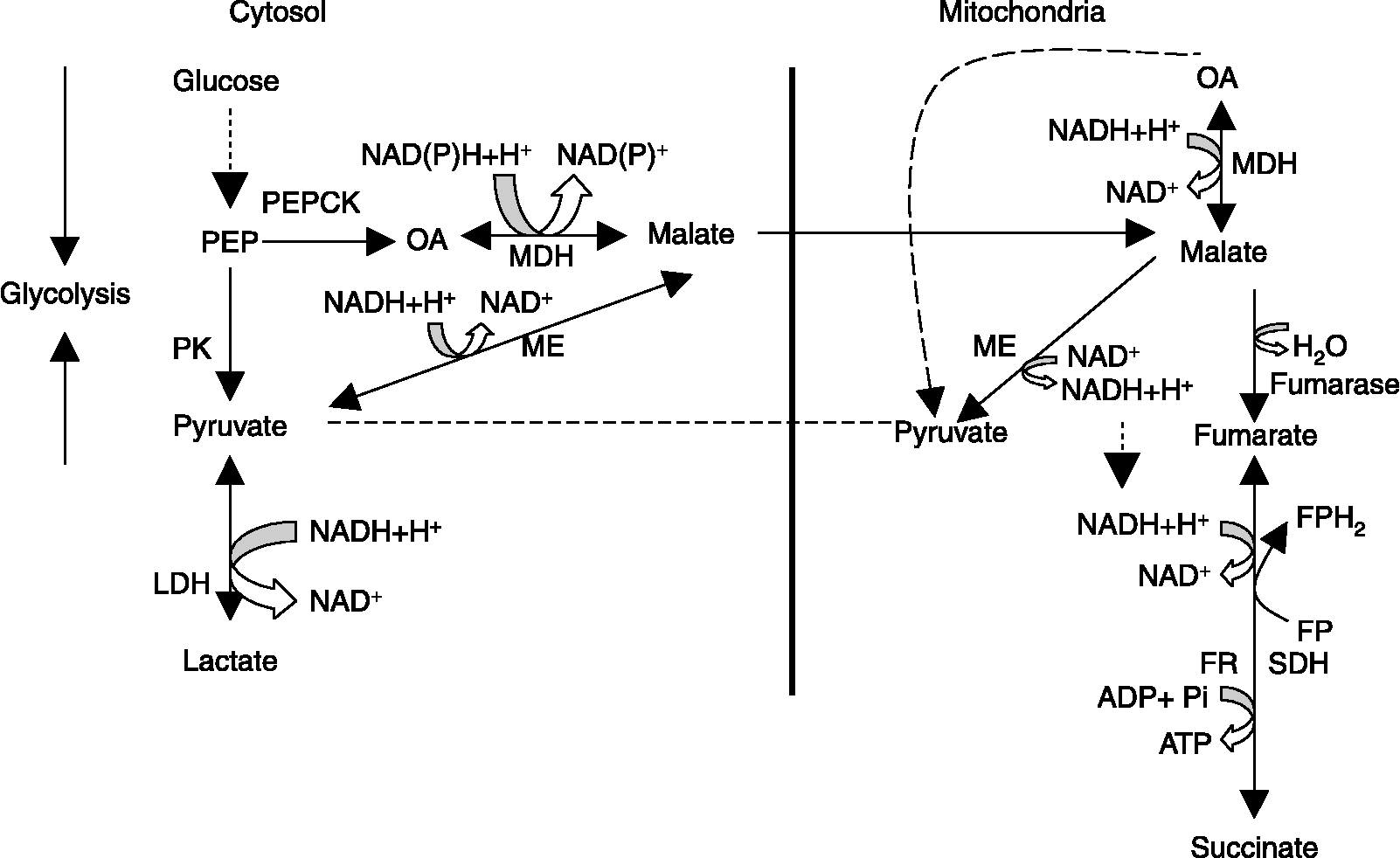
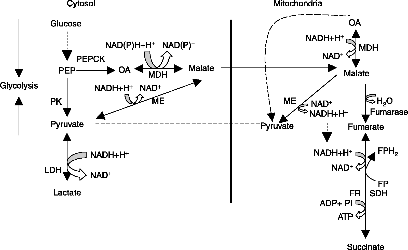
It can be asserted categorically that S. digitata has evolved mechanisms with mixed metabolic pathways, which enable it to depend on lactate formation and carbon dioxide fixation for energy release and survival, as depicted in fig. 2. Hence, the retention of the last part of the tricarboxylic acid (TCA) cycle in S. digitata can be accounted for in that it not only plays a role in energy metabolism but also interconverts the carbon skeleton (Barrett, Reference Barrett, Taylor and Müller1977).
Proposed catabolic pathway of carbohydrates in S. digitata. → , ↔ Pathways studied; ––– pathways not established; see Banu et al. (Reference Banu, Dhandayuthapani and Nellaiappan1991). ADP, Adenosine diphosphate; ATP, adenosine triphosphate; FP, flavoprotein; FPH2, reduced flavoprotein; FR, fumarate reductase; LDH, lactate dehydrogenase; MDH, malate dehydrogenase; ME, malic enzyme; NAD+, nicotinamide adenine dinucleotide; NADH, reduced nicotinamide adenine dinucleotide; NAD(P)+, nicotinamide adenine dinucleotide phosphate; NAD(P)H, reduced nicotinamide adenine dinucleotide phosphate; OA, oxaloacetate; PEP, phosphoenolpyruvate; PEPCK, phosphoenolpyruvate carboxykinase; Pi, inorganic phosphorus; PK, pyruvate kinase; SDH, succinate dehydrogenase.
Pentose phosphate pathway
The enzymes of the pentose phosphate pathway, glucose-6-phosphate dehydrogenase and 6-phosphogluconate dehydrogenase, have been detected in both mf and adult stages (Anwar et al., 1977; Rathaur et al., Reference Rathaur, Anwar, Saxena and Ghatak1982). The significance of this pathway is not clearly understood in the physiology of S. cervi but it may be related to the need for a large amount of riboses. Elucidation of this pathway might be useful in the development of novel chemotherapeutic agents in the future.
Glycogen metabolism and gluconeogenesis pathways
Setaria cervi has been found to contain almost all the enzymes involved in glycogen degradation (Anwar et al., Reference Anwar, Ansari, Ghatak and Murti1977). Glycogen phosphorylase has been found to be localized predominantly in the particulate-free cytosolic fraction, although small amounts of glycogen phosphorylase have also been detected in the mitochondrial fraction (Anwar et al., 1977).
Glucose-6-phosphatase, the last and most important enzyme of gluconeogenesis, has been localized histochemically in the mf of S. cervi (Khatoon et al., Reference Khatoon, Ullah, Baqui and Ansari1984). PEPCK activity has also been demonstrated in both mf and adult worms (Anwar et al., 1977; Rathaur et al., Reference Rathaur, Anwar, Saxena and Ghatak1982; Hussain et al., Reference Hussain, Shukla, Ghatak and Kaushal1990).
Anaplerotic reactions
All major enzymes catalysing anaplerotic reactions, namely pyruvate carboxylase, PEPCK and ME have been observed in Setaria species (Anwar et al., 1977; Rathaur et al., Reference Rathaur, Anwar, Saxena and Ghatak1982; Hussain et al., Reference Hussain, Shukla, Ghatak and Kaushal1990; Banu et al., Reference Banu, Dhandayuthapani and Nellaiappan1991). In S. digitata, it has been observed that the pyruvate carboxylation reaction and the OA reduction reaction are more active than their respective opposite reactions, suggesting that the pathway proceeds in the direction of malate formation. It has been reported by many workers that the malate formed as a result of cytosolic MDH activity enters into the mitochondria for further catabolic processes (Barrett, Reference Barrett1981; Saz, Reference Saz and Braude1981; Ward, Reference Ward1982). Similarly, the malate formed by cytosolic malic enzyme might also enter the mitochondria to take part in oxidative processes.
Phosphoenol pyruvate–succinate pathway
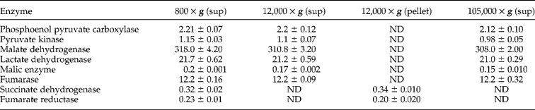
Enzymes of the phosphoenol pyruvate–succinate (PEP–succinate) pathway have been demonstrated in mf (Rathaur et al., Reference Rathaur, Anwar, Saxena and Ghatak1982) as well as in adults (Anwar et al., 1977). Succinate dehydrogenase (SDH) and fumarate reductase (FR) are localized in the mitochondria-rich particulate fraction while all other enzymes are cytosolic (Hussain et al., Reference Hussain, Shukla, Ghatak and Kaushal1990). The distribution pattern of the enzymes of the PEP–succinate pathway and other PEP-metabolizing enzymes analysed in different subcellular fractions of S. cervi is shown in table 3. The in vitro effect of certain antifilarial/anthelmintic agents on these enzymes has also been investigated. Suramin, at low concentration, has been shown to cause a marked inhibition of most of the enzymes of this pathway. Centperazine, an antifilarial drug, has shown significant inhibitory action on PK, LDH, fumarase and SDH, suggesting that this antifilarial probably exerts microfilaricidal action by blocking the PEP–succinate pathway (Rathaur et al., Reference Rathaur, Anwar, Saxena and Ghatak1982). DEC and levamisole, however, have been found to be more or less ineffective at low concentrations against all the enzymes of this pathway (Hussain et al., Reference Hussain, Shukla, Ghatak and Kaushal1990).
Distribution of the enzymes of S. cervi in different subcellular fractionsa; reproduced according to Hussain et al. (Reference Hussain, Shukla, Ghatak and Kaushal1990).
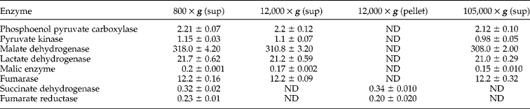
a The enzymes were assayed according to standard assay system of each enzyme. Enzyme units are defined as the amount of enzyme required to catalyse the transformation of one μmole of the substrate or the formation of one μmole of product min− 1 under specified experimental conditions. Enzyme activity is expressed as units g− 1 wet worms.
Values are mean ± SD of three experiments.
sup, Supernatant; ND, not detected.
Oxidative and respiratory metabolism
TCA cycle
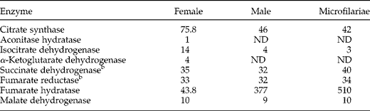
Enzymes of the TCA cycle have also been demonstrated in adult and mf stages of S. cervi and S. digitata (Anwar et al., 1977; Agarwal et al., Reference Agarwal, Saxena and Ghatak1986; Banu et al., Reference Banu, Dhandayuthapani and Nellaiappan1991). It is quite surprising to note that in S. digitata the enzymes related to the initial part of the TCA cycle are undetectable while in other filarial worms, such as Brugia pahangi and Dirofilaria immitis, a full complement of the enzymes is present although the enzymes of the initial part of the cycle are found with very low activity (Barrett, Reference Barrett1983). The absence of the enzymes of the initial part of the TCA cycle reduces the possibility of entry of OA, formed as a result of malate oxidation, into the TCA cycle. So the malate that enters the mitochondria probably undergoes dismutation, as has been reported in Ascaris (Saz, Reference Saz and Braude1981).The NADH required for the reduction of fumarate to succinate is obtained from the dismutation of malate (Barrett, Reference Barrett, Taylor and Müller1977).
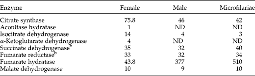
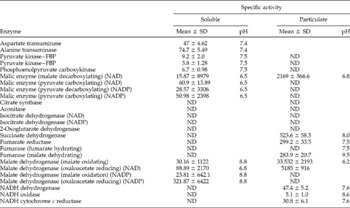
It is generally believed that the TCA cycle is not of much importance in helminths as it plays little role in the breakdown of carbohydrates in helminths (Barrett, Reference Barrett1981). Specific activities of the TCA cycle enzymes in adults and microfilariae of S. cervi are shown in table 4. Only a few of the enzymes involved in the TCA cycle have been found in the particulate fraction of S. digitata, unlike the findings in Onchocerca gutturosa and O. lienalis (Dunn et al., Reference Dunn, Rainess, Barrett and Butterworth1988). Fumarase has been found to be active only in one direction (malate dehydration) and malic enzyme is found to be present only in the malate decarboxylating direction. The soluble fraction has been found to contain MDH and ME activity in both directions and, surprisingly, both are NADH and NADP dependent. The results are presented in table 5.
Specific activitiesa of the enzymes of the TCA cycle in cell-free extracts of S. cervi; reproduced and modified according to Agarwal et al. (Reference Agarwal, Saxena and Ghatak1986).
a Measured as n moles substrate utilized min− 1 mg− 1 protein.
b Assayed in mitochondrial preparation.
Values are means of four separate observations.
ND, not detected.
Activities of enzymes in the soluble and particulate fractions of S. digitata (nmol substrate or co-factor min−1 mg−1 protein); reproduced according to Banu et al. (Reference Banu, Dhandayuthapani and Nellaiappan1991).

ND, not detected; FBP, fructose-1,6-bisphosphate.
Among the TCA cycle enzymes, the ratio of SDH : FR is said to give an idea as to the aerobic or anaerobic status of the organisms (Barrett, Reference Barrett and Van Den Bossche1976). S. digitata has been found to exhibit a ratio of 1.75 : 1, which is close to the value of facultative anaerobes, as reported in Ascaridia galli (2.5 : 1) and Nippostrongylus brasiliensis (3 : 1) (Fry & Brazeley, Reference Fry and Brazeley1983).
Excepting fumarate reductase, all other enzymes have been found to be localized in the particulate-free cytosolic fraction, although small amounts of glycogen phosphorylase, aldolase and lactate dehydrogenase were also detected in the mitochondrial fraction (Anwar et al., 1977). The presence of cytosolic MDH and ME is a common feature among parasites.
Mitochondrial malate dehydrogenase (mMDH) and malic enzyme (mME) of S. digitata have also been studied in detail. Mitochondrial malate dehydrogenase has been found to exhibit the highest activity for the oxidation reactions at pH 9.5 and reduction reactions at pH 6.2, while mitochondrial malic enzyme exhibits maximal activity in the malate decarboxylation reaction at pH 6.8. DEC citrate has been found to be more or less ineffective in inhibiting mMDH and mME from S. digitata, while filarin, a drug of herbal origin, effectively inhibits mMDH. The leaf extracts of Ocimum sanctum, Lawsonia inermis and Calotropis gigantea and leaf and flower extracts of Azadirachta indica, however, have been found to inhibit both mMDH and mME (Banu et al., Reference Banu, Nellaiappan and Dhandayuthapani1992).
Microfilariae of S. cervi resemble the adults in their metabolic pattern. MDH has been found to be the most active enzyme in mf, followed by LDH and fumarase, while phosphoglucoisomerase, PEPCK and FBP-aldolase are comparatively less active.
Oxidative phosphorylation
The mitochondria-like particles (MLPs) of the filarial parasite, S. digitata have shown NAD reduction with sodium lactate. This study has provided new dimensions to the study of metabolism in filarial parasites (Sivan & Raj, Reference Sivan and Raj1994). In S. digitata, the MLPs have been shown to possess site I-associated oxidative phosphorylation and rotenone sensitive and insensitive pathways for the dehydrogenation of NADH. Thus, activities of complex I and fumarate reductase (FR) are linked to quinone. The FR system, consisting of NADH–coenzyme Q reductase, cytochrome b like component(s) and succinate dehydrogenase/fumarate reductase, is thus very important, and hence specific inhibitors of the system may prove useful in the effective control of filariasis (Unnikrishnan & Raj, Reference Unnikrishnan and Raj1992).
A lipid quinone, possibly with a short side-chain and related to ubiquinone, has been detected in adult worms. The results suggest the existence of two cyanide-insensitive oxygen-consuming reactions in Setaria; one respiratory substrate-independent lipid peroxidation and a second substrate-dependent reaction that requires an auto-oxidizable quinone (Raj et al., Reference Raj, Puranam, Kurup and Ramasarma1989).
Setaria digitata is unique in having two ubiquinones, Q6 and Q8, in the adult stage, in place of one, namely Q10, in the host. These quinones are synthesized within the parasite itself and are not of host origin (Abhilash et al., Reference Abhilash, Mohan, Jayakumar and Kaleysa2001). Maximum concentration is found in the mitochondria of the parasite. While both Q6 and Q8 are formed and present in the adult stage, the microfilariae (mf), as well as the electron transfer complexes from adult mitochondria, have been recently shown to contain only Q6. The second ubiquinone Q8 is present only in the adult and absent in the mf (Mohan et al., Reference Mohan, Pradeep, Abhilash, Jayakumar and Kaleysa2001). Setaria digitata is unique in having peculiarities such as hydrogen peroxide (H2O2) production, cyanide insensitivity and absence of cytochromes and presence of quinones (Santhamma & Raj, Reference Santhamma and Raj1991a). Hence inhibitors at the level of quinones might prove to be effective in designing drugs for filariasis (Santhamma & Raj, Reference Santhamma and Raj1990, Reference Santhamma and Raj1991b, Reference Santhamma and Raj1993). 2, 3-Dimethoxy-5-methyl-1, 4-benzoquinone, an analogue of ubiquinone, irreversibly paralyses the adults and mf of the cattle filarial parasite S. digitata. Thus, an analogue of quinone can inactivate the filarial parasites and may prove to be an effective drug to control filariasis (Sivan & Raj, Reference Sivan and Raj1992, Reference Sivan and Raj1999).
A mitochondria-rich fraction isolated from the cuticle–hypodermis–muscle system of S. cervi, has been found to possess substrate-coupled cytochrome c reductases and cytochrome c oxidase in appreciable activities (Goyal & Srivastava, Reference Goyal and Srivastava1995). NADH-coupled cytochrome c reductase is more prominent than NADPH- and succinate-coupled reductases. All three reductases exhibit marked sensitivity to rotenone and antimycin A.
Setaria digitata, like the human filarial parasites, possesses significant activities of the four transhydrogenases, namely NADH-NAD+, NADPH-NAD+, NADH-NADP+ and NADPH-NADP+ in sonicated MLPs (Unnikrishnan & Raj, Reference Unnikrishnan and Raj1995). The transhydrogenases appear to regulate the metabolic pathways of the parasite in response to the presence of adenyl nucleotides and are non-energy linked. Hence, specific drugs targeted against this interconnected complex enzyme system could prove to be very effective in the control of filarial parasites (Unnikrishnan & Raj, Reference Unnikrishnan and Raj1995).
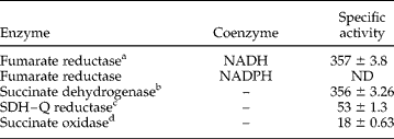
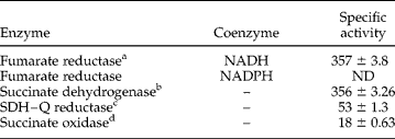
The presence of only two activities of the succinate oxidase system, namely SDH and succinate–UQ reductase, has so far been detected in S. digitata. The third enzyme of the system, namely cytochrome oxidase, is absent and it is proposed that an alternative oxidase is responsible for completing succinate oxidation (Unnikrishnan & Raj, Reference Unnikrishnan and Raj2000). Activities of FR, SDH, SDH–UQ reductase and succinate oxidase in MLP of S. digitata are shown in table 6.
Activities of fumarate reductase, succinate dehydrogenase, SDH–Q reductase and succinate oxidase in mitochondria-like particles of S. digitata; reproduced according to Unnikrishnan & Raj (Reference Unnikrishnan and Raj2000).
a Activity expressed as n moles of NADH oxidized min− 1 mg− 1 protein.
b Activity expressed as n moles of 2, 6-dichlorophenol indophenol (DCIP) oxidized min− 1 mg− 1 protein.
c Activity expressed as μmoles of succinate oxidized min− 1 mg− 1 protein.
d Activity expressed as n atoms of oxygen consumed min− 1 mg− 1 protein.
Values are ± SD of six different experiments.
ND, not detected; SDH–Q reductase, succinate dehydrogenase–ubiquinone Q reductase.
Setaria cervi has also been found to contain NADPH oxidase activity. The system is predominantly located in the mitochondrial membranes, with very little activity detectable in the soluble fraction of the organelle. The oxidase activity was inhibited by all the respiratory inhibitors examined, with the greatest sensitivity to rotenone, a site I-specific inhibitor. The system has also been found to be susceptible to exposure to anthelmintics, amongst which levamisole proved the most effective (Goyal & Srivastava, Reference Goyal and Srivastava1990).
ATPases
A Mg2+-independent, Ca2+-ATPase requiring high concentrations of Ca2+(5 mM) for activation has been identified in S. cervi (Agarwal et al., Reference Agarwal, Shukla and Tekwani1992). Trifluoperazine (phenothiazine derivative) has been observed to be the most potent inhibitor, followed by promethazine and chlorpromazine. Some anthelmintics, namely DEC and centperazine, were found to be highly potent inhibitors of the enzyme; significant inhibition of filarial Ca2+-ATPase was also observed with levamisole and suramin. These studies indicate Ca2+-ATPase of S. cervi as a potential chemotherapeutic target.
Particulate-bound Ca2+-ATPase and Na+,K+-ATPase activities have been demonstrated in S. cervi, while Mg2+-ATPase has been detected in traces (Agarwal et al., Reference Agarwal, Tekwani, Shukla and Ghatak1990a). Adult female parasites and mf exhibit higher Ca2+-ATPase and Na+,K+-ATPase activities than the male adults, and the enzyme Na+,K+-ATPase is mainly concentrated in the gastrointestinal tract (GIT) of the filarial parasite. Na+,K+-ATPase of the filariid is ouabain-sensitive, while Ca2+-ATPase activity is regulated by concentration of Ca2+ ions and inhibited by EGTA. Phenothiazines, namely trifluoperazine, promethazine and chlorpromazine, have been found to cause significant inhibition of Ca2+-ATPase and Na+K+-ATPase of S. cervi. DEC is a potent inhibitor of these ATPases. Mebendazole, levamisole and centperazine also cause significant inhibition of the filarial ATPases, indicating that this enzyme system is a common target for the action of anthelmintic drugs (Agarwal et al., Reference Agarwal, Tekwani, Shukla and Ghatak1990a).
Amino acid and protein metabolism
Little work has been done on the amino acid metabolism of the filarial parasites; amino acid compositions have been studied for S. cervi. Protein constitutes over 63 and 57% of the dry weight of adults and mf of S. cervi (Rathaur et al., Reference Rathaur, Anwar and Ghatak1980). Amino acids are essential for maintaining the viability of helminth worms (Srivastava & Ghatak, Reference Srivastava and Ghatak1990).
The high rate of incorporation of exogenous amino acids into proteins has been demonstrated in intact adult S. cervi (Anwar et al., Reference Anwar, Srivastava and Ghatak1978). Setaria cervi was found to synthesize proteins actively in vitro. The synthesis in S. cervi was susceptible to puromycin, chloramphenicol, cycloheximide, neomycin and polymyxin B (Wani & Srivastava, Reference Wani and Srivastava1993). Very little is known about the ability of adult S. cervi to interconvert amino acids. Transamination between α-ketoglutarate and aspartate or alanine could be detected and glutamate dehydrogenase has been partially purified and characterized (McManus, Reference McManus1987). Other enzymes present in crude extracts of adult S. cervi were l-alanine dehydrogenase, l-threonine dehydratase, l-ornithine transcarbamoylase and arginase.
DEC has been found to inhibit activities of monoamine oxidase, aspartate amino transferase and alanine amino transferase, and enhanced those of cathepsin and glutamate dehydrogenase (Vijayanathan & Raj, Reference Vijayanathan and Raj1991).
A 175 kDa antigen fraction with collagenase activity has been isolated and purified from somatic extracts of adult S. cervi females, using column chromatography. Further analysis has been undertaken on the immunoprophylactic potential of 175 kDa collagenase in inducing immunity against B. malayi in jirds (Meriones unguiculatus) in vitro and in situ (Srivastava et al., Reference Srivastava, Rathaur, Bhandari, Reddy and Harinath2004). Release of mf from adult S. digitata has been shown to be reversibly arrested in the presence of 2 mM l-glutamine. DEC also showed a similar effect, but at a lower concentration (0.26 mM). Both compounds have been found to inhibit activities of protease and leucine amino peptidase enzymes (Murugan & Raj, Reference Murugan and Raj1995).
In vitro released products of adult S. cervi females, mf and extracts have shown considerable amounts of collagenase activity (Singh & Rathaur, Reference Singh and Rathaur2003). Two collagenase enzymes with molecular masses of 50 and 70 kDa have been resolved. The 50 and 70 kDa collagenases exhibit pH optima of 5.2 and 7.0, respectively. An inhibition study revealed obvious differences between them. Thiol group inhibitors such as N-ethylmaleimide and leupeptin have been found to inhibit the 50 kDa enzyme, but this was strongly activated by dithiothreitol (DTT), a thiol group stabilizer. Alternatively, the 70 kDa enzyme showed sensitivity to a metal chelator and a serine group inhibitor, indicating its metalloserine protease nature.
Chlorpromazine (CPZ) also causes suppression of the release of mf in vitro from S. cervi at 10− 5 M concentration. The inhibitory effect of CPZ has been found to be further stimulated by Ca2+(2 mM), whereas addition of EGTA (2 mM) results in partial reversal. Addition of dopamine (1 mM) along with CPZ (10− 5 M) causes similar inhibition as observed in the presence of CPZ (10− 5 M) alone (Tekwani et al., Reference Tekwani, Tripathi, Shukla and Ghatak1988).
A protease isolated from adult S. digitata has revealed a single, 110-kDa band in a gelatin-impregnated substrate gel. Analysis with specific inhibitors has indicated that it is a metalloprotease (Bal & Das, Reference Bal and Das1999). A zinc-dependent cysteine proteinase has also been characterized from a low molecular weight fraction (30 kDa) of S. digitata (Beuria et al., Reference Beuria, Bal and Das1995).
The investigation of protein kinase activity in different lifecycle stages of S. cervi indicates the presence of different protein kinases and of dissimilarities between the distribution patterns of these enzymes during development (Saxena et al., 1984). The presence of protein kinases phosphorylating a variety of proteins and peptides has been demonstrated in adults and mf, and the dissimilarities between the distribution patterns of these enzymes during development have also been noted (Saxena et al., 1984). Isolation, partial purification and characterization of protein kinase I, protein kinase II and protein kinase III from S. cervi females has also been reported (Srivastava et al., Reference Srivastava, Saxena, Singh, Singh, Chatterjee and Ghatak1994a).
Excretory–secretory products (ES) of S. cervi, prepared by in vitro maintenance of the worms, have revealed 11–15 antigens, most of which are common to somatic antigenic preparations from both adult worms and mf. The ES products have also been found to contain 3–4 host proteins, including serum albumin (Malhotra et al., Reference Malhotra, Kaushal, Kaushal and Ghatak1987). ES material isolated from the culture fluid of S. digitata is highly antigenic. The amniotic fluid appears to be the most important source of ES material released by the filarial parasites (Thilagavathy et al., Reference Thilagavathy, Prabha and Raj1990). Analysis of ES products, using anti-ES serum, has shown the presence of 10–14 immunogenic proteins in S. cervi (Mustafa et al., Reference Mustafa, Srivastava, Kaushal and Kaushal1996). Similar proteins secreted by human filarial parasites could be targets for combating the disease by cure or control (Sugunan & Raj, Reference Sugunan and Raj1990; Murugan & Raj, Reference Murugan and Raj1991). ES that are released along with mf from S. digitata have shown immunosuppression in BALB/c mice on immunization, while those released in the presence of a sublethal concentration of 0.25 mM DEC have shown initial potentiation followed by suppression. These observations suggest a change in the nature of materials of embryonic origin before and after the release of mf (Devi & Raj, Reference Devi and Raj1994).
High activities of hydrolytic enzymes such as protease, collagenase, alkaline phosphatase and lipase have been detected in the ES products and adult worm preparations. The activity of collagenase has not been detected in the male worms under experimental conditions (Murugan & Raj, Reference Murugan and Raj1997).
Osmoregulation
Studies on osmotic and ionic regulation in S. cervi indicate that it can osmoregulate in hypertonic solutions but not in hypotonic solutions (Singh & Sharma, Reference Singh and Sharma1984).
Lipid metabolism
Nothing is known about the details of lipid synthesis except the ability of the adult worms to incorporate [14C]glucose into their lipid fraction. Microfilariae as well as adult males and females of S. cervi are able to synthesize several important classes of lipids and thus exhibit a functional lipid metabolism (Kumar, Reference Kumar1987a). The enzymes involved in lipid catabolism have been studied only in adult females.
Investigations of lipid metabolism in helminths are few and are mainly restricted to analyses of lipid composition and studies of lipid synthesis (Zachariah & Raj, Reference Zachariah and Raj1986; McManus, Reference McManus1987). Analysis of lipid composition has been done in S. cervi and was found to be in the range from 1.3 to 8% (Ansari et al., Reference Ansari, Yusufi and Siddiqi1973; Khatoon et al., Reference Khatoon, Ullah, Baqui and Ansari1984). Triacylglycerols and sterols have been demonstrated in adult S. cervi (Khatoon et al., Reference Khatoon, Ullah, Baqui and Ansari1984). All the major phospholipid classes have also been identified in adult filarial worms, phosphatidylcholine being the predominating one.
The biological significance of isoprenoid compounds such as ubiquinones, prenols and sterols has been well established in Setaria species. Inhibition of the function of quinone has already been shown to be an effective means of controlling these filarial parasites. Blocking of the biosynthesis of these useful compounds may prove to be an additional means of control of these parasites (Kumari et al., Reference Kumari, Santhamma and Raj1994).
Nucleic acid metabolism
Genomic in situ hybridization (GISH) has been used to investigate genomic relationships between different Setaria species (Benabdelmouna et al., Reference Benabdelmouna, Shi, Abirached-Darmency and Darmency2001). Ribonucleotide biosynthesis from 14C-labelled glucose has been observed in mf (Kumar, Reference Kumar1987c). It has been shown that DNA topoisomerase II is an essential enzyme, which displays an important role in DNA replication, repair and transcription. Recently, DNA topoisomerases II have been identified as targets for the development of antifilarial compounds (Tripathi et al., Reference Tripathi, Rastogi, Kundu, Saxena, Reddy, Srivastava, Chandra and Bhaduri2001). 7-O-Acetamidyl-4-alkyl-2H-1-benzopyran-2-ones, glycosylated beta-amino acid derivatives and Ru (II) poly-pyridyl hydridocarbonyl complexes have been prepared and screened against DNA topoisomerase II of S. cervi (Tripathi et al., Reference Tripathi, Tripathi, Bhaduri, Singh, Chatterjee and Murthy2000, Reference Tripathi, Tiwari, Misra-Bhattacharya, Tyagi, Srivastava and Murthy2003; Chandra et al., Reference Chandra, Sahay, Pandey, Tripathi, Saxena, Reddy, Puerta and Valerga2004). The uptake of [14C]uracil and thymidine has been demonstrated in adult S. cervi and an appreciable amount of radioactive uracil was found to be associated with the nucleic acid fraction, mainly with RNA.
The interaction of DNA from the filarial parasite S. cervi with polyamines has been monitored by melting temperature (T m) profile, condensation and B to Z DNA transition, and compared with DNA of Escherichia coli, Micrococcus luteus and calf thymus, having a different GC content (Pandya et al., Reference Pandya, Saxena and Shukla1997). The antifilarial compound suramin has been found to significantly decrease T m of filarial DNA as compared to GC-rich DNAs of other parasites. Suramin adversely affects the condensation and B to Z DNA transition of various DNAs.
It has been shown that S. digitata possesses the ability to respond to mammalian epidermal growth factor (EGF), which is probably responsible for upregulation of a DP-1 family transcription factor. From this it can be concluded that mammalian growth factors may regulate developmental maturation of filarial parasites (Dissanayake, Reference Dissanayake2000).
Two repetitive sequences (IpSdM and IpSdS) have been cloned and sequenced from the genome of S. digitata (Wijesundera et al., Reference Wijesundera, Chandrasekharan, Karunanayake and Dharmasena1996). A high degree of homology (85%) has been observed between the sequences, indicating that they belong to the same family of repeats. IpSdM represents a complete repeating element while IpSdS consists of two partial repeating elements arranged in tandem. The elements are present in about 10,000 copies, comprising 2.8% of the S. digitata genome.
A series of 21 compounds of trisubstituted pyrimidine derivatives has been synthesized and evaluated for their in vitro topoisomerase II inhibitory activity against filarial parasite S. cervi (Katiyar et al., Reference Katiyar, Bansal, Saxena and Chauhan2005). All the above-mentioned compounds have shown better topoisomerase II inhibitory activity than the standard antifilarial drug (DEC) and enzyme topoisomerase II inhibitors such as novobiocin and nalidixic acid.
A series of 7-O-acetamidyl-4-alkyl-2H-1-benzopyran-2-ones has been synthesized and screened for their filarial DNA topoisomerase inhibitory activity under in vivo conditions in S. cervi. The compounds were tested in vitro against B. malayi. A few of the compounds have been found to possess promising antifilarial activity (Katiyar et al., Reference Katiyar, Tiwari, Tripathi, Reddy, Bhattacharya and Saxena2003).
Very little is currently known about the biology of this parasite at the molecular level. One hsp70 gene has been cloned and characterized and Southern blot analysis has revealed the presence of two or three additional hsp70-related genes in the S. digitata genome (Jayasena et al., Reference Jayasena, Chandrasekharan and Karunanayake1999).
5S rRNA intergenic regions of S. digitata and S. labiatopapillosa have been amplified by polymerase chain reaction (PCR) with primers designed from the 5S rRNA gene of B. malayi. By means of random polymorphic DNA (RAPD)-PCR analyses, 107 bands have been identified, of which 86 were polymorphic. The polymorphic loci identified by the RAPD-PCR analyses can be studied further to develop species-specific probes/PCR primers for the identification of each species (Jayasinghe & Wijesundera, Reference Jayasinghe and Wijesundera2003).
Phosphate metabolism
Distribution of acid and alkaline phosphomonoesterases in the parasitic nematode has been studied in S. cervi (Sharma & Singh, Reference Sharma and Singh1985). Acid phosphatase activity has been demonstrated in smears of S. labiatopapillosa mf and compared with those of Dirofilaria immitis, D. repens and an equine Setaria species (Rossi & Abate, Reference Rossi and Abate1990).
Filarial receptors, channels and neurotransmitters
Nematodes possess a variety of receptors, receptor subunits and channels, and their expression can be developmentally regulated. By targeting such receptors we can selectively obstruct the parasite nervous system (Lazdins & Kron, Reference Lazdins and Kron1999).
The total and free acetylcholine and acetylcholinesterase contents of adult S. cervi have been estimated (Singhal et al., Reference Singhal, Zehra, Singhal and Saxena1978a). It is possible that there might exist a well-developed system responsible for the synthesis, storage, release and destruction of acetylcholine, and that acetylcholine may be acting as an excitatory neurohormone in S. cervi. Acetylcholine, levamisole and pyrantel pamoate cause stimulation of spontaneous rhythmic movements of whole worms and nerve muscle preparations of S. cervi (Singhal & Singhal, Reference Singhal and Singhal2001). The findings indicate that the excitatory neurotransmitter acetylcholine and the anthelmintics levamisole and pyrantel pamoate produce their stimulant effect by triggering entry of Ca2+ into the muscle cell. Further, blocking the calcium channels by nifedepine blocks the stimulant effect of acetylcholine, levamisole and pyrantel pamoate (Singhal & Singhal, Reference Singhal and Singhal2001).
Setaria cervi secretes acetylcholinesterase during in vitro cultivation. The mf stage has been shown to possess a higher level of acetylcholinesterase activity than adult worms, with females considerably more active than males. The secretory acetylcholinesterases from S. cervi mf may be utilized for diagnosis of early filarial infections (Sharma et al., Reference Sharma, Misra and Rathaur1998).
Nerve muscle preparations of S. cervi have been found to exhibit spontaneous rhythmical movements in modified Ringer's solution (Ganguly et al., Reference Ganguly, Awasthi and Singhal1996). Deprivation of Ca2+ results in a gradual reduction in the amplitude and rate of contraction, until the movements cease completely. The evidence clearly indicates that the presence of Ca2+ is essential for the stimulant effect of acetylcholine on S. cervi which, like mammalian tissues, contain calcium channels that can be blocked by specific blocking agents (Singhal et al., Reference Singhal, Ganguly and Awasthi1995).
Synthetic compounds of 5/6/7/8 mono- or di-substituted 1H/1-phenyl-9H pyrido-(3,4)-indoles have been screened for their in vitro antifilarial activity by observing their effect on the spontaneous movements of the whole worm and nerve–muscle preparations of S. cervi (Kapoor & Singhal, Reference Kapoor and Singhal1998).
Redox metabolism
Glutathione (GSH) is of major importance in filarial species as it has been proposed to constitute the antioxidant system responsible for the long-term existence of filarial worms in their respective mammalian hosts, by protecting them from the reactive oxygen species (ROS) produced by the normal metabolism and the immune cells of the host (Callahan et al., Reference Callahan, Crouch and James1988; Brophy & Pritchard, Reference Brophy and Pritchard1992). It therefore seems useful to develop drugs that could selectively deplete or distort GSH stores in these parasites. Pharmacological depletion of GSH in the cells is known to be useful in the development of antiparasitic agents (Arrick et al., Reference Arrick, Griffith and Cerami1981; Gupta & Srivastava, Reference Gupta and Srivastava2006). Many inhibitors of enzymes involved in GSH metabolism are known to be associated with antiparasitic activities and are potential chemotherapeutic targets (Srivastava et al., Reference Srivastava, Tripathi, Khan, Bhaduri, Singh and Chatterjee1995; Mishra et al., Reference Mishra, Tewari, Arora, Ahmad, Tripathi, Tiwari, Walter and Srivastava2003; Tiwari et al., Reference Tiwari, Tewari, Katiyar, Tripathi, Arora, Gupta, Ahmad, Srivastava, Khan and Murthy2003; Ahmad et al., Reference Ahmad, Mishra, Tewari, Tripathi, Srivastava and Walter2004; Arora et al., Reference Arora, Mishra, Tripathi, Srivastava and Walter2004; Gupta et al., Reference Gupta, Arora, Tiwari, Katiyar, Tripathi, Srivastava and Walter2004). GSH either acts in solo or as a substrate to various enzymes, such as glutathione peroxidase (GPX), phospholipid hydroperoxide GPX and glutathione-S-transferase (GST) (Zhang et al., Reference Zhang, Maiorino, Roveri and Ursini1989; Lomaestro & Malone, Reference Lomaestro and Malone1995), to quench the free radicals. After GSH has been oxidized to GSSG (oxidized glutathione) by playing its role, the enzyme glutathione reductase (GR) accomplishes the recycling of GSSG back to GSH (Carlberg & Mannervik, Reference Carlberg and Mannervik1985; Schirmer et al., Reference Schirmer, Schollhammer, Eisenbrand and Krauth-Siegel1987).
GSH has been analysed in S. cervi (Singh et al., Reference Singh, Srivastava and Chatterjee1997a; Gupta et al., Reference Gupta, Tripathi, Srivastava, Iqbal, Callaos, He and Perez-Perez2002). It has been shown that interference with the filarial GSH metabolism has a curative effect on filarial infections in mice (Bhargava et al., Reference Bhargava, LeTrang, Cerami and Eaton1983). Glutamate cysteine ligase (GCL), the rate-limiting enzyme of GSH biosynthesis which catalyses the ligation of l-glutamate and l-cysteine to form l-γ-glutamyl-l-cysteine (Plummer et al., Reference Plummer, Smith, Sies and Bend1981) has been detected in S. cervi. The GSH hydrolysing enzyme, γ-glutamyl transpeptidase (γ-GT), has also been detected in S. cervi (Singh et al., Reference Singh, Khan and Singhal1996; Gupta et al., Reference Gupta, Srivastava and Banu2005a).
Setaria cervi parasites have been shown to possess great susceptibility to the oxidants generated in vitro by the xanthine–xanthine oxidase system (Batra et al., Reference Batra, Chatterjee and Srivastava1992). In order to counteract such injurious effects, S. cervi parasites possess an active antioxidant enzyme system. Superoxide dismutase (SOD), catalase and GPX have been detected in appreciable amounts, but GR and glucose-6-phosphate dehydrogenase in very low quantities. 2,2’-Dicarbomethoxylamino-5,5’-dibenzimidazolylketones have been shown to markedly inhibit catalase and GPX of S. cervi. This class of compounds, therefore, appears to render the filariids prone to H2O2 toxicity, leading to penultimate damage.
Glutathione reductase (GR) activity has been detected in crude extracts of S. cervi (Singh et al., Reference Singh, Srivastava and Chatterjee1997a). GR from the cattle filariid S. digitata has been isolated and its properties compared to those of human erythrocyte GR. Although the physical and kinetic properties of the parasitic enzyme are very similar to those of human GR, studies on the inhibition of the enzyme by arsenical melarsen oxide have revealed that human GR is less susceptible to inhibition by the arsenical than the filarial enzyme (Müller et al., Reference Müller, Walter and Fairlamb1995). Moreover, the mechanism of inhibition has been found to be different in host and filarial enzyme. These differences between host and parasite enzyme might reflect differences in the primary and secondary structure of the proteins that might be exploitable for the design of new specific macrofilaricidal drugs (Müller et al., Reference Müller, Walter and Fairlamb1995).
Setaria cervi has been found to have an abundance of glutathione synthetase (GS) activity, the enzyme being involved in catalysing the final step of GSH biosynthesis (Gupta et al., Reference Gupta, Srivastava and Banu2005b). Subcellular fractionation of the enzyme has confirmed it to be a soluble protein residing mainly in cytosolic fraction. Attempts to determine the K m value for substrate l-γ-glutamyl-l-cysteine have given a distinctly non-linear double-reciprocal plot. The effect of various amino acids on S. cervi GS activity has been studied at 1.0 mM concentration. l-Cystine caused a significant 11% inhibition of the enzyme. Pre-incubation with N-ethylmaleimide (NEM) also resulted in significant inhibition of GS activity.
Glutathione-S-transferase (GST); a phase-II drug detoxification enzyme has been detected in S. cervi (Srivastava et al., Reference Srivastava, Tripathi, Khan, Bhaduri and Singh1994b). GST activity has also been determined in adult stages of S. digitata. It is absent in other stages of Setaria species and also in infective larval stages of Wuchereria bancrofti and B. malayi. It has been observed that DEC treatment of microfilaraemic sera results in considerable reduction in enzyme activity (Bal & Das, Reference Bal and Das1996).
In vitro effects of DEC, butylated hydroxyanisole (BHA) and phenobarbitone on the GST of adult female S. cervi have been assayed by the addition of these compounds to the maintenance medium (Gupta & Rathaur, Reference Gupta and Rathaur2005). The results suggest that S. cervi GST is inducible in response to the antifilarial drug DEC, and may play an important role in parasite's survival, and thus could be a potential drug target (Gupta et al., Reference Gupta, Bhandari, Reddy, Harinath and Rathaur2005c). The role of S. cervi GST antigen in inducing immunity in the host against B. malayi mf and infective larvae has been studied by in vitro antibody-dependent cell-mediated reaction, as well as in situ inoculation of filarial parasites within a microchamber in Mastomys (Gupta et al., Reference Gupta, Bhandari, Reddy, Harinath and Rathaur2005c). Native GST from S. cervi is effective in inducing protection against heterologous B. malayi filarial parasite and thus has potential in immunoprophylaxis.
It has also been reported that S. cervi secretes a selenium glutathione peroxidase (GPX) during in vitro cultivation. A significant amount of enzyme activity has been detected in the somatic extract of different developmental stages of the parasite. Among different stages, mf have shown a higher level of selenium-dependent GPX activity, followed by males then females. Further, inhibition of enzyme activity by potassium cyanide (KCN) suggests the presence of selenium at the active site of the enzyme (Singh & Rathaur, Reference Singh and Rathaur2005).
Biogenic amine metabolism
Various biogenic amines, namely norepinephrine (noradrenaline; NE), dopamine (DA), 5-hydroxytryptamine (5-HT) and histamine (Hm), have been reported to be present in mf and adults of S. cervi (Rathaur et al., Reference Rathaur, Kaushal and Ghatak1985; Gupta & Srivastava, Reference Gupta and Srivastava2005). The role of biogenic amines in neuromuscular activity (Gainutsos & Bennett, Reference Gainutsos and Bennett1977) and behavioural co-ordination (Croll, Reference Croll1975) of nematodes is well documented. Dopamine-β-hydroxylase has been detected in S. cervi (Agarwal et al., Reference Agarwal, Mishra, Rathaur, Chatterjee, Katiyar and Ghatak1982). Monoamine oxidase (MAO) and acetylcholinesterase, which play a role in neuromuscular transmission, are present in both mf and adults of S. cervi. MAO from the parasites has been shown to exhibit unusually high K m values for various monoamines (Agarwal et al., Reference Agarwal, Shukla, Ghatak and Tekwani1990b). The enzyme oxidizes dopamine at the maximum rate while putrescine is not utilized as the substrate. MAO activity is predominantly associated with the mitochondrial fraction. The enzyme has been found to be most active around pH 7.5 and 37 °C, and relatively stable in the frozen state but is thermolabile.
Centperazine, DEC and levamisole are known to strongly inactivate MAO and acetylcholinesterase from S. cervi females (Rathaur, Reference Rathaur1980). Setaria cervi MAO can be differentiated from the host enzyme on the basis of substrate specificity, pH optimum and K m value (Rathaur, Reference Rathaur1980). Levels of serotonin, dihydroxyphenyl alanine and epinephrine increase, while that of histamine has been found to remain the same, when adult S. digitata are incubated in DEC (Vijayanathan & Raj, Reference Vijayanathan and Raj1991).
Polyamine metabolism
The polyamines putrescine, spermidine and spermine have been found in all living organisms and are involved in growth, differentiation and macromolecular synthesis (Tabor & Tabor, Reference Tabor and Tabor1984; Pegg et al., Reference Pegg, Kameji, Shirahata, Stanley, Madhubala and Pajunen1988; Svensson et al., Reference Svensson, Kockum and Persson1993). Histamine, 5-hydroxytryptamine, norepinephrine and dopamine have been detected in both mf and adults of S. cervi. Dopamine is the major monoamine of S. cervi, followed by norepinephrine and 5-hydroxytryptamine (Agarwal et al., Reference Agarwal, Shukla, Ghatak and Tekwani1990b). 5-Hydroxyindoleacetic acid and tryptophan have also been detected in significant amounts. The specific MAO inhibitors, clorgyline and deprenyl, inhibited the enzyme with K i values of 2 × 10− 7 M and 5 × 10− 6 M, respectively. These characteristics of the enzyme indicate that it may have a role in host–parasite interactions (Agarwal et al., Reference Agarwal, Shukla, Ghatak and Tekwani1990b).
Polyamine levels of S. cervi have been analysed by reverse-phase high-performance liquid chromatography (HPLC) of benzoyl derivatives. Setaria cervi contained higher levels of spermine than spermidine, whereas putrescine and n-acetylated polyamines have been observed in very low amounts (Srivastava et al., Reference Srivastava, Roy and Shukla1980; Wittich et al., Reference Wittich, Kilian and Walter1987). The enzymes of polyamine biosynthesis, namely ornithine decarboxylase, S-adenosyl methionine (SAM) decarboxylase and arginine decarboxylase, were present in very low to negligible amounts in S. cervi. Significant activity of polyamine oxidase and appreciable activity of ornithine aminotransferase have been detected in S. cervi (Singh et al., Reference Singh, Saxena, Ghatak, Shukla, Wittich and Walter1989). The questionable presence of polyamine biosynthesis enzymes such as ornithine decarboxylase and arginine decarboxylase and, on the other hand, an effective uptake mechanism for polyamines indicates that the parasite may depend on the host for its polyamine requirement, thereby indicating a possible target for chemotherapy (Sharma et al., Reference Sharma, Tekwani, Saxena, Gupta, Katiyar, Chatterjee, Ghatak and Shukla1991).
Haem metabolism
There is currently renewed interest in the biological significance of haem proteins. It has been demonstrated that S. digitata is devoid of typical cytochrome systems. However, studies have shown the presence of δ-aminolevulinate synthase (ALAS) and δ-aminolevulinate dehydratase (ALAD) as well as haem oxygenase in appreciable amounts, suggesting the presence of the necessary equipment for the biosynthesis of haem. This is further confirmed by the end-product inhibition of ALAS by haem and the observation of the death of the parasite caused by succinyl acetone, an inhibitor of haem biosynthesis. Although typical cytochrome systems are absent, microsomal cytochrome P450 and elevated levels of haem-containing enzymes, such as catalase and peroxidase, have been shown to be present in the parasite. A unique haemoglobin has also been detected which shows a difference in biological functions from the host system and that of the much-studied nematode parasite Ascaris suum (Kumar & Raj, Reference Kumar and Raj1998).
Immunochemical studies
The protein and antigenic patterns of adult (female/male) and mf stages of S. cervi have been studied using certain immunochemical techniques (Malhotra et al., Reference Malhotra, Kaushal and Ghatak1986). Setaria cervi antigens have been evaluated for the immunodiagnosis of human filariasis (Almeida et al., Reference Almeida, Bhopale and Renapurkar1990). Immunoelectrophoresis has revealed the presence of 9–10 precipitin lines in adults and only four precipitin lines in microfilarial antigenic preparations. Crossed immunoelectrophoresis has resolved these antigenic preparations further and has revealed the presence of about 22–24 and 12–14 antigens in adults and mf, respectively.
Identification and characterization of antigenic proteins of S. cervi adults and mf have been done by immunoblotting technique using hyperimmune rabbit sera against S. cervi and B. malayi. Fifteen different antigens have been observed to be common between adult and mf stages of the parasite. Some stage-specific antigens have also been identified. Many antigens of S. cervi adults and mf have also been recognized by rabbit anti-B. malayi serum, showing the existence of common antigenic determinants between the bovine and human filarial parasites (Kaushal et al., Reference Kaushal, Kaushal and Ghatak1987). An enzyme-linked immunosorbent assay (ELISA) has been developed using cross-reacting antigens of Litomosoides carinii and S. cervi to detect antibodies in filarial patients (Tandon et al., Reference Tandon, Srivastava, Saxena, Saxena and Saxena1983).
SXP-1, an immunodominant filarial protein identified from W. bancrofti and previously exploited for diagnosis of human lymphatic filariasis, has been shown to be well conserved across several filarial species, including S. digitata (Sasisekhar et al., Reference Sasisekhar, Suba, Sindhuja, Sofi and Narayanan2005). Serological studies on animal and human filariasis with antigens from Dirofilaria immitis and S. cervi have also been done (Minning & Mcfadzean, Reference Minning and Mcfadzean1956). Human Onchocerca volvulus infection sera have been found to cross-react with those of other parasitic nematodes (Ascaris suum, S. digitata and Litomosoides sigmodontis). By the use of an epitope-specific monoclonal antibody, zwitterionic glycolipids of all these nematode species have been observed to contain the antigenic determinant phosphocholine (Wuhrer et al., Reference Wuhrer, Rickhoff, Dennis, Lochnit, Soboslay, Baumeister and Geyer2000). Setaria digitata has shown strong antigenic reactivity with sera of several filaria species (Dalai et al., Reference Dalai, Das and Kar1998).
Filariasis is one of the typical parasitic infections that cause immune suppression during the course of infection in both humans and experimental animals. A 29 kDa protein isolated from detergent-soluble antigen of S. digitata has shown maximum inhibition of the cell-mediated immune response, thereby contributing to the factors responsible for the survival of filarial parasites in hosts (John & Raj, Reference John and Raj1998).
The presence of cuticle-specific antigens (CSAs) has been demonstrated in S. digitata. As the cuticle of the parasite is in contact with the host system, an antigenic preparation from it might prove a useful tool for the detection of filariasis (Bright & Raj, Reference Bright and Raj1990).
The surface antigens of S. digitata have been isolated by treatment with Triton X-100/EDTA extraction and purified by affinity chromatography using Sepharose-bound W. bancrofti antibodies obtained from chronic human filarial sera (Bright & Raj, Reference Bright and Raj1990, Reference Bright and Raj1992; Theodore & Kaliraj, Reference Theodore and Kaliraj1990). In non-SDS PAGE, the surface antigen preparation resolved into more than six protein bands (Theodore & Kaliraj, Reference Theodore and Kaliraj1990). Analysis of surface antigens by SDS-PAGE has shown that the proteins with molecular weights 17, 29 and 36 kDa are the three major polypeptides, and different combinations of these give rise to the six native surface proteins (Theodore & Kaliraj, Reference Theodore and Kaliraj1990). The 29 kDa protein exists as a monomer as well as cross-linked with the 17 and 36 kDa proteins. All surface antigen fractions have shown antigenicity, whereas the 29 kDa protein remains as a high avidity surface antigen (Theodore & Kaliraj, Reference Theodore and Kaliraj1990). The release of 29 kDa surface protein during in vitro culture of adult parasites, and its cross-reactivity with an antiserum to surface antigens, has revealed the possible natural shedding of surface molecules into the host system (Bright & Raj, Reference Bright and Raj1990). The purified and crude antigens have been used in ELISA for the detection of serum antibodies in bancroftian filariasis (Bright & Raj, Reference Bright and Raj1991; Sharma & Upadhyay, Reference Sharma and Upadhyay1993).
The aqueous suspension of trehalose-6,6’-dimycolate (TDM) has been used successfully as an effective immunomodulator in experimental studies on filariasis. The effectiveness of such an antigen–TDM combination has been demonstrated by enhanced humoral and cellular immunity. It has been shown that antigens obtained from adult female S. cervi are more protective than those obtained from males against the development of microfilaraemia in guinea-pigs transplanted with S. cervi (Kumar, Reference Kumar1987d).
The antifilarial drug DEC citrate is known to mediate in vivo microfilaricidal activity in conjunction with the host immune system. Active immunization of microfilaraemic animals with methyl piperazine carboxylic acid (MPCA) coupled to bovine serum albumin (BSA), followed by administration of subcurative doses of DEC, has been reported to result in rapid clearance of microfilaraemia in both S. digitata and B. malayi infected M. coucha, indicating the synergistic activity of DEC and the antibodies to the drug. Since some of the filarial antibodies are known to react with DEC, it is proposed that such antibodies may potentiate the microfilaricidal activity of the drug in vivo (Mukhopadhyay & Ravindran, Reference Mukhopadhyay and Ravindran1997).
Neurophysiological studies
A number of neurohumors have been identified and their role in regulating the movements of the parasite has been studied in detail. As has been mentioned earlier, acetylcholine has been found to stimulate the movements of adult worms (Singhal & Singhal, Reference Singhal and Singhal2001). This effect has been found to be imitated by nicotine, xylocaine and procaine, and is antagonized by γ-aminobutyric acid (GABA) but not by tubocurarine, hexamethonium or atropine. Adrenaline (epinephrine), histamine, 5-hydroxytrptamine (5-HT), picrotoxin, strychnine, eserine and glycine have no effect on the active movements of the worm. Pentobarbitone and GABA have been shown to cause depression of the worm's movements (Singhal et al., Reference Singhal, Madan and Saxena1977). The antifilarial drug DEC has been found to produce an initial stimulation followed by depression of the movements of the intact worm and nerve–muscle preparations of S. cervi (Singhal et al., Reference Singhal, Madan and Saxena1978b).
As mentioned earlier, acetylcholinesterase (AChE), a key enzyme of neuromuscular transmission, has been demonstrated and characterized in mf and adult S. cervi (Kaushal et al., Reference Kaushal, Khan, Srivastava and Kaushal1996) and compared with acetylcholinesterase of B. malayi and W. bancrofti (Rathaur et al., Reference Rathaur, Misra, Mohapatra and Taneja1992). The enzyme has been found to be present in both the soluble and particulate fractions of the cell-free homogenate. The S. cervi acetylcholinesterase preferentially utilizes acetylcholine iodide as substrate and is strongly inhibited by eserine and decamethonium. These results suggest the presence of true AChE in S. cervi (Kaushal et al., Reference Kaushal, Khan, Srivastava and Kaushal1996).
The effects of alcoholic and aqueous extracts of Streblus asper (Parveen et al., Reference Parveen, Singhal, Khan and Singhal1989; Singh et al., Reference Singh, Raina, Chatterjee and Srivastava1998), Centratherum anthelminticum (Singhal et al., Reference Singhal, Sharma and Mehta1992), fruits of Ficus racemosa (Mishra et al., Reference Mishra, Khan and Singhal2005a), flowers of Azadirachta indica (Mishra et al., Reference Mishra, Parveen, Singhal and Khan2005b), leaves and seeds of Psoralea corylifolia (Uddin et al., Reference Uddin, Parveen, Khan and Singhal2003a), fruits and leaves of Pongamia pinnata (Uddin et al., Reference Uddin, Parveen, Khan and Singhal2003b), roots of Saxifraga stracheyi (Singh et al., Reference Singh, Singhal and Khan2000), leaves of Mallotus philippensis (Singh et al., Reference Singh, Singhal and Khan1997b), roots of Asparagus adscendens (Singh et al., Reference Singh, Khan and Singhal1997c), leaves of Sencio nudicaulis (Singh et al., Reference Singh, Khan and Singhal1996) and seed extracts of C. anthelminticum (Singhal et al., Reference Singhal, Sharma and Mehta1992) on the spontaneous movements of both the whole worm and nerve–muscle preparations of S. cervi and on the survival of mf in vitro have been studied. Alcoholic as well as aqueous extracts have shown inhibition of spontaneous motility of whole worm and nerve–muscle preparations of S. cervi, characterized by an increase in amplitude and tone of contractions. All the above alcoholic and aqueous extracts have an almost similar lethal effect on the mf of S. cervi. Two triterpenoid saponins, acaciaside A and acaciaside B, isolated from Acacia auriculiformis, have been demonstrated to possess potent microfilaricidal effects in vitro (Ghosh et al., Reference Ghosh, Babu, Sukul and Mahato1993).
In vitro studies on the effect of neurotransmitter amino acids and amines on the motility of S. digitata have also been done. It has been reported that inhibition of acetylcholine esterase activity by DEC causes an accumulation of acetylcholine in the synapses, resulting in receptor desensitization which, after momentary stimulation, causes paralysis of the parasite (Singhal et al., Reference Singhal, Madan and Saxena1978b; Vijayanathan & Raj, Reference Vijayanathan and Raj1992). The effects of DEC on the levels of neurotransmitter amino acids and on the activities of related enzymes of S. digitata have also been studied in detail (Vijayanathan & Raj, Reference Vijayanathan and Raj1992). Substances known to have neurotransmitter effects have been found to increase when the worms were incubated in DEC. Results have indicated that reversible paralysis caused by DEC is due to the accumulation of neurostimulants and associated decrease in the concentration of inhibitors.
Conclusions
Limited knowledge of the biochemical pathways of Setaria species is still the major impediment to the identification of promising and novel chemotherapeutic targets. Excluding carbohydrate, folate and glutathione metabolism, which have been studied in some detail, practically no advances have been made in understanding the basic metabolic processes of S. cervi upon which the selective toxicity of the existing chemotherapeutic targets are directed. To date, no attempts have been made to develop culture media in which Setaria species can grow and progress from the infective third stage (L3) to the sexually mature adult.
All major pathways of filarial lipid, amino acid and nucleic acid metabolism need to be elucidated if new generations of anthelmintics are to be developed. Detailed investigations of the effects of currently or previously used antifilarial drugs on the biochemical mechanisms of Setaria worms should be undertaken, which probably would provide an insight into the possible sites of attack for novel inhibitors. Parasite-specific differences in the areas of carbohydrate energy metabolism, phosphoprotein phosphatases and the release or synthesis of neurotransmitters should also be studied in depth, as their inhibition might result in drastic consequences for the worms. Therefore, to continue to exploit filarial metabolism as a tool for a rational approach to antifilarial drug development and chemotherapy, all major steps in metabolic pathways that are essential and crucial to the survival of Setaria worms must first be identified.
Acknowledgements
Financial assistance from the Council for Scientific and Industrial Research (CSIR), New Delhi (India), in the form of a Senior Research Fellowship to R.A., is gratefully acknowledged.