INTRODUCTION
Zoonotic cestodiases, including echinococcoses caused by several species of the genus Echinococcus, cysticercosis caused by Taenia solium, and taeniases caused by T. solium and T. saginata are globally distributed. However, these infections and the diseases they cause are regarded as neglected due to the lack of tools for their detection and because they are given a low priority in most countries (Ito et al. Reference Ito, Urbani, Qiu, Vuitton, Qiu, Heath, Craig, Feng and Schantz2003a; Budke et al. Reference Budke, Deplazes and Torgerson2006, Reference Budke, White and Garcia2009; Craig et al. Reference Craig, Budke, Schantz, Li, Qiu, Yang, Zeyhle, Rogan and Ito2007). Echinococcus spp. requires herbivorous or omnivorous mammals as intermediate hosts and carnivorous mammals as definitive hosts. By contrast, T. solium and T. saginata require omnivorous or herbivorous mammals, mainly swine and cattle, respectively, as intermediate hosts, and humans as definitive hosts (human taeniases). Recently, a third human taeniasis, caused by T. asiatica, was reported from Asia (Fan, Reference Fan1988; Fan et al. Reference Fan, Soh and Kosin1990; Eom and Rim, Reference Eom and Rim1993, Simanjuntak et al. Reference Simanjuntak, Margono, Okamoto and Ito1997; Ito et al. Reference Ito, Nakao and Wandra2003b; Eom, Reference Eom2006; Flisser et al. Reference Flisser, Craig, Ito, Palmer, Lord, Torgerson and Brown2011). While T. solium cysticercosis can affect humans, other Taenia spp., such as T. saginata and T. asiatica, cause cysticercosis solely in livestock (Ito, Reference Ito1992vs Ito et al. Reference Ito, Nakao and Wandra2003b). Furthermore, cysticercoses in domestic animals are caused by numerous other non-human Taenia spp. such as T. hydatigena (Euzeby, Reference Euzeby and Soulsby1974). Complicated and various life cycles and differences in pathogenicity in humans and domestic animals also lead to these conditions being considered neglected (Budke et al. Reference Budke, White and Garcia2009). How to best evaluate human cysticercosis is still a complicated question and more data are required to better assess risk factors associated with disease transmission.
In order to move towards improved treatment and control or eradication of cestode zoonoses, we need to establish highly reliable indirect tools for detection of parasite carriers. Since Echinococcus spp. require mainly wild animals for the completion of the life cycle, with the exception of E. granulosus (=E. granulosus sensu stricto, Nakao et al. Reference Nakao, Yanagida, Konyaev, Lavikainen, Ondokurtsev, Zaikov and Ito2013a) which predominately has a domestic dog-livestock cycle, establishing control interventions can be quite difficult. Due to the severity of disease caused by some species of Echinococcus, such as E. multilocularis, which causes alveolar echinococcosis (AE) and can resemble hepatic cancer, some endemic countries, such as China, have started to give echinococcosis a higher priority.
By contrast, cysticercosis, due to T. solium, is commonly neglected because asymptomatic taeniasis carriers are not routinely detected and do not receive treatment. In addition, cysticercosis is mainly endemic in poor villages where people eat pork without meat inspection, with these populations having little political will (Ito et al. Reference Ito, Ishikawa, Kitada, Nakaya and Sasajima2003c). It should be possible to eradicate human cysticercosis since transmission is based on hygiene and food preparation practices (Schantz et al. Reference Schantz, Cruz, Sarti and Pawlowski1993). Side effects may occur when cases of human cysticercosis are treated with praziquantel (PZQ); however, the severity and extent of these side effects is still largely unknown due to lack of data (Pawlowski, Reference Pawlowski2006; Takayanagui et al. Reference Takayanagui, Odashima, Bonato, Lima and Lanchote2011; Jung-Cook, Reference Jung-Cook2012; Baird et al. Reference Baird, Wiebe, Zunt, Halperin, Gronseth and Roos2013; Ito et al. Reference Ito, Li, Chen, Long, Yanagida, Nakao, Sako, Okamoto, Wu, Raoul, Giraudoux and Craig2013).
In this review article, I will summarize the importance of the application of modern diagnostic tools for detection of infections in humans and animals harbouring these zoonotic cestodes. I will also discuss how to evaluate the tools themselves, and address the importance of real-time detection of taeniasis carriers. My purpose in writing this personal perspective is to stress the importance in conducting less biased work through some cautionary tales from my own work (Ito, Reference Ito1992), remembering that nothing is ever perfect.
INDIRECT VS DIRECT EVIDENCE OF INFECTION
Diagnostic imaging, antibody responses and clinical background are very important to help clinicians come to a definitive diagnosis of cysticercosis or echinococcosis before treatment. However, advanced imaging (for example, ultrasound, computer tomography and magnetic resonance imaging) for cysticercosis and echinococcoses are not always readily available and clinicians require specialized training to interpret available images adequately. Neurocysticercosis (NCC) cases are often asymptomatic in endemic areas. Cases of subcutaneous cysticercosis (SCC) often have visible or palpable lesions, with reports of SCC common in Asia (Ito et al. Reference Ito, Sako, Yamasaki, Mamuti, Nakaya, Nakao and Ishikawa2003d; Kobayashi et al. Reference Kobayashi, Nakamura-Uchiyama, Nishiguchi, Isoda, Kokubo, Ando, Katurahara, Sako, Yanagida, Ito, Iwabuchi and Ohnishi2013). The only truly pathognomonic advanced imaging feature for cysticercosis is visualization of an invaginated scolex in a cyst wall (Ito et al. Reference Ito, Takayanagui, Sako, Sato, Odashima, Yamasaki, Nakaya and Nakao2006; Nash and Garcia, Reference Nash and Garcia2011; Del Brutto, Reference Del Brutto2012). In AE cases, imaging may look similar to other space-occupying diseases, including hepatic cancers and other hepatic conditions, including fascioliasis or amoebiasis (Eckert et al. Reference Eckert, Gemmell, Meslin and Pawlowski2001; Bresson-Hadni et al. Reference Bresson-Hadni, Delabrousse, Blagosklonov, Bartholomot, Koch, Miguet, Mantion and Vuitton2006, Reference Bresson-Hadni, Blagosklonov, Knapp, Grenouillet, Sako, Delabrousse, Brientini, Richou, Minello, Antonino, Gillet, Ito, Mantion and Vuitton2011; Yang et al. Reference Yang, Craig, Ito, Vuitton, Giraudoux, Sun, Williams, Huang, Li, Wang, Teng, Li, Huang, Wen, Jones and McManus2007; Brunetti et al. Reference Brunetti, Kern and Vuitton2010; Li et al. Reference Li, Chen, Zhen, Qiu, Qiu, Xiao, Ito, Wang, Giraudoux, Sako, Nakao and Craig2010a, Reference Li, Ito, Chen, Sako, Qiu, Xiao, Qiu, Nakao, Yanagida and Craigb).
More than one decade ago, major newspapers in Japan, reported a single AE case on the main island of Honshu. These reports stated that this killer parasite had invaded the main island from the endemic island of Hokkaido. However, this information was based solely on serology using crude antigens, which were not evaluated for cross reactions with more common parasites such as Fasciola spp. If the researchers had checked antibody responses using a panel of other parasitic infections, they would have determined that there was no real evidence to support a diagnosis of AE (Ito et al. Reference Ito, Sako, Ishikawa, Nakao, Nakaya, Yamasaki, Craig and Pawlowski2002a, Reference Ito, Ishikawa, Kitada, Nakaya and Sasajima2003c). At that time, I personally believed that there was no chance that E. multilocularis would become established on the main island of Honshu, even though there were several confirmed reports of accidental infections in pigs (Kimura et al. Reference Kimura, Toukairin, Tatezaki, Tanaka, Harada, Araiyama, Yamasaki, Sugiyama, Morishima and Kawanaka2010), horses (Kaji et al. Reference Kaji, Taniyama, Matsukawa, Okada, Tsunoda, Tagami and Akita1993; Goto et al. Reference Goto, Sato, Yahagi, Komatsu, Hoshina, Abiko, Yamasaki and Kawanaka2010; Ueno et al. Reference Ueno, Kuroda, Yahagi, Ohtaki and Kawanaka2012) and dogs (Yamamoto et al. Reference Yamamoto, Morishima, Kon, Yamaguchi, Tanno, Koyama, Maeno, Azuma, Mizusawa, Kimura, Sugiyama, Arakawa and Kawanaka2006) imported from the endemic island of Hokkaido. After a tsunami hit Japan and the subsequent atomic power station explosions in Fukushima occurred in March of 2011, I changed my mind. I now urge caution due to the escape of numerous livestock during post-tsunami flooding as well as food sources for livestock on the main island being provided from Hokkaido. As shown in Konyaev et al. (Reference Konyaev, Yanagida, Nakao, Krivopalov, Abramov, Karpenko, Lopatina, Dupal, Lukmanova, Ingovatova, Odnokurtsev, Loskutova, Dokuchaev, Spiridonov, Tatyana, Andreyanov, Sako and Ito2013), numerous Galagos (or bush babies) in the Moscow Zoo died of AE in 2010 and 2011 due to contaminated food and mulch being brought in from an endemic area. In cystic echinococcosis (CE) cases, diagnostic imaging findings are highly variable during the different developmental stages of the cysts (Eckert et al. Reference Eckert, Gemmell, Meslin and Pawlowski2001; Brunetti et al. Reference Brunetti, Kern and Vuitton2010). Therefore, clinicians often require additional information to confirm a diagnosis, including serology and a working knowledge of the epidemiology and clinical manifestations associated with this condition.
General problems in serology
There are many review articles reporting serological studies on cestode zoonoses (Gottstein, Reference Gottstein1992; Craig et al. Reference Craig, Rogan and Allan1996; Siles-Lucas and Gottstein, Reference Siles-Lucas and Gottstein2001; Ito Reference Ito2002; Ito and Craig, Reference Ito and Craig2003; Ito et al. Reference Ito, Takayanagui, Sako, Sato, Odashima, Yamasaki, Nakaya and Nakao2006, Reference Ito, Nakao and Sako2007; Schantz, Reference Schantz2006; Deckers and Dorny, Reference Deckers and Dorny2010; Nash and Garcia, Reference Nash and Garcia2011; Bames et al. Reference Bames, Deplazes, Gottstein, Jenkins, Mathis, Siles-Lucas, Torgerson, Ziadinov and Heath2012; Del Brutto, Reference Del Brutto2012). Serodiagnostic tools have been greatly improved based on new advanced knowledge and technology in immunology. For example, the technology originally used for the indirect haemagglutination (IHA) test has now been applied to a nano-magnetic particle agglutination test (Handali et al. Reference Handali, Klarman, Gaspard, Dong, LaBorde, Noh, Lee, Rodriguez, Gonzalez, Garcia, Gilman, Tsang and Wilkins2010). Newly available tools have another benefit in that they use recombinant antigens or synthetic peptides which can increase test specificity. However, these new tools are expensive and often under patent, which restricts their use in poor developing countries where neglected tropical diseases (NTDs) such as echinococcosis and cysticercosis are prevalent (Handali et al. Reference Handali, Klarman, Gaspard, Dong, LaBorde, Noh, Lee, Rodriguez, Gonzalez, Garcia, Gilman, Tsang and Wilkins2010; Lee et al. Reference Lee, Handali, Hancock, Pattabhi, Kovalenko, Levin, Lin, Scheel, Gonzalez, Gilman, Garcia and Tsang2011).
When we used IHA two to three decades ago, our ability to purify and apply diagnostic antigens was still in the early stages. IHA or enzyme-linked immunosorbent assays (ELISA) using hydatid cyst fluid (HCF) from E. granulosus s.s. still lack specificity for the detection of CE, but may be ‘better or much better than nothing in mass screening where CE is highly endemic’ (Mamuti et al. Reference Mamuti, Yamasaki, Sako, Nakaya, Nakao, Lightowlers and Ito2002; Mohammadzadeh et al. Reference Mohammadzadeh, Sako, Sadjjadi, Sarkari and Ito2012). In contrast, HCF is of little use for screening or detection of AE cases (Yu et al. 2008). There is a report that describes specific antibody responses in horses naturally infected with E. multilocularis (Ueno et al. Reference Ueno, Kuroda, Yahagi, Ohtaki and Kawanaka2012). However, the quality of the Western blot (WB) results from the one AE sample used as a positive control was poor compared to the results by Tappe et al. (Reference Tappe, Grüner, Kern and Frosch2008). The findings strongly suggest that the quality of the commercially available WB membrane was also very poor or expired. It is my belief that a positive result might have been obtained regardless of the infection status of the horse. Nonetheless, these serum samples appear to be useful or informative for further studies.
Nowadays, we have improved skills for preparing highly purified antigens using new biotechnological tools for the production of recombinant and synthetic proteins. There are those with the opinion that personnel in endemic areas should buy highly reliable test kits for mass screening or identification of individual patients. In the past, when such kits were widely available commercially, the quality of the kits often was very inadequate with poor quality control. These unreliable diagnostic kits can still be found for sale today. One solution is that people in endemic areas or countries use their own skills and knowledge to set up reference centres for the detection of infected humans and animals. Experts in developed countries can be called upon to lend their expertise to help develop appropriate diagnostic strategies in developing countries. There are many good diagnostic materials which are readily available with which to conduct serology in endemic areas. Therefore, we need to consider simple but reasonably reliable tools which are easily introduced into these areas (Sako et al. Reference Kobayashi, Nakamura-Uchiyama, Nishiguchi, Isoda, Kokubo, Ando, Katurahara, Sako, Yanagida, Ito, Iwabuchi and Ohnishi2013).
ELISA vs WB
It is widely believed that ELISA is highly useful for serological screening, but WB is only helpful for confirmative serology. Is this really correct? It might be correct only when we use crude antigens, including cyst fluid from T. solium or other related species, such as T. hydatigena or T. crassiceps, or HCF or crude antigens from Echinococcus spp. These cyst fluids are, nonetheless, much better than crude antigens extracted from the whole intact parasite, due to the lower quantities of non-specific components. When we used crude antigens for ELISA, it was impossible for us to differentiate specific antigen-antibody responses. These specific responses are more useful for diagnosis than non-specific responses or antigen-antibody responses, which are read based on a change in colour of the ELISA solution and do not correlate as well with diagnosis. However, if we use the same antigens for WB, we can visualize the specific antigen-antibody responses as unique band(s) among a myriad of other bands that are not helpful in diagnosis. Therefore, when we use such crude antigens with both specific and non-specific components, it is essential to be able to recognize and confirm the specific band(s) that are diagnostically meaningful. This might not always be straightforward since it is possible to misidentify multiple components with similar molecular weights as a single band, through insufficient running time on sodium dodecyl sulfate polyacrylamide gel electrophoresis (SDS–PAGE). Furthermore, if one dimensional SDS–PAGE for routine WB is not sufficient to differentiate two or more components with the same or very similar molecular weights, two dimensional SDS–PAGE must be employed.
When we used highly purified antigen(s) which showed specific responses, there was virtually no difference between ELISA and WB (Ito et al. Reference Ito, Ma, Paul, Stefaniak and Pawlowski1998a, Reference Ito, Plancarte, Nakao, Nakaya, Ikejima, Piao, Kanazawa and Margono1999a, Reference Ito2007; Sako et al. Reference Sako, Nakao, Ikejima, Piao, Nakaya and Ito2000, Reference Sako, Nakao, Nakaya, Yamasaki, Gottstein, Lightowlers, Schantz and Ito2002; Müller et al. Reference Müller, Frei, Nuñez and Gottstein2007). Dot-ELISA on a nitrocellulose membrane may be sufficient for detection of specific antibody responses to the purified antigen and this should be the same for the development of rapid imunochromatography (ICT) tests (Sako et al. Reference Sako, Tappe, Fukuda, Kobayashi, Itoh, Frosch, Grüner, Kern and Ito2011). Therefore, identification of specific components for diagnosis, purification of these components, and production of these components as recombinant proteins or synthetic peptides is suggested. A more important objective may be to keep or establish serum banks of confirmed human patients or animals, since it is difficult to keep a sufficient number of confirmed serum samples, especially representing different stage of these diseases. The development and maintenance of these serum banks should be facilitated by the World Health Organization (WHO) and/or the Food and Agriculture Organization of the United Nations (FAO).
Is Em18 perfect for detection of active AE?
As shown by Sako et al. (Reference Sako, Nakao, Nakaya, Yamasaki, Gottstein, Lightowlers, Schantz and Ito2002), Em18, ezrin-radixin-moesin (ERM)-like protein (ELP), encoded by the gene elp (Brehm et al. Reference Brehm, Jensen, Frosch and Frosch1999), is a degradation product of cysteine proteases. Four components of ELP (EMII/3 (Gottstein et al. Reference Gottstein, Muller and Seebeck1988), EM10 (Frosch et al. Reference Frosch, Frosch, Pfister, Schaad and Bitter-Suemann1991), EM4 (Hemmings and McManus, Reference Hemmings and McManus1991) and Em18 (Ito et al. Reference Ito, Nakao, Kutsumi, Lightowlers, Itoh and Sato1993a)) have been reported by four different groups. Em18 corresponds to a region with very limited homology between the host and parasite ERM factors, which indicates that serology using Em18 as an antigen might lead to more specific responses when compared with full-length ELP (Ito et al. Reference Ito, Nakao and Sako2007). This strongly suggests that degradation products may be detected, not from the early stage, but rather from the later stage of AE. If this is true, it might suggest that detection of antibody responses to recombinant Em18 (RecEm18) is useful for AE cases with active and advanced lesions (Ito et al. Reference Ito, Schantz and Wilson1995; Tappe et al. Reference Tappe, Grüner, Kern and Frosch2008, Reference Tappe, Sako, Itoh, Frosch, Grüner, Reuter, Nakao, Ito and Kern2009, Reference Tappe, Sako, Itoh, Frosch, Grüner, Kern and Ito2010). According to Li et al. (Reference Li, Ito, Chen, Sako, Qiu, Xiao, Qiu, Nakao, Yanagida and Craig2010b), 67% of AE1 cases were positive to RecEm18, whereas 80, 90 and 97% of AE2, AE3 and AEf cases, respectively were positive to RecEm18. Therefore, the question arises ‘How should we interpret these findings for AE1’? Is it possible for us to look for other components for serodiagnosis of the early stage of AE? E. multilocularis metacestode vesicle fluid (EmVF) producing bands at 20–22 kDa (Müller et al. Reference Müller, Frei, Nuñez and Gottstein2007) and E. multilocularis major vault protein (MVP) (Goto et al. Reference Goto, Kouguchi, Yamano and Sawada2013) may be alternative candidate antigens for diagnosis of early stage AE cases if they prove to be more sensitive and specific than RecEm18.
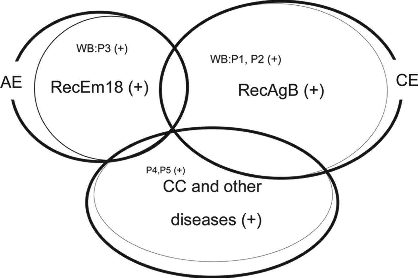
Recently, we were presented with an early stage AE case, with a hepatic lesion measuring approximately 1 cm in diameter. The patient was sero-negative by both confirmative WB using crude antigen carried out at the Hokkaido Institute of Public Health and RecEm18-WB carried out at Asahikawa Medical University (AMU). However, when tested using crude antigen-WB at AMU, I found one very strong band with a high molecular weight. It completely differs from any diagnostic components of a commercially available WB kit (Liance et al. Reference Liance, Janin, Bresson-Hadni, Vuitton, Houin and Piarroux2000). Therefore, crude antigen-WB might also be useful for the serological detection of early stage of AE (Hasegawa et al. unpublished). Thus further studies are necessary. The use of highly purified antigen can detect most, but not 100% of true cases, with very few false positives. However, when we use crude antigens, we may detect 100% of cases with substantial numbers of false positives (Fig. 1). Approximately one decade ago, serology applied in Hokkaido, Japan using crude antigen-ELISA, showed that approximately 99% of cases, positive by crude antigen-ELISA, were in fact false positives. Unfortunately, this serological test sometimes also failed in the detection of true AE cases which were easily confirmed by RecEm18-WB (Ito et al. Reference Ito, Nakao and Wandra2003b, Reference Ito, Ishikawa, Kitada, Nakaya and Sasajimac). Using crude antigens, we estimate that we obtain a very large number of false positives and, possibly more importantly, false negatives. It is not clear whether false negatives are rare or not (Ito et al. Reference Ito, Sako, Ishikawa, Nakao, Nakaya, Yamasaki, Craig and Pawlowski2002a, Reference Ito, Ishikawa, Kitada, Nakaya and Sasajima2003c; Aoki et al. Reference Aoki, Kino, Yamazaki, Obara, Kasai, Yamasaki and Ito2006). In contrast, Em18-serology has proven to be much better for the detection of AE cases, with almost no false positive cases in Japan (Ito et al. Reference Ito, Nakao, Kutsumi, Lightowlers, Itoh and Sato1993a, Reference Ito, Ishikawa, Kitada, Nakaya and Sasajima2003c, Aoki et al. Reference Aoki, Kino, Yamazaki, Obara, Kasai, Yamasaki and Ito2006), China (Ito et al. Reference Ito, Wang and Liu1993b), USA (Ito et al. Reference Ito, Schantz and Wilson1995), Poland (Ito et al. Reference Ito, Ma, Paul, Stefaniak and Pawlowski1998a), France (Bart et al. Reference Bart, Abdukader, Zhang, Lin, Wang, Nakao, Ito, Craig, Piarroux, Vuitton and Wen2006) and Germany (Tappe et al. Reference Tappe, Grüner, Kern and Frosch2008, Reference Tappe, Sako, Itoh, Frosch, Grüner, Reuter, Nakao, Ito and Kern2009, Reference Tappe, Sako, Itoh, Frosch, Grüner, Kern and Ito2010). There are a few cases of early stage AE that have shown no antibody response to Em18. However, I expect these cases will become positive over time. There is no way to determine the number of false negative AE cases other than to utilize different antigenic components or tools on stocked serum samples. Therefore, we must decide on what is our true objective: Detection of 96–97% of AE cases with no false positives or detection of 98% of AE cases with high numbers of false positives. An alternative idea is to utilize fine needle aspiration for histopathological and molecular confirmation of all stages of AE, since the risk of anaphylaxis is believed to be low for AE cases (Kern et al. Reference Kern, Frosch, Helbig, Wechsler, Usadel, Beckh, Kunz, Lucius and Frosch1995; Kawakami et al. Reference Kawakami, Kuwatani and Sakamoto2013).
Schematic figure of AE, CE and non-echinococcal cases including cysticercosis (CC) detectable by Western blots (WB) using RecEm18, RecAgB (Ito et al. Reference Ito, Wang and Liu1993b, Reference Ito, Schantz and Wilson1995, Reference Ito, Wen, Craig, Ma, Nakao, Horii, Pang, Okamoto, Itoh, Osawa, Wang and Liu1997, Reference Ito, Ma, Paul, Stefaniak and Pawlowski1998a) and commercially available crude antigen-WB P1-P5 (Liance et al. Reference Liance, Janin, Bresson-Hadni, Vuitton, Houin and Piarroux2000). The majority of AE and CE cases except some early or abortive stages of AE and CE are easily detected by RecEm18 and RecAgB, respectively (Li et al. Reference Li, Chen, Zhen, Qiu, Qiu, Xiao, Ito, Wang, Giraudoux, Sako, Nakao and Craig2010a, Reference Li, Ito, Chen, Sako, Qiu, Xiao, Qiu, Nakao, Yanagida and Craigb) and by WB:P3 (WB:P3 (+) in Fig. 1) for detection of mainly Em18 (Ito et al. Reference Ito, Wang and Liu1993b, Reference Ito, Schantz and Wilson1995, Reference Ito, Wen, Craig, Ma, Nakao, Horii, Pang, Okamoto, Itoh, Osawa, Wang and Liu1997, 1998a) and WB:P1, P2 (WB:P1, P2 (+) in Fig. 1) for detection of mainly AgB (8 kDa), respectively (Ito et al. Reference Ito, Wen, Craig, Ma, Nakao, Horii, Pang, Okamoto, Itoh, Osawa, Wang and Liu1997, Reference Ito, Ma, Paul, Stefaniak and Pawlowski1998a; Liance et al. Reference Liance, Janin, Bresson-Hadni, Vuitton, Houin and Piarroux2000). WB:P4, P5 (P4, P5 (+) in Fig. 1) may include echinococcosis, either CE or AE but they are positive under blind test for CC and some other diseases and no use under a blind test or screening (Liance et al. Reference Liance, Janin, Bresson-Hadni, Vuitton, Houin and Piarroux2000). The real confirmative WB for AE and CE are based on detection of AgB, Em16, Em18 at least and P1, P2, P3 but not P4, P5 (Liance et al. Reference Liance, Janin, Bresson-Hadni, Vuitton, Houin and Piarroux2000). Based on the banding patterns using crude antigens, we can identify AE by detection of antibody responses to Em18 or both Em18 and Em16, and CE by those responses to Em16 and AgB (Ito et al. Reference Ito, Nakao, Kutsumi, Lightowlers, Itoh and Sato1993a, Reference Ito, Wang and Liub, Reference Ito, Schantz and Wilson1995, Reference Ito, Wen, Craig, Ma, Nakao, Horii, Pang, Okamoto, Itoh, Osawa, Wang and Liu1997, Reference Ito, Sako, Ishikawa, Nakao, Nakaya, Yamasaki, Craig and Pawlowski2002a, Reference Ito, Xiao, Liance, Sato, Sako, Mamuti, Ishikawa, Nakao, Yamasaki, Nakaya, Bardonnet, Bresson-Hadni and Vuittonb; Furuya et al. Reference Furuya, Kawanaka, Yamano, Sato and Honma2004; Tappe et al. Reference Tappe, Grüner, Kern and Frosch2008). However, we cannot identify AE and CE by crude antigen-ELISA, since crude antigens have many non-specific components shared between the host and parasite. More important is that these serological tools cannot always detect early stages of AE or CE.

There have been a few reports stating that RecEm18 does not detect 100% of active AE cases, but that other commercially available WB kits (Liance et al. Reference Liance, Janin, Bresson-Hadni, Vuitton, Houin and Piarroux2000) could detect 100% of active AE cases (Furuya et al. Reference Furuya, Kawanaka, Yamano, Sato and Honma2004; Yamano et al. Reference Yamano, Yagi, Furuya, Sawada, Honma and Sato2005). These reports did not include other infectious disease samples for evaluation of test specificity (Fig. 1). Furthermore, the authors did not use RecEm18, but instead used crude antigens for identification of Em18-WB. It is necessary to run the SDS-PAGE for a sufficient amount of time to have adequate separation of the various components (Ito et al. Reference Ito, Nakao, Kutsumi, Lightowlers, Itoh and Sato1993a, Reference Ito, Wang and Liub, Reference Ito, Schantz and Wilson1995, Reference Ito, Wen, Craig, Ma, Nakao, Horii, Pang, Okamoto, Itoh, Osawa, Wang and Liu1997, Reference Ito, Sako, Ishikawa, Nakao, Nakaya, Yamasaki, Craig and Pawlowski2002a, Reference Ito, Xiao, Liance, Sato, Sako, Mamuti, Ishikawa, Nakao, Yamasaki, Nakaya, Bardonnet, Bresson-Hadni and Vuittonb; Liance et al. Reference Liance, Janin, Bresson-Hadni, Vuitton, Houin and Piarroux2000; Tappe et al. Reference Tappe, Grüner, Kern and Frosch2008) and use some specific markers, such as monoclonal or polyclonal antibodies to Em16 or Em18, or any other diagnostic components (Ito et al. Reference Ito, Nakao, Kutsumi, Lightowlers, Itoh and Sato1993a, Reference Ito, Wang and Liub, Reference Ito, Schantz and Wilson1995, Reference Ito, Ma, Paul, Stefaniak and Pawlowski1998a; Jiang et al. Reference Jiang, Wen and Ito2001).
In South America, three Echinococcus spp., E. granulosus sensu stricto, E. canadensis and E. vogeli are distributed. As the genes of Em18 and AgB are shared among Echinococcus spp. (Nirmalan and Craig, Reference Nirmalan and Craig1997; Nakao et al. Reference Nakao, Xiao, Okamoto, Yanagida, Sako and Ito2009) and the expression of these genes is expected to be variable among the different pathological feature of echinococcoses (Wen and Craig, Reference Wen and Craig1994), we may expect that antibody responses in E. vogeli infections (polycystic echinococcosis, PE) may be somewhere between that of AE and CE (Knapp et al. Reference Knapp, Chirica, Simonnet, Grenouillet, Bart, Sako, Itoh, Nakao, Ito and Millon2009; Ito et al. Reference Ito, Yanagida, Sako, Nakao, Nakaya, Knapp, Ishikawa and Liu2011a).
How rapidly do antibody responses to RecEm18 decline after curative surgery?
Most recent serological follow-up studies of cured post-surgical hepatic AE cases in Japan showed unexpected antibody responses (Fig. 2). For example, we were asked to follow-up one AE case for 6 months after surgery. After 6 months, antibody responses to RecEm18 were already negative (data not shown). RecEm18 has been known to be a good marker to follow-up the progression of AE especially in resected AE cases (Xiao et al. Reference Xiao, Mamuti, Yamasaki, Sako, Nakao, Nakaya, Gottstein, Schantz, Lightowlers, Craig and Ito2003; Bart et al. Reference Bart, Piarroux, Sako, Grenouillet, Bresson-Hadni, Piarroux and Ito2007; Ishikawa et al. Reference Ishikawa, Sako, Itoh, Ohtake, Kohgo, Matsuno, Ohsaki, Miyokawa, Nakao, Nakaya and Ito2009; Tappe et al. Reference Tappe, Sako, Itoh, Frosch, Grüner, Reuter, Nakao, Ito and Kern2009, Reference Tappe, Sako, Itoh, Frosch, Grüner, Kern and Ito2010; Bresson-Hadni et al. Reference Bresson-Hadni, Blagosklonov, Knapp, Grenouillet, Sako, Delabrousse, Brientini, Richou, Minello, Antonino, Gillet, Ito, Mantion and Vuitton2011), but recently it was shown that antibody responses start to decline within only a few days post-surgery (Fig. 1a and c) (Akabane et al. Reference Akabane, Nakano, Inagaki, Yanagida, Shoumura, Kudo, Shonaka, Orimo, Oikawa, Aiyama, Shibaki, Sako, Itoh and Ito2012). We have had several AE cases show similar drastic declines in antibody responses within one week of curative surgery, with all becoming negative within 6 months (Fig. 1b and d) (Akabane et al. unpublished). This rapid decline in antibody response may be true not only in AE cases, but also for other helminthic diseases or other conditions requiring hepatic surgery. If this is confirmed, there might be some unknown mechanism for inactivation of antibody responses after hepatic surgery. Otherwise, we will need to re-evaluate the immune memory itself using purified antigen(s). At present, AE is the only parasitic disease where surgical resection of the entire lesion is recommended as the first therapeutic choice. Very active homeostatic responses in patients after surgery, anergy or some unknown mechanism to induce a rapid drop in antibody titres after surgery may exist and need to be evaluated further.
The rapid decline in antibody responses in two resected hepatic AE cases (PNM stage I) (a and c: modified from Akabane et al. Reference Akabane, Nakano, Inagaki, Yanagida, Shoumura, Kudo, Shonaka, Orimo, Oikawa, Aiyama, Shibaki, Sako, Itoh and Ito2012; b and d: modified from Akabane et al. unpublished). ELISA (a and b) and WB (c and d) using RecEm18 (Sako et al. Reference Sako, Nakao, Nakaya, Yamasaki, Gottstein, Lightowlers, Schantz and Ito2002) before surgery until 60 months (a and c) and until 17 months after surgery (b and d).
Comparative studies using different tools including commercially available kits
There are many reports that compare the specificity and sensitivity of diagnostic tools. Studies where the test developers apply the various diagnostic tools are optimal in order to reduce bias (Ito et al. Reference Ito, Sako, Ishikawa, Nakao, Nakaya, Yamasaki, Craig and Pawlowski2002a), unless the test(s) being evaluated are commercially available (Ito et al. Reference Ito, Plancarte, Ma, Kong, Flisser, Cho, Liu, Kamhawi, Lightowlers and Schantz1998b; Tappe et al. Reference Tappe, Grüner, Kern and Frosch2008; Carod et al. Reference Carod, Randrianarison, Razafinahefa, Ramahefarisoa, Rakotondrazaka, Debruyne, Dautigny, Cazal, Andriantseheno and Charles2012). Comparative studies for serological tools that are not commercialized should be carried out by the two or more independent groups that have been involved in the establishment of the tools (Dorny et al. Reference Dorny, Phiri, Vercruysse, Gabriel, Willingham, Brandt, Victor, Speybroeck and Berkvens2004). Usually, such joint work should be carried out under blind or double blind conditions (Ito et al. Reference Ito, Nakao, Kutsumi, Lightowlers, Itoh and Sato1993a, Reference Ito, Wang and Liub, Reference Ito, Schantz and Wilson1995, Reference Ito, Xiao, Liance, Sato, Sako, Mamuti, Ishikawa, Nakao, Yamasaki, Nakaya, Bardonnet, Bresson-Hadni and Vuitton2002b). Otherwise, researchers who are interested in using or evaluating another group's diagnostic tool should attempt to collaborate with the test's developer (Li et al. Reference Li, Zhang, Wilson, Ito and McManus2003; Bart et al. Reference Bart, Piarroux, Sako, Grenouillet, Bresson-Hadni, Piarroux and Ito2007). In all cases when serology is used, it is important to consider ancillary findings (for example, diagnostic imaging and clinical manifestations) to support the diagnosis since no test is 100% reliable (Tappe et al. Reference Li, Zhang, Wilson, Ito and McManus2008).
Detection of specific antibodies vs detection of circulating antigens
Most serological approaches are based on detection of specific antibody responses but an alternative is to detect circulating antigens. The former cannot differentiate current and previous infections due to immunological memory after cure. However, there are no detailed follow-up studies on how long antibody responses remain after surgical resection in cysticercosis or echinococcosis cases when using purified antigens. Usually, clinicians will try to follow disease progression via imaging and antibody responses 6 months to one year after treatment. As an example, we were asked to check antibody responses just prior to and one year after the surgical treatment of an NCC case with a solitary lesion. The patient was seropositive before surgery, but completely seronegative one year later (Ito et al. Reference Ito, Nakao, Ito, Yuzawa, Morishima, Kawano and Fujii1999b). The original serology applied at the hospital was not sensitive and, therefore, the patient was believed to have a malignant brain tumor. The lesion was surgically resected and later confirmed to be NCC. A seronegative result one year post-surgery was not able to provide information on how long the antibody response remained (Deckers and Dorny, Reference Deckers and Dorny2010). In order to determine length of antibody response, follow-up studies are required for both surgically treated and chemotherapeutically managed cases (Kobayashi et al. Reference Kobayashi, Nakamura-Uchiyama, Nishiguchi, Isoda, Kokubo, Ando, Katurahara, Sako, Yanagida, Ito, Iwabuchi and Ohnishi2013).
In the past, clinicians in Asia used to perform surgery for lung paragonimiasis which, in Japan, was often misdiagnosed as pulmonary tuberculosis (TB). However, after serology for paragonimiasis was established and the drug, Bithionol, was commercially produced, clinicians preferred chemotherapy to surgery. Currently, we have no serum samples from surgically treated paragonimiasis cases. If it is possible to acquire samples from such cases and perform weekly or monthly post-surgical follow-up, we should be able to determine if there is also a rapid decline in antibody titres with this parasitic disease. Another interesting research topic could be to determine how rapidly antibody responses in NCC cases with surgery become negative post-surgery (Deckers and Dorny, Reference Deckers and Dorny2010).
Serology to detect circulating antigens is expected to show evidence of an ongoing infection but the specificity of these tests is still not entirely known. Almost all antigen tests have been designed to detect certain components of metacestodes of T. saginata, but not of T. solium. This means that the current antigen-based tools for T. solium cysticercosis are based on cross reactions. The question is, ‘Why has no one tried to produce specific antibodies to specific components of the metacestodes of T. solium?’. T. solium-based tests would be useful for detection of human cysticercosis, since T. solium is essentially the only species which can infect humans other than very rare cysticercosis cases caused by non-human Taenia spp. common in wild animals (Euzeby, Reference Euzeby and Soulsby1974). If this tool is used for the detection of pigs infected with T. solium, there is no doubt that positive results would occur not only for pigs infected with T. solium, but also for pigs infected with other taeniid cestodes, including T. hydatigena. A joint project to evaluate antibody-ELISA and antigen-ELISA under blind testing resulted in reasonably reliable results for the antibody-ELISA, with all pigs harbouring 16 or more cysticerci at necropsy 30 days after egg inoculation confirmed antibody positive (Sato et al. Reference Sato, Yamasaki, Sako, Nakao, Nakaya, Plancarte, Kassuku, Dorny, Geerts, Benitz-Orgiz, Hashiguchi and Ito2003). In contrast, the sera from one uninfected pig became antigen-ELISA-positive based on the utilized cut-off.
Which is better to use – experimentally infected or naturally infected animal sera?
Experimental infections to establish specific serodiagnosis are widely used. The candidate diagnostic antigens are then applied for the detection of specific antibody responses in patients or animals infected with the parasite species of interest. As endemic areas for T. solium tend to be located in poor regions of developing countries, people and animals are often infected with multiple pathogens, including several other helminths. Therefore, serum samples from endemic areas are very useful for the establishment of better diagnostic tests with higher specificity (Ito et al. Reference Ito, Plancarte, Nakao, Nakaya, Ikejima, Piao, Kanazawa and Margono1999a). It is for this reason that we often discuss how to establish negative controls. Ideally, negative controls from endemic areas are better than negative controls from non-endemic countries. If the antigens applied are specific enough, there may be no difference between negative controls from endemic and non-endemic areas (Nkouawa et al. Reference Nkouawa, Sako, Itoh, Kouojip-Mabou, Nganou, Saijo, Knapp, Yamasaki, Nakao, Nakaya, Moyou-Somo and Ito2011; Mohammadzadeh et al. Reference Mohammadzadeh, Sako, Sadjjadi, Sarkari and Ito2012).
Pigs in developing countries are commonly infected with T. hydatigena and infection with this parasite appears to be more common than infection with T. solium in Asia (China, Thailand and Indonesia). There is a report that T. hydatigena is not common in Africa (Dorny et al. Reference Dorny, Phiri, Vercruysse, Gabriel, Willingham, Brandt, Victor, Speybroeck and Berkvens2004) but no one knows if it is always and everywhere true. Although we had no data on T. hydatigena from pigs that were confirmed to be co-infected with T. solium, we applied our serological test, developed to detect human cysticercosis, and found pigs naturally infected and confirmed positive for T. solium infection in Indonesia (Papua), China and Mexico (Ito et al. Reference Ito, Plancarte, Nakao, Nakaya, Ikejima, Piao, Kanazawa and Margono1999a). Such results from pigs confirmed to have been naturally infected with T. solium were ideal. Therefore, we should use serum samples from pigs grown in endemic areas for evaluation of serology.
There has been a push to use a WB kit using glycoproteins (GP-WB) established for detection of human cysticercosis (Tsang et al. 1989) for swine cysticercosis (Garcia et al. Reference Garcia, Gonzalez, Gavidia, Falcon, Bernal, Verastegui, Rodriguez, Tsang and Gilman2003; DeGiorgio et al. 2005; Mwape et al. Reference Mwape, Phiri, Praet, Speyboreck, Muma, Dorny and Gabriël2013). The GP-WB may only be specific in humans who are infected with T. solium exclusively. This means that there is no real control for evaluation of these GPs in animals which can be infected with other taeniid cestodes. There are several reports discussing ‘transient antibodies’ detected by the GP-WB in swine cysticercosis (Garcia et al. 2001, Reference Garcia, Gonzalez, Gavidia, Falcon, Bernal, Verastegui, Rodriguez, Tsang and Gilman2003; DeGiorgio et al. 2005; Mwape et al. Reference Mwape, Phiri, Praet, Speyboreck, Muma, Dorny and Gabriël2013). Therefore, it is suspected that some of the GPs are shared with other non-human Taenia species (Lightowlers, Reference Lightowlers2013).
Evaluation of serological results
Use of advanced active cases of echinococcosis or cysticercosis to evaluate serodiagnostic tools should increase test sensitivity. Therefore, serum samples with good clinical background information are essential in the evaluation of serological studies. In general, advanced cases are much easier to detect by any tool. If we use purified antigens, the specificity becomes very high, but simultaneously the sensitivity tends to be lower. In contrast, if we want a test that is 100% sensitive, we usually sacrifice specificity. For a disease such as AE, it is essential to use a panel of serum samples from other diseases such as hepatic cancers, CE, cysticercosis, fascioliasis, toxocariasis and amoebiasis. The most important prerequisite for such panels is that the panel sera have been confirmed antibody positive to the homologous parasite or pathogen's antigen. However, even with such careful analysis, we may find shared epitopes among cestode or platyhelminthes, especially in terms of hydrophobic ligand binding proteins including Antigen B family (Iopposo et al. Reference Iopposo, Notargiacomo, Profumo, Franchi, Ortona, Rigano and Siracusano1996; Ito, Reference Ito2002; Mamuti et al. Reference Mamunti, Sako, Bart, Nakao, Ma, Wen and Ito2007; Jiang et al. Reference Jiang, Zhang, Liu and Feng2012; Mohammadzadeh et al. Reference Mohammadzadeh, Sako, Sadjjadi, Sarkari and Ito2012; Obal et al. Reference Obal, Ramos, Silva, Lima, Battyany, Bessio, Ferreira, Salinas and Ferreira2012; Santivañez et al. Reference Santivañez, Arias, Portocarrero, Rodriguez, Gonzalez, Gilman, Gavidia and Garcia2012).
Serology for detection of taeniasis
There are serological tools available for the detection of taeniasis carriers due to T. solium but, to my knowledge, there is no scientifically sound work on how species specific these tests are. Some groups used no or few samples from T. saginata or T. asiatica, but stressed that their newly developed serological test was 100% specific for T. solium. If sera from people infected with other Taenia species were seronegative to the homologous antigens, it resulted in a negative test. It is, therefore, difficult to evaluate such work without blind tests with greater numbers of samples. Nonetheless, even if the test cannot differentiate species, it may still be useful for the detection of taeniasis carriers. It should also be noted that such a serological tool for the detection of taeniasis carriers cannot differentiate ongoing infection from past infection due to immunological memory. Therefore, confirmation is needed on how long the antibody responses remain after treatment. This problem pushes us to develop better tools for molecular identification. Better parasite identification is also needed to assess anthelminthic drug therapy. Salim et al. (Reference Salim, Ang, Handali and Tsang2009) used commercially available serological tools in Papua, Indonesia to perform serological screening of the local population. The study reported the proportion of people who were taeniasis carriers or cysticercosis patients but there was no direct evidence to confirm which Taenia species were causing infection. We require better direct evidence of these parasites in people (Wandra et al. Reference Wandra, Subahar, Simanjuntak, Margono, Suroso, Okamoto, Nakao, Sako, Nakaya, Schantz and Ito2000), pigs (Subahar et al. Reference Subahar, Hamid, Purba, Wandra, Karma, Sako, Margono, Craig and Ito2001; Margono et al. Reference Margono, Ito, Sato, Okamoto, Subahar, Yamasaki, Hamid, Wandra, Purba, Nakaya, Ito, Craig and Suroso2003) and even dogs (Ito et al. 2002c) in Papua (Wandra et al. Reference Wandra, Ito, Swastika, Dharmawan, Sako and Okamoto2013). Nonetheless, there are still better data on human cases of echinococcoses, cysticercosis and taeniases compared with data on infections in animals.
Problems with animal surveys
Detection of animals parenterally infected with metacestodes of Echinococcus spp. or Taenia spp. is difficult due to the cost of performing diagnostic tests. There are no practical tools for the detection of animals infected with E. granulosus sensu lato. The same is true for other Echinococcus spp. infections in South America (Knapp et al. Reference Knapp, Chirica, Simonnet, Grenouillet, Bart, Sako, Itoh, Nakao, Ito and Millon2009; Santos et al. Reference Santos, Soares, Brito, Rodrigues, Siqueira, Gomes-Gouvêa, Alves, Carneiro, Malheiros, Póvoa, Zaha and Haag2012). If echinococcoses were more lethal in livestock and other high value animals, the animal sector would be more likely to set up sustainable screening for infections in animals.
In contrast, cattle infected with metacestodes of T. saginata are very rare in developed countries; with most cases the result of infected employees from developing countries contaminating farming areas by defaecating in fields containing livestock (Dorny and Praet, Reference Dorny and Praet2007; McFadden et al. Reference McFadden, Heath, Morley and Dorny2011; Yanagida et al. Reference Yanagida, Sako, Nakao, Nakaya and Ito2012; Yamasaki, Reference Yamasaki2013). If we introduce stool examination for all employees working in farming areas, it might be cheaper than introducing a new serological tool to detect infected cattle.
Copro-ELISA for detection of adult tapeworm carriers
Copro-ELISA has been useful for detection of adult worms in definitive hosts, including humans with taeniases or dogs and/or foxes infected with Echinococcus spp. (Allan et al. Reference Allan, Avila, Garcia Noval, Flisser and Craig1990, Reference Allan, Velasquez-Tohom, Torres-Alvarez, Yurrita and Garcia-Noval1996; Deplazes et al. Reference Deplazes, Jimenez-Palacios, Gottstein, Skaggs and Eckert1994, Reference Deplazes, Alther, Tanner, Thompson and Eckert1999; Allan and Craig, Reference Allan and Craig2006). There are several groups which have been working on copro-ELISA tests. Most of them use polyclonal antibodies to capture antigens in faecal samples. These tests performed well in laboratory-based studies, especially when known positives were compared with uninfected negative controls. In field studies in endemic areas where other parasitic infections were common, it became much more difficult to interpret test results (Raoul et al. Reference Raoul, Deplazes, Nonaka, Piarroux, Vuitton and Giraudoux2001). Copro-ELISA appeared to work better in earlier projects, indicating that expired capturing antibodies prepared approximately 2 decades ago may be affecting the test's outcome in more recent studies (Hartnack et al. Reference Hartnack, Budke, Craig, Jiamin, Boufana, Campos-Ponce and Torgerson2013). In order to address the problem with specificity, some groups have applied monoclonal antibodies to capture the antigens (Nonaka et al. Reference Nonaka, Iida, Yagi, Ito, Ooi, Oku and Kamiya1996; Morel et al. Reference Morel, Lasssabe, Elola, Bondard, Herrera, Mari, Last, Jensen and Gonzalez-Sapienza2013). Most of the antigenic components in faeces were not proteins, but rather glycoproteins or lipoproteins, which may result in non-specific responses. Even if such antibodies for capturing proteins, sugars or lipids were specific, there might be blockers or inhibitors in the faecal samples. Therefore, it is not possible to know if these copro-ELISA tests are useful without direct evidence of infection (for example, eggs or tapeworms) from copro-ELISA-positive humans or animals.
Molecular identification of cestode species
Molecular identification is almost 100% reliable when applied to parasites collected from human patients or infected animals but contamination of tools with other DNA may cause erroneous results. Therefore, repeated analyses of the same samples at different laboratories may be important for evaluation of the results (Hüttner et al. Reference Hüttner, Nakao, Wassermann, Siefert, Boomker, Dinkel, Sako, Mackenstedt, Romig and Ito2008; Knapp et al. Reference Knapp, Chirica, Simonnet, Grenouillet, Bart, Sako, Itoh, Nakao, Ito and Millon2009; Snabel et al. Reference Snabel, Altintas, D'Amelio, Nakao, Romig, Yolasigmaz, Gunes, Turk, Busi, Huttner, Sevcova, Ito, Altintas and Dubinsky2009). There is also the risk of contamination, especially by beginners, which may even result in unreliable sequences being inputted into GenBank.
Eggs, metacestodes and adult tapeworms are all targets for molecular identification. Even though eggs of taeniid species cannot be differentiated morphologically, they can be differentiated using molecular tools. Based on the molecular data, we should treat tapeworm carriers and collect adult worms. Morphology and molecular information from adult worms should also be compared with molecular data obtained from parasite eggs.
The merit of using coproDNA detection is that DNA of immature worms can be amplified. Therefore, coproDNA detection may be more sensitive and reliable for all stages of the worm's development in the host intestine. Polymerase chain reaction (PCR) has widely been applied for coproDNA detection (Yamasaki et al. Reference Yamasaki, Allan, Sato, Nakao, Sako, Nakaya, Qiu, Mamuti, Craig and Ito2004) but inhibitors in the faeces may interfere with the test. Recent approaches using loop-mediated isothermal amplification (LAMP) for copro-tests, coproLAMP, are more reliable, with almost no influence from such inhibitors (Nkouawa et al. Reference Nkouawa, Sako, Nakao, Nakaya and Ito2009, Reference Nkouawa, Sako, Li, Chen, Wandra, Swastika, Nakao, Yanagida, Nakaya, Qiu and Ito2010, Reference Nkouawa, Sako, Li, Chen, Nakao, Yanagida, Okamoto, Giraudoux, Raoul, Nakaya, Xiao, Qiu, Qiu, Craig and Ito2012).
Recent molecular studies of Echinococcus spp. have revealed that E. granulosus sensu lato consists of 5 independent species: E. granulosus, E. equinus, E. ortleppi, E. canadensis and E. felidis (Nakao et al. Reference Nakao, McManus, Schantz, Craig and Ito2007, Reference Nakao, Yanagida, Okamoto, Knapp, Nkouawa, Sako and Ito2010, Reference Nakao, Yanagida, Konyaev, Lavikainen, Ondokurtsev, Zaikov and Ito2013a; Hüttner et al. Reference Hüttner, Nakao, Wassermann, Siefert, Boomker, Dinkel, Sako, Mackenstedt, Romig and Ito2008; Knapp et al. Reference Knapp, Chirica, Simonnet, Grenouillet, Bart, Sako, Itoh, Nakao, Ito and Millon2009). Furthermore, the hermaphroditic nature of cestodes allows them to reproduce without sexual reproduction. Infection with multiple worms may result in outcrossing. Such data may be obtained for Echinococcus spp. when we compare mitochondrial (haploid) and nuclear (diploid) DNA from Echinococcus worms in a single definitive host co-infected with different species of Echinococcus or different genotypes of worms. There are also reports stressing that E. granulosus and E. multilocularis parasitize different parts of the dog intestine, while other reports state that the two species parasitize the same regions of the intestine (Thompson and Eckert, Reference Thompson and Eckert1983; Kumaratilake et al. Reference Kumaratilake, Thompson, Eckert and D'Alessandro1986; Lymbery et al. Reference Lymbery, Hobbs and Thompson1989) but it is not known if these reports can be generalized to all dogs and other canids. There might be hybrids of, or at least introgression between, E. granulosus s.s. and E. canadensis (Bart et al. Reference Bart, Abdukader, Zhang, Lin, Wang, Nakao, Ito, Craig, Piarroux, Vuitton and Wen2006) or other species of E. granulosus s.l. where these species are co-endemic. Therefore, it is possible that morphologically indistinguishable hybrid species may be found in the small intestine, but additional data are needed.
Evidence of outcrossing has been suggested from E. multilocularis, in Hokkaido, Japan, using microsatellite DNA (Nakao et al. Reference Nakao, Sako and Ito2003) as well as from T. saginata and T. asiatica. In these cases, although the mtDNA indicated T. asiatica, nuclear DNA indicated T. saginata, or highly variable heterozygotes of both species, and vice versa (Okamoto et al. Reference Okamoto, Nakao, Blair, Anantaphruti, Waikagul and Ito2010; Yamane et al. Reference Yamane, Suzuki, Tachi, Li, Chen, Nakao, Nkouawa, Yanagida, Sako, Ito, Sato and Okamoto2012, Reference Yamane, Yanagida, Li, Chen, Dekumyoy, Waikagul, Nkouawa, Nakao, Sako, Ito, Sato and Okamoto2013). Experimental infection with eggs of T. asiatica by Fan et al. (Reference Fan, Soh and Kosin1990) resulted in T. asiatica metacestodes developing in the viscera of pigs, but also produced metacestodes in cattle and other domestic animals. This led to additional studies on this new Asian Taenia species (Simanjuntak et al. Reference Simanjuntak, Margono, Okamoto and Ito1997; Wandra et al. Reference Wandra, Ito, Swastika, Dharmawan, Sako and Okamoto2013; Yamane et al. Reference Yamane, Yanagida, Li, Chen, Dekumyoy, Waikagul, Nkouawa, Nakao, Sako, Ito, Sato and Okamoto2013), which leads to the question of ‘Are there any other human Taenia species’? In Ethiopia, cysticerci from a new Taenia species have recently been confirmed from cattle (Hailemariam et al. Reference Hailemariam, Nakao, Menkir, Lavikainen, Iwaki, Yanagida, Okamoto and Ito2013). We, therefore, are facing a more complicated world in the area of taeniid taxonomy (Nakao et al. Reference Nakao, Lavikainen, Iwaki, Haukisalmi, Konyaev, Oku, Okamoto and Ito2013b). Coevolution of cestodes and host animals are other emerging topics.
Pathogenicity of Echinococcus spp. to humans
Recent molecular studies have revealed that E. granulosus s.s. and E. felidis (Hüttner et al. Reference Hüttner, Nakao, Wassermann, Siefert, Boomker, Dinkel, Sako, Mackenstedt, Romig and Ito2008), and E. multilocularis and E. shiquicus (Xiao et al. Reference Xiao, Qiu, Nakao, Li, Yang, Chen, Schantz, Craig and Ito2005) are sister species, respectively, with the former species, of both pairs, known to be highly pathogenic to humans (Nakao et al. Reference Nakao, Yanagida, Okamoto, Knapp, Nkouawa, Sako and Ito2010; Knapp et al. Reference Knapp, Nakao, Yanagida, Okamoto, Saarma, Lavikainen and Ito2011). Therefore, it is hypothesized that these two newer species, E. felidis in Africa and E. shiquicus in Tibet, China can also infect humans but we should not jump to conclusions until we have concrete evidence of human infections. All CE samples due to E. granulosus s.s. in Africa, especially where E. felidis has been confirmed, should be re-evaluated for the possibility of E. felidis.
The importance of the real-time detection of taeniasis carriers and cysticercotic pigs
We have been working on taeniasis and cysticercosis in several Asian countries. Since 2004, the Ministry of Education, Japan, has sponsored numerous seminars to help transfer technology to scientists in Asia and Africa. Parasite materials and human samples from endemic areas have been analysed at AMU (Japan) after obtaining ethical approval. Molecular identification of Taenia species using eggs, metacestodes, adult worms, faecal samples and serology have been carried out by junior colleagues from endemic countries. Unfortunately, it has been very difficult to locate identified taeniasis carriers for treatment because many had moved in the time between sampling and diagnosis. Therefore, we have decided to establish a real-time detection system in order to identify and treat carriers during a single visit as well as immediately identify pigs that show evidence of being infected (Ito et al. Reference Ito, Okamoto, Li, Wandra, Dharmawan, Swastika, Dekumyoy, Kusolsuk, Davaajav, Davaasuren, Dorjsuren, Meconnen, Negasi, Yanagida, Sako, Nakao, Nakaya, Lavikainen, Nkouawa and Mohammadzadeh2011b). This method was first employed in Bali in 2011 (Swastika et al. Reference Swastika, Dewiyani, Yanagida, Sako, Sudamaja, Sutisna, Wandra, Dharmawan, Nakaya, Okamoto and Ito2012). To use the ELISA for pigs, in the field, an ELISA reader is not required since a colour change indicates a positive result. Thus far, all ELISA-positive pigs examined in Bali, Indonesia, were confirmed to have T. solium cysticerci, with or without T. hydatigena. When we used an ELISA-reader to check the cut-off border line samples in the laboratory, some of these samples were weak positives and some were confirmed to harbour T. hydatigena on necropsy (Dharmawan et al. unpublished). Therefore, we believe that the use of a colour change ELISA may be sufficient or better for screening and identifying pigs infected with T. solium under field conditions (Fig. 3). However, more work is necessary. A LAMP test that can be used in the field and does not require electricity has also been developed (Nkouawa et al. Reference Nkouawa, Sako, Li, Chen, Nakao, Yanagida, Okamoto, Giraudoux, Raoul, Nakaya, Xiao, Qiu, Qiu, Craig and Ito2012). Real-time identification of taeniasis carriers and pigs infected with T. solium, in endemic areas, is essential to demonstrate risk factors for human cysticercosis.
A one year old pig full of cysticerci of T. solium in Bali, Indonesia, suspected to be infected based on ELISA in the field and based on our naked eyes ELISA in the field in Jan 2013. Such a pig is sufficient for local personnel to prepare huge amount of diagnostic antigens.
Dilemma for intervention of cysticercosis or echinococcoses in developing countries: What is the contribution of commercially available kits?
A serious problem with the application of modern diagnostic tools is that we often use such tools but do not work to obtain direct evidence of the infection itself. This oversight should be avoided. As mentioned above, serology for the detection of cysticercosis in endemic areas of Asia is not difficult even when applying simple purification tools (Sako et al. Reference Kobayashi, Nakamura-Uchiyama, Nishiguchi, Isoda, Kokubo, Ando, Katurahara, Sako, Yanagida, Ito, Iwabuchi and Ohnishi2013). Therefore, we are strongly recommending keeping metacestodes from pigs (Fig. 3) and trying to purify the diagnostic antigens using a simple and inexpensive method (Sako et al. Reference Kobayashi, Nakamura-Uchiyama, Nishiguchi, Isoda, Kokubo, Ando, Katurahara, Sako, Yanagida, Ito, Iwabuchi and Ohnishi2013). Commercial kits may be more useful in developed countries, especially in the USA, which has many cysticercosis cases due to refugees or immigrants from endemic areas. In addition, US citizens may bring back the parasite to USA, after visiting countries where T. solium infections are endemic (Yanagida et al. Reference Yanagida, Yuzawa, Joshi, Sako, Nakao, Nakaya, Kawano, Oka, Fujii and Ito2010, Reference Yanagida, Sako, Nakao, Nakaya and Ito2012; Jongwietiwes et al. Reference Jongwietiwes, Yanagida, Ito and Kline2011; Serpa et al. Reference Serpa, Graviss, Kass and White2011; Sorvillo et al. Reference Sorvillo, Wilkins, Shafir and Ebenbard2011).
One serious disadvantage of the use of commercially available kits is the possibility of discouraging researchers in endemic developing countries to establish tools for their own use. Another serious issue is a lack of knowledge on how to evaluate potentially erroneous data from the kits. There are many kits on the market, with many of the tests appearing to lose reliability after being commercialized. Based on this view point, in 2004 I started to encourage personnel in Asia and Africa to understand the mechanism of antigen-antibody responses using their own samples and develop purified antigens to establish their own ELISA or IB serological tests (Ito, Reference Ito2007). At the same time, I was encouraged to produce rapid serological kits for AE, CE and cysticercosis by the Ministry of Education, Japan (ADAMU-AE, -CE and -CC: ICST Co. Ltd., Saitama, Japan). Evaluation of commercially available kits is recommended by the WHO, and numerous other organizations.
No test is 100% reliable, but we should challenge ourselves to obtain better results using confirmed patients’ serum. After this, we can apply the tools on suspected cases or utilize them in epidemiological surveys for confirmation of the infection itself.
CHEMOTHERAPY OF TAENIASIS IN REMOTE AND RURAL AREAS IN ASIA
In Southeast Asian countries, eggs of Taenia spp. may be found through stool examination for soil transmitted helminths (STH) but the number of samples with Taenia eggs will be very small compared with eggs of the major STHs. Therefore, there is usually no further analysis for the identification of Taenia species, since identification of Taenia spp. is time consuming and morphological identification is dependent on the adult worm being expelled. Therefore, we need molecular tools for identification of the species. As a result of the inability in adequately identify Taenia species, these cestodes have been further neglected in relation to the other STHs and/or fish-borne trematodiases (FBTs). If the eggs of Taenia spp. are identified as T. solium, it means that there is a risk of cysticercosis to both the carrier and his or her family members and others in the community (Montresor and Palmer, Reference Montresor and Palmer2006). WHO has recommended mass treatment with PZQ even though there can be safety issues with mass treatment. There are records of individuals dying within days of treatment as part of mass treatment campaigns against schistosomiasis and/or FBTs (Ito et al. Reference Ito, Li, Chen, Long, Yanagida, Nakao, Sako, Okamoto, Wu, Raoul, Giraudoux and Craig2013), but there have been no analyses of the cause of these sudden deaths. If the areas endemic for schistosomiasis and/or other trematodiases are also endemic for T. solium, these sudden deaths could be due to NCC cases who succumb due to a side effect of PZQ treatment since, in these cases, PZQ can result in acute seizures or convulsions when given without a steroid (Pawlowski Reference Pawlowski2006; Wandra et al. Reference Wandra, Sudewi, Swastika, Sutisna, Dharmawan, Yulfi, Darlan, Kapti, Samaan, Sato, Okamoto, Sako and Ito2011). Therefore, we need to reconsider the danger of PZQ for treatment of NCC, especially during mass treatment campaigns where there could be numerous asymptomatic cases. In Asia, we need to establish a better strategy for the detection of taeniasis and cysticercosis using highly reliable immunological and molecular tools.
CONCLUSIONS
Taeniases are neglected due to the small number of cases detected via screening for STHs and the fact that Taenia eggs in human faeces are impossible to identify to the species level. The highly pathogenic T. solium should be differentiated from the two less pathogenic species (T. saginata and T. asiatica) by morphology of the tapeworm's scolex or by molecular tools, including copro tests. Serology and imaging are still necessary for evaluation of human cases of echinococcosis and cysticercosis. A more complicated situation remains in terms of identification of animal infections. There are many acute and chronic infectious diseases. Due to the small population of known patients or carriers of cestode zoonoses, these conditions will continue to be neglected until an outbreak occurs in a developed country. As the risk of infection in people is primarily from individuals living in remote areas of developing countries, we are faced with numerous challenges for controlling these neglected cestode zoonoses. We have to keep in mind that ‘Nothing is perfect without direct evidence of the infection’. In this article, I did not discuss vaccination trials due to a lack of personal experience. There are numerous references that describe this topic in detail (Lightowlers, Reference Lightowlers2006, Reference Lightowlers2010a, 2010b, 2010c, 2013; Bethony et al. Reference Bethony, Cole, Guo, Kamhawi, Lightowlers, Loukas, Petri, Reed, Valenzuela and Hotez2011; Gauci et al. Reference Gauci, Jayashi and Lightowlers2013).
ACKNOWLEDGEMENTS
I sincerely thank Christine Budke for her amendment of this article and valuable comments and suggestions, and Hiromitsu Akabane who has treated AE cases at Asahikawa Kosei Hospital. I greatly appreciate the Ministry of Education, Japan for its sustained support that has allowed me to establish a network with the goal of controlling cestode zoonoses in Asia and in Africa from 1994 onwards. Over the last decade, my research team has invited more than 100 researchers from Asia and the Pacific (Korea, Taiwan, China, Mongolia, Kazakhstan, Philippines, Vietnam, Lao PDR, Thailand, Nepal, India, Indonesia, Papua New Guinea, Australia, New Zealand), Africa (Tanzania, Mozambique, Kenya, Sudan, Egypt, Senegal, Cameroon, Ethiopia), the Middle East (Iran, Jordan), the Americas (USA, Mexico, Brazil, Ecuador, Peru, Argentina) and Europe (UK, France, Germany, Poland, Switzerland, Italy, Slovenia, Finland, Russia) to conduct joint collaborative work for the establishment and evaluation of serological and molecular tools for diagnosis, as well as organize international workshops and symposiums (Ito et al. Reference Ito, Ishikawa, Kitada, Nakaya and Sasajima2003c, Reference Ito, Okamoto, Li, Wandra, Dharmawan, Swastika, Dekumyoy, Kusolsuk, Davaajav, Davaasuren, Dorjsuren, Meconnen, Negasi, Yanagida, Sako, Nakao, Nakaya, Lavikainen, Nkouawa and Mohammadzadeh2011b, Reference Ito, Li, Chen, Long, Yanagida, Nakao, Sako, Okamoto, Wu, Raoul, Giraudoux and Craig2013; Ito, Reference Ito2007). I have enjoyed international projects with more than 250 collaborators from 45 countries. I am grateful to all of those who worked on these topics and encouraged me, especially Schantz, P. M., Craig, P. S., Mitchell, G. F., Rickard, M. D., Lightowlers, M. W., Pawlowski, Z. S., Lord Soulsby, Smyth, J. D., Urbani, C., Fan, P. C., Cross, J., Rausch, L. R. and all my Japanese collaborators.
I dedicate this article to my wife, Hikari Ito who continuously encouraged my international research collaboration for over 30 years but passed away in Kyoto on 30 May 2012.
FINANCIAL SUPPORT
The studies by the author's research team were supported by Grant-in-Aid for scientific research (1994–2015), Asia-Africa Scientific Platform Funds (2006–2008, 2009–2011), Challenging Exploratory Research Fund (2010–2011) from the Japan Society for the Promotion of Science, and Infection Matrix Fund (2007–2008), the Hokkaido Translational Research Fund (2007–2011) and the Special Coordination Fund for Promoting Science and Technology (2003–2005, 2010–2012) from the Ministry of Education, Culture, Sports, Science & Technology in Japan (MEXT), and from the Fogarty International Centre of the National Institutes of Health (Grants RO1 TW001565, RO1 TW001665)(2000–2008)(PI: Craig, P.S.). The content is solely the author's responsibility and does not necessarily represent the official views of the funders.