Implications
To our knowledge, this is the first study to evaluate the effects of erythropoietin (EPO) gene therapy on mammalian semen characteristics. Our results demonstrated that EPO gene therapy did not affect the characteristics of rabbit sperm when compared with the control group. This result suggests that EPO gene therapy did not alter the reproductive capacity of the male rabbits studied, thus advocating that cloned EPO could be used as gene therapy that would not affect male reproduction. Further studies are needed to confirm these results.
Introduction
The successful development of gene therapy has provided the concepts, tools, opportunity and, in some cases, the justification for genetic modification of functions that affect normal human aspects, including athletic performance (Friedmann, Reference Friedmann2010; Friedmann et al., Reference Friedmann, Rabin and Frankel2010). The potential misuse of this type of therapy is regarded as gene doping. Gene doping is based on the introduction and subsequent expression of a target gene into a host. It also involves the modulation of the expression of endogenous genes. In vivo or ex vivo methods can be used for the introduction of the target genes into an athlete (Azzazy et al., Reference Azzazy, Mansour and Christenson2009). The World anti-Doping Agency (WADA) prohibits this as the non-therapeutic use of cells, genes, genetic elements or the modulation of gene expression, having the capacity to enhance athletic performance (Wells, Reference Wells2009; McKanna and Toriello, Reference McKanna and Toriello2010).
Many genes with the potential to enhance athletic performance are available, such as insulin-like growth factor, vascular endothelial growth factor, growth hormone, myostatin and erythropoietin (EPO). These genes not only have the potential to improve human athletic performance, but they can also be applied to animal sports, such as horse racing (Marklund et al., Reference Marklund, Sandberg and Andersson1996; Haisma and de, Reference Haisma and de2006). EPO is an essential growth factor for the red cell production, controlling the survival, proliferation and differentiation of erythroid precursors. Deficient EPO production, as observed in patients with end-stage renal disease, results in anemia (Lacombe and Mayeux, Reference Lacombe and Mayeux1998). Genetic engineering has enabled the production of recombinant human EPO (rHuEpo) to treat this and other diseases. Unfortunately, rHuEpo can be misused by athletes to stimulate erythropoiesis, thereby increasing the blood oxygen capacity and hence endurance of the athlete (Jelkmann, Reference Jelkmann2003; McKanna and Toriello, Reference McKanna and Toriello2010).
There are several risks associated with EPO gene therapy, including cancer, severe autoimmune reaction to the EPO and/or reaction to the virus construct (Sharp, Reference Sharp2010). Overexpression of EPO has a number of potential safety risks, such as an increase in the hematocrit, making the blood more viscous and increasing the load on the heart. Potential consequences include microcirculation blockages, stroke and heart failure (Wells, Reference Wells2008). With EPO doping, it is unlikely that adequate medical supervision is provided, potentially risking the health of the athlete.
With regard to the reproductive system, EPO receptors are expressed by a number of tissues, including female and male genital organs (Yasuda et al., Reference Yasuda, Fujita, Musha, Tanaka, Shiokawa, Nakamatsu, Mori, Matsuo and Nakamura2001; Kobayashi et al., Reference Kobayashi, Yanase, Iwanaga, Sasaki and Nagao2002). Studies with cultured Leydig cells have shown that EPO stimulates steroidogenesis, triggering an increase in testosterone production (Yamazaki et al., Reference Yamazaki, Kanzaki, Kamidono and Fujisawa2004), suggesting that EPO plays a role in the reproductive status of an individual. EPO is present in the seminal plasma and has been suggested to play a role in spermatogenesis in humans, however, no association between EPO concentrations and sperm parameters were found (Temma et al., Reference Temma, Shimoya, Hashimoto, Zhang, Koyama and and Murata2004). Recently, it was demonstrated that media supplementation with EPO could improve sperm motility in humans, however, the mechanisms involved are unknown (Tug et al., Reference Tug, Altunkaynak, Aktas, Kilic, Yilmaz, Cam and Karateke2010). In addition, the intravenous administration of rHuEPO leads to an increase in testosterone levels in the testis, suggesting that systemic EPO can affect testicular function (Foresta et al., Reference Foresta, Mioni, Bordon, Miotto, Montini and Varotto1994) in humans. However, the effect of EPO gene therapy on in vivo reproductive status has not yet been described.
The white New Zealand rabbit (Oryctolagus cuniculus) is frequently used as a model for in vivo studies, including studying the effects of EPO, and for in vivo reproduction studies, such as transgenesis, artificial insemination and embryo development. Therefore, their reproductive physiology is very well understood, including sperm characteristics such as sperm motility, viability, concentration and morphology. For reproductive studies, they are more appropriate than mice, as they are easier to handle and collect sperm samples from, and it is not necessary to euthanize the animal to collect semen.
The aims of this study were to construct an EPO expression vector, transfer it to male rabbits via intramuscular injection and to evaluate the toxicological effects of EPO gene transfer and the intravenous administration rHuEpo on sperm characteristics including motility, viability, morphology and concentration.
Material and methods
Rabbit EPO cDNA amplification
Total RNA from rabbit kidney cells was isolated with TRIzol Reagent (Invitrogen, Carlsbad, CA, USA), according to the recommended protocol. DNase treatment of RNA samples was conducted with a DNA-free Kit (Ambion, Foster City, CA, USA) by following the manufacturer's protocol. First-strand cDNA was performed with 200 ng of RNA using SuperScript III Reverse Transcriptase (Invitrogen) according to the manufacturer's protocol. The primers were designed using Primer Express version 3.0 software (Applied Biosystems, USA) to amplify the rabbit EPO coding region (GenBank accession no. AF290943) as follows: FOR 5′-ATGGGGGCGCGCGGACGC and REV 5′-TCACCTGTCCCCTCTCCTGCAGGC. The PCR parameters were 35 cycles of 94°C for 30 s, 55°C for 30 s and 72°C for 1 min, with an additional initial 5 min denaturation at 95°C and a 5 min final extension at 72°C. PCR amplification produced a 588 bp product. To confirm EPO cDNA amplification, PCR products were sequenced using a MegaBACE 1000 automatic sequencer (Amersham Biosciences, Piscataway, NJ, USA).
EPO mammalian expression vector construction
The PCR products were purified with the illustra GFX PCR DNA and Gel Band Purification Kit (GE Healthcare, Buckinghamshire, UK) and were cloned using the pTarget Mammalian Expression Vector System (Promega, Madison, WI, USA) according to the manufacturer's instructions. After transformation into Escherichia coli TOP10F, recombinant colonies were screened for correct insert direction and length by colony PCR and digestion with EcoRI. The selected clone was sequenced by automatic sequencer as described above. Large-scale purification of the plasmid was carried out using the Perfectprep Plasmid Maxi Kit (Eppendorf, Köln, Germany).
Evaluation of pTarget/EPO expression vector
Cell culture
The HeLa cell line (ATCC CCL-2) was cultured in Dulbecco's modified Eagle's medium (DMEM), supplemented with 10% fetal bovine serum (FBS), purchased from Vitrocell Embriolife (Campinas, Brazil) and Gibco (Grand Island, NY, USA), respectively. Cells were grown at 37°C in an atmosphere of 95% humidified air and 5% CO2. The experiments were conducted with cells in the logarithmic growth phase.
Transfection, RNA extraction and cDNA synthesis
Before transfection, 0.25 × 10⁴ cells/cm2 were seeded in a 6-well plate and incubated until 80% confluence. The pTarget/EPO plasmid construct and a control plasmid without the EPO insert (pTarget) were prepared for transfection using Lipofectamine 2000 Transfection Reagent (Invitrogen) according to the manufacturer's instructions. The cells were washed twice with phosphate buffer saline (PBS; Invitrogen) and incubated for 4 h with the lipoplex mixture. After 4 h, the lipoplexes were removed and the cells were washed twice with PBS. Consequently, the cells were incubated for 48 h in culture medium to confirm EPO expression. Then, the cells were washed twice with PBS, and TRIzol Reagent (Invitrogen) was added for subsequent RNA extraction as described above. DNase treatment and cDNA synthesis were conducted as previously described (item 2.1).
RT-PCR
In order to detect the EPO mRNA in the transfected HeLa cells, the same primer pair used for cloning the EPO cDNA was used in the RT-PCR using the same conditions described above (item 2.1). Amplification of the endogenous control, GAPDH (GenBank accession no. NM_002046), was carried out using primers (5′ GGATTTGGTCGTATTGGG 3′ and 5′ TCGCTCCTGGAAGATGG 3′) that produced a 201 bp fragment. The PCR parameters were 30 cycles of 94°C for 30 s, 60°C for 30 s and 72°C for 1 min, with an additional initial 5 min denaturation step at 95°C and a 5 min final extension at 72°C. The EPO and GAPDH PCR products were analyzed by horizontal gel electrophoresed on a 1% agarose gel and stained using 0.5 μg/ml ethidium bromide.
Animals and experimental design
Adult male New Zealand White rabbits weighing 4.5-5 kg were kept under conventional housing conditions. The animals were maintained in appropriate bedding and provided free access to drinking water and food. Rabbits were kept in standard single cages under controlled temperature and light conditions (12L: 12D). The study protocol was approved (No. 200935) and maintained in accordance with the guidelines of the Ethics Committee in Animal Experimentation, UFPel. A total of 18 rabbits were randomly divided into three different groups: group I received subcutaneously 25 UI/kg of rHuEpo three times a week for 5 weeks; group II received a single dose of the pTarget/EPO construct; and group III (Control) received a single dose of saline solution. Intramuscular DNA injection was administered as previously described, with the following modifications (Maruyama et al., Reference Maruyama, Ataka, Gejyo, Higuchi, Ito, Hirahara, Imazeki, Hirata, Ichikawa, Neichi, Kikuchi, Sugawa and Miyazaki2001): 400 μg of plasmid DNA was injected into the lateral sides of each lower leg (200 μg per site). The rHuEpo used in this study was Eprex (Issy-les-Moulinaux, France).
Blood collection and hematocrit evaluation
In order to evaluate the effect of the EPO treatments on the hematocrit, blood samples were collected on the first day of the experiment (before EPO administration) and once a week over the 5 weeks of the experiment. Blood samples (1.0 ml) were collected from the auricular artery. The hematocrit analyses were performed using the ABX Micros 60 automated method.
Sperm evaluation
Sperm collection
An ejaculate was collected from each animal on the first day of the experiment (before EPO administration) and once a week over the 5 weeks of the experiment using an artificial vagina. The volume of fresh semen was measured in a graduated conical tube.
Sperm motility, vigor and viability
Sperm motility and viability were evaluated as previously described (Campos et al., Reference Campos, de Leon, Komninou, Dellagostin, Deschamps, Seixas and Collares2011a and Reference Campos, Komninou, Urtiaga, de Leon, Seixas, Dellagostin, Deschamps and Collares2011b). Progressive sperm motility (0 to 100%) was visually assessed in four to six fields of view, each containing ∼100 spermatozoa, using a phase contrast microscope. Motility was expressed as the average percentage of forward motile spermatozoa in each sample. Sperm vigor (0 to 5) was graded by the intensity of movement of the sperm tail. All evaluations were made by three independent researchers and the data were averaged. Sperm viability was evaluated using the LIVE/DEAD Sperm Viability Kit (Invitrogen) according to the manufacturer's protocol. The number of red (dead) and green cells (live) in a total of 100 sperm cells was counted in triplicate for each sample. Viability was expressed as the average percentage of viable sperm cells. All evaluations were made by three independent researchers and the data were averaged.
Sperm morphology and concentration
In order to evaluate the morphology of each sample, sperm smears were prepared and stained with hematoxylin–eosin (HE). Sperm morphology was analyzed using an optical microscope at 200× magnification; 4–6 fields of view, containing ∼100 spermatozoa per sample, were evaluated. Individual spermatozoa were classified as having normal or abnormal morphology (including head, neck and tail defects). Sperm concentration was measured using a Neubauer counting cell chamber.
Histological analysis
The animals were euthanized at the end of by administration of 200 mg/kg intravenous Pentobarbital. The testes were dissected out, fixed in 10% buffered formaldehyde for 24 h, embedded in paraffin and then subjected to standard histological procedures. Sections with a thickness of 5 to 6 μm from each testis were stained by HE. Three regions were evaluated and three slides per region were evaluated to detect any possible tissue injury.
Data analyses
Data sets from hematocrit, sperm motility, vigor, viability, morphology and concentration were compared using two-way ANOVA, followed by the Tukey test for multiple comparisons. The two considered factors were: treatment (three levels) and week analysis (six levels), and P < 0.05 were considered significant.
Results
EPO expression vector construction and evaluation
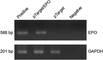
An EPO PCR 588 bp product was successfully amplified and confirmed by sequence analysis. The PCR product was cloned into pTarget vector, and the recombinant plasmid was produced in large scale for the subsequent experiments. In addition, RT-PCR of transfected HeLa cells demonstrated that the pTarget/EPO plasmid construct was functional (Figure 1).
RT-PCR gene expression analyses of transfected HeLa cells. The upper panel shows the amplification of the erythropoietin (EPO) CDS and the lower panel shows the amplification of the GAPDH endogenous control. Positive = Rabbit kidney cDNA; pTarget/EPO = pTarget/EPO vector; pTarget =HeLa cell transfected with empty pTarget vector; and Negative = No template control (NTC) reaction.

Effects of EPO on the rabbit hematocrit
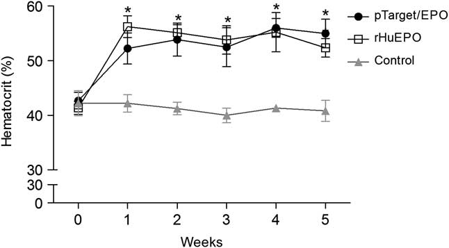
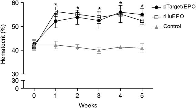
The EPO gene transfer and the rHuEPO treatments significantly increased the hematocrit levels (P < 0.05) 1 week after the first treatment, and these levels remained significantly higher (P < 0.05) than the control levels over the 5 weeks of the experiment (Figure 2). The hematocrit levels of the control group were similar throughout the experiment.
Effects of erythropoietin (EPO) treatment on the hematocrit of male rabbits. *Indicates significant differences between week and control group. Data are expressed as means ± s.e.m. (n = 6). Significance was considered at P < 0.05. pTarget/EPO = pTarget/EPO vector; rHuEpo = recombinant human EPO.
Effects of EPO on sperm motility, vigor, viability, morphology and concentration
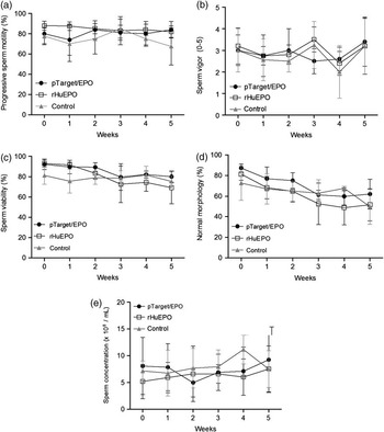
Neither the administration of rHuEPO nor the EPO gene transfer had an effect (P > 0.05) on sperm motility, vigor, viability, concentration or morphology (Figure 3). In addition, no alteration in sperm volume production or any visual or olfactory differences were observed.
Effects of erythropoietin (EPO) treatment on sperm characteristics. (a) sperm motility, (b) sperm vigor, (c) sperm viability, (d) sperm with normal morphology and (e) sperm concentration. Data are expressed as means ± s.e.m. (n = 6). Significance was considered at P < 0.05. pTarget/EPO = pTarget/EPO vector; rHuEpo = recombinant human EPO.

Effects of EPO on testis cells
The testis from each group was histologically inspected at the conclusion of the experiment. In the control group, the EPO gene transfer group and the rHuEPO group, no testicular damage was observed.
Discussion
The current study demonstrated the use of a non-viral EPO gene transfer in male rabbits. A single intramuscular injection of a mammalian expression vector containing EPO cDNA resulted in an increase in the production of red blood cells in comparison with a control group. This is in agreement with a previous report that demonstrated the long-term production of EPO following a single, intramuscular, dose of a plasmid construct similar to that described herein (Maruyama et al., Reference Maruyama, Ataka, Gejyo, Higuchi, Ito, Hirahara, Imazeki, Hirata, Ichikawa, Neichi, Kikuchi, Sugawa and Miyazaki2001). In addition, they demonstrated that a single dose increased hematocrit levels for at least 11 weeks. This is in agreement with our findings that the hematocrit levels increased during the first week and remained at peak levels for at least 5 weeks. As the EPO vector was administered intramuscularly, we believe that the recombinant plasmid was only incorporated into the muscle cells; the EPO was expressed in these cells and subsequently liberated into circulation. The EPO plasmid did not appear to be incorporated into the testicular cells, as it was not expressed by these cells.
The characteristics of the rabbit sperm were not affected by EPO gene transfer. Indeed, in all sperm analyses, no significant differences were found among the control group and the EPO gene transfer group. Previously, Tug et al. (Reference Tug, Altunkaynak, Aktas, Kilic, Yilmaz, Cam and Karateke2010) demonstrated that media supplementation used for sperm preparation techniques with EPO led to increased sperm motility in humans. In contrast, Temma et al. (Reference Temma, Shimoya, Hashimoto, Zhang, Koyama and and Murata2004) detected EPO in human seminal plasma and found no association between the EPO seminal plasma levels and sperm concentration, morphology, leukocyte count or cytoplasmic droplets. Our results for sperm motility, viability, sperm morphology and concentration are in broad agreement with these results, although the effect of EPO in the seminal plasma was not evaluated. Previously, Foresta et al. (Reference Foresta, Mioni, Bordon, Miotto, Montini and Varotto1994) demonstrated that testosterone production was increased by intravenous EPO administration in patients with renal failure, suggesting that this systemic effect of EPO might act directly on human Leydig cell function, which requires activation of a protein kinase-C-dependent pathway. Therefore, the increase in EPO levels following intravenous rHuEPO administration or EPO gene transfer could have affected the testicular function in the rabbits evaluated in this study. Physiologically, spermatozoa are always under the influence of EPO, as they swim in semen and body fluids of the female genitalia until fertilization, and all these environments contain EPO (Yamamoto et al., Reference Yamamoto, Sofikitis and and Miyagawa1997; Yasuda et al., Reference Yasuda, Fujita, Musha, Tanaka, Shiokawa, Nakamatsu, Mori, Matsuo and Nakamura2001; Kobayashi et al., Reference Kobayashi, Yanase, Iwanaga, Sasaki and Nagao2002; Tug et al., Reference Tug, Altunkaynak, Aktas, Kilic, Yilmaz, Cam and Karateke2010). To the best of our knowledge, this study is the first to investigate the effects of EPO gene transfer or rHuEPO administration on sperm characteristics.
Previous studies found that EPO has other functions on reproduction in mammals, such as the induction of testosterone secretion by Leydig cells, spermatogenesis, epididymal sperm maturation and the fertilizing capacity of sperm in rats with chronic renal failure (Yamamoto et al., Reference Yamamoto, Sofikitis and and Miyagawa1997; Tug et al., Reference Tug, Altunkaynak, Aktas, Kilic, Yilmaz, Cam and Karateke2010). Köseoğlu et al. (Reference Köseoğlu, Yilmaz, Ceylan, Uzun, Bayram and Hizli2009) demonstrated that rHuEPO administration significantly influenced the rescue of testicular function by preserving the intact seminiferous tubular morphology, lowering the percentage of necrotic seminiferous tubules and significantly reducing histological damage after induced testicular torsion/detorsion in male rats. In the current study, no histological damage was observed in the rabbit testis upon completion of the experiment, suggesting that EPO gene transfer or rHuEPO administration did not damage the rabbit testis.
In summary, the present study was the first to report the evaluation of sperm characteristics in response to the use of EPO gene therapy and rHuEpo administration. EPO gene transfer and rHuEpo administration did not appear to affect sperm motility, viability, concentration or morphology. Further studies are needed to clarify the precise mechanisms underlying this process.
Acknowledgments
T.F. Collares is a graduate student in the Biotechnology Program at Federal University of Pelotas and is supported by a grant from CNPq. A.J. McBride, J.C. Deschamps and O.A. Dellagostin are CNPq research fellows. Conflicts of Interest: The authors declare that there are no conflicts of interest.