Lignins and their untapped potential
Wood fibers are widely utilized in mature industries that take advantage of unique chemical and structural features of cellulose. However, the same cannot be said about lignin, another important component in wood. Lignin is a highly branched and amorphous biomacromolecule that displays different inter-unit linkages and is derived from complex phenylpropanoid pathways. In plants, lignin provides protection against various biotic/abiotic stresses and support to sap-conducting elements. The existence of lignin has contributed to the capacity of land-based plants to dominate terrestrial environments. Reference Weng and Chapple1,Reference Uzal, Gómez Ros, Pomar, Bernal, Paradela, Albar and Ros Barceló2
The main source of lignin, readily available for use on a large scale, comprises so-called “black liquors” produced as side streams during fractionation of wood fibers for use in pulp and paper industries. Reference Toledano, García, Mondragon and Labidi3,Reference Doherty, Mousavioun and Fellows4 Lignin extracted from the most typical process, so-called kraft pulping (a method that uses alkaline liquors for lignin solubilization), has a low degree of polymerization, with a weight-averaged molecular weight of 1–5 kDa (1–5 Kg/mol), lower than other commercially available lignins such as lignosulfonates (5–400 kDa [5–400 kg/mol]). Other lignins, such as those extracted with organic solvents (organosolv lignins) are in the range of 0.5–3 kDa ([0.5–3 kDa]; note that the polydispersity of lignosulfonate and kraft lignins is high compared to organosolv lignins). These technical lignins comprise low quantities of oxygen-containing functional groups (such as phenolic hydroxyl, carboxyl, and sulfonate groups, depending on the type), making them a viable feedstock for biomaterials production. Reference Norgren and Edlund5 Water-soluble lignosulfonates are, by far, the most used technical lignin today. Reference Norgren and Edlund5 Lignin represents a suitable source of bioenergy, considering its density and calorific value (similar to that of ethanol, 27 KJ/g). Reference Mendu, Harman-Ware, Crocker, Jae, Stork, Morton, Placido, Huber and DeBolt6,Reference Mendu, Shearin, Campbell, Stork, Jae, Crocker, Huber and DeBolt7 For this and other practical reasons, most industrial lignins are burned, on-site, for energy cogeneration; only a small fraction are used for materials.
Assessments performed on extracted kraft lignin have determined that it may be a preferable precursor compared to synthetic organic counterparts of similar molecular complexity. This is due to the energy efficiency of lignin extraction over organic synthesis. Recent environmental life cycle cradle-to-gate analyses of the impact of integrating a commercial-scale lignin extraction process and lignin co-product to an existing kraft pulp mill indicated a considerable reduction in global warming. Reference Culbertson, Treasure, Venditti, Jameel and Gonzalez8 Thus, lignin isolated from pulp mills represents an emerging opportunity to create new co-products and novel biomaterials. Lignin is biodegradable, antimicrobial, antioxidative, and CO2 neutral. Reference Doherty, Mousavioun and Fellows4,Reference Laurichesse and Avérous9–Reference Thakur and Thakur11 In this article, we discuss the use of lignin for its colloidal or supramolecular assembly (e.g., in particles), one of its many promising possibilities.
Micro- and nanoparticles from lignins
In materials science, nano- and colloidal particles (herein, referred to as microparticles) are used to achieve properties that are otherwise difficult to realize (i.e., to impart color, thermal and light stability, or to improve material strength). Reference Stark, Stoessel, Wohlleben and Hafner12 Typically, nanoparticle isolation is less energy demanding than chemical-synthesis routes. This is a direct result of nanoparticles possessing surface energies that are comparatively smaller than typical reaction enthalpies (e.g., dissolution, reduction). Reference Stark, Stoessel, Wohlleben and Hafner12 In particular, polymer nanoparticles are playing a pivotal role in a wide spectrum of areas. Reference Rao and Geckeler13 Compared to other biomolecules, Reference Guo, Tardy, Christofferson, Dai, Richardson, Zhu, Hu, Ju, Cui, Dagastine, Yarovsky and Caruso14 lignin superstructures are not an exception since they can also be converted into functional nanoparticles. Industrial or technical lignins have been proposed for the development of value-added particulate materials that preserve the structure, composition, and colloidal features of the macromolecule. They are of high interest for controlled release, encapsulation, and protection. Reference Campos, Branquinho, Carreira, Carvalho, Coimbra, Ferreira and Gil15 Some of the functions are already bestowed to lignin by nature, to fight against (bio)degradation (microbial, UV, heat), oxidation, and dehydration.
Lignin assemblies extruded or coalesced from fibers and self-assembly
The tendency of lignin to form particles is exemplified by the coagulation process that occurs, in situ, in the cell walls of stems of plants subjected to heating or mild hydrolytic treatments. This was observed, accidentally, in efforts aimed at enhancing the access of cellulose to enzymes. Reference Zeng, Ximenes, Ladisch, Mosier, Vermerris, Huang and Sherman16–Reference Xiao, Sun, Shi, Xu and Sun18 The temperature onset for particle formation was identified to be above 175°C after a 30-min hydrothermal treatment, with a large number of particles already formed at 205°C. Reference Araya, Troncoso, Mendonça and Freer19,Reference Araya, Tronscoso, Mendonca, Freer, Rencoret and Del Rio20 The lignin particles were revealed on or inside the fibers, presumably after cleavage of the carbohydrate–lignin bonds and formation of a continuous lignin phase above its glass-transition temperature.
Particles ranging in size between 5 nm and 10 μm have been reported, depending on the thermochemical treatment type at 150–220°C. Reference Donohoe, Decker, Tucker, Himmel and Vinzant21 The particles were noted to be smooth (by scanning electron microscopy [SEM]), Reference Castillo, Araya, Troncoso, Vinet and Freer22 while a significant increase in roughness occurred for pseudo-lignins or lignin–carbohydrate complexes. Reference Donohoe, Decker, Tucker, Himmel and Vinzant21,Reference Sannigrahi, Kim, Jung and Ragauskas23 The particle size was reduced when the fibers were treated with ionic liquids. Reference You, Zhang, Guo, Shao and Xu24 Fourier transform infrared analysis highlighted strong absorption from the aromatic character of lignin; however, no bands were observed for the alcohol C-O stretching, typical of carbohydrates attached to lignin. Reference Donohoe, Decker, Tucker, Himmel and Vinzant21,Reference Castillo, Araya, Troncoso, Vinet and Freer22 This was further confirmed by nuclear magnetic resonance analysis and cytochemical staining. When alkali-extracted lignin was heat treated, it formed highly porous grains. Reference Wang, Zhuang, Yuan, Qi, Yu and Wang25 The lignin particles showed a strong affinity to cellulose (Avicel Reference Li, Pu, Kumar, Ragauskas and Wyman26 or filter paper Reference Donohoe, Decker, Tucker, Himmel and Vinzant21 ).
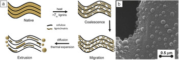
The suggested mechanism for the formation of lignin particles ( Figure 1 ) involves lignin coalescence into a fluid phase at high temperature and migration toward the cell surface, leading to collapse of the underlying fiber and particle formation by extrusion. This migration occurred mostly toward the cell wall rather than cell corners. Reference Ji, Zhang, Ling, Zhou, Ramaswamy and Xu27 Similar phenomena may occur upon acid pretreatment at relatively lower temperatures (130°C). Reference Selig, Viamajala, Decker, Tucker, Himmel and Vinzant28
(a) Schematic illustration of the process of lignin particle formation in the fiber upon heat or mild hydrolytic treatments. The fibers coalesce at or above the melting temperature of lignin (T m), followed by cleavage of the carbohydrate-lignin bond, resulting in free-flowing lignin that migrates to the cell wall with phase separates forming the particles on the surface. (b) Appearance of lignin particles as observed on the fibers. Adapted with permission from Reference Reference Donohoe, Decker, Tucker, Himmel and Vinzant21. © 2008 Wiley.

While the in situ formation of lignin particles is a fascinating observation, their isolation in such a state is a challenging task. In fact, scalable production of lignin particles is more likely to rely on lignin that has been extracted and processed in the dissolved state, as in the “black liquors.” Such particle formation strategies may take advantage of the self-assembling and self-polymerizing abilities already noted for lignin, combined with solvent exchange, heat, or enzymatic treatment, as well as acid hydrolysis or condensation in alkali. Reference Norgren, Edlund and Wågberg29–Reference Frangville, Rutkevičius, Richter, Velev, Stoyanov and Paunov33
The kinetics of assembly or aggregation of lignin into particles can be controlled by tuning salinity, temperature, pH, Reference Norgren, Edlund and Wågberg29 or solvency. Reference Lievonen, Valle-Delgado, Mattinen, Hult, Lintinen, Kostiainen, Paananen, Szilvay, Setälä and Österberg34 When the particles form by precipitation, small aggregates initially form, clustering into larger entities. Reference Qian, Deng, Qiu, Li and Yang32 Alternatively, fractal networks of lignin that display a high surface area can be obtained via sol-gel processing. Reference You, Zhang, Guo, Shao and Xu24,Reference Norgren, Edlund and Wågberg29
Within each lignin precursor there is usually a large diversity of structures, chemical functionalities, and molecular masses. Despite this, it is remarkable that the formation of such networks closely follows the aggregation behavior observed for systems that use only a few, well-defined components. Reference Norgren and Edlund5,Reference Norgren, Edlund and Wågberg29 This suggests supramolecular aggregation of lignin into clusters and then into particles of known composition and properties. In addition to factors previously discussed that affect particle homogeneity upon self-aggregation, the interactions between functional groups with the solvent are expected to control clustering, especially in the initial stages of particle formation.
The nonhomogeneous charge distribution as well as the molecular stiffness of the aromatic groups suggest π-π stacking of lignin molecules, which plays an important role in their self-assembly into clusters and particles. Reference Sarkanen, Teller, Stevens and McCarthy35 It has been shown that such stacking can be favored when the particles are formed in a solvent exchange process, in the absence of alkali groups. In such cases, better particle stability has been noted compared to that observed for particles formed (e.g., by precipitation). Reference Frangville, Rutkevičius, Richter, Velev, Stoyanov and Paunov33
Routes for the synthesis of lignin nano- and microparticles
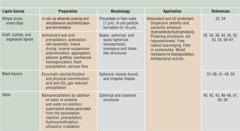
The large interest in fabricating polymeric particles has triggered recent efforts to use lignin in this context. The fabrication methods reported to date (summarized in Table I Reference Castillo, Araya, Troncoso, Vinet and Freer22,Reference Qian, Deng, Qiu, Li and Yang32,Reference Sarkanen, Teller, Stevens and McCarthy35–Reference Gevorkyants, Chernoberezhskii and Lotentsson61 ) include precipitation with acid Reference Frangville, Rutkevičius, Richter, Velev, Stoyanov and Paunov33,Reference Wei, Yang, Yang and Wang36–Reference Moreva, Alekseeva and Chernoberezhskii38 or gas saturated with CO2, Reference Kannangara, Marinova, Fradette and Paris39 ultrasonic irradiation, Reference Gilca, Popa and Crestini40 mechanical homogenization, Reference Nair, Sharma, Pu, Sun, Pan, Zhu, Deng and Ragauskas41 chemical modification, Reference Gilca, Ghitescu, Puitel and Popa42–Reference Deng, Zhao, Qian, Lü, Wang and Qiu45 antisolvent exchange, emulsion inversion, a combination of methods, Reference Qian, Deng, Qiu, Li and Yang32,Reference Yang, Kenny and Puglia46–Reference Richter, Bharti, Armstrong, Brown, Plemmons, Paunov, Stoyanov and Velev49 as well as procedures involving enzymes Reference Castillo, Araya, Troncoso, Vinet and Freer22,Reference Shikinaka, Fujii, Egashira, Murakami, Nakamura, Otsuka, Ohara and Shigehara50 or simply by drying. Reference Barakat, Gaillard, Lairez, Saulnier, Chabbert and Cathala51–Reference Chen, Liu, Seyed Shahabadi, Xu and Lu53 Among these, acid precipitation, antisolvent exchange, and their combination have been the most popular. In the acid-precipitation process, lignin is initially dissolved in alkaline aqueous media (pH > 11), which favors electrostatic stabilization by deprotonation of carboxylic groups and phenolic OH. Upon reduction of pH (∼3), the carboxylic and hydroxyl groups partially protonate, decreasing the surface charge density, resulting in destabilization of the lignin colloids. This triggers association and favors particle formation.
Methods reported to synthesize lignin nano- and microparticles.

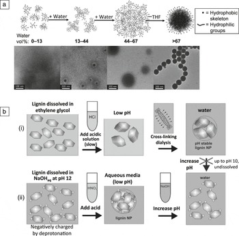
Only two generic methods from Table I will be discussed here, the antisolvent and the aerosol drying methods (for a complete list, please refer to Table SI, available online). In the antisolvent precipitation method, lignin is first dissolved in an appropriate solvent such as tetrahydrofuran (THF). Then spherical colloidal particles are formed by the addition of an antisolvent (e.g., water Reference Qian, Deng, Qiu, Li and Yang32 ) or by slow solvent exchange to water via dialysis. Reference Lievonen, Valle-Delgado, Mattinen, Hult, Lintinen, Kostiainen, Paananen, Szilvay, Setälä and Österberg34 A simple mechanism for colloidal formation has been proposed ( Figure 2a). Reference Qian, Deng, Qiu, Li and Yang32
(a) Schematic representation of the formation process of lignin colloids after solvent exchange (tetrahydrofuran [THF]–H2O) and respective transmission electron microscope images from the dispersions at different water content, as indicated. Adapted with permission from Reference Reference Qian, Deng, Qiu, Li and Yang32. © 2014 Royal Society of Chemistry. (b[i]) Proposed mechanism for particle formation by precipitation with HCl (aq.) from lignin dispersions in ethylene glycol and subsequent cross-linking with glutaraldehyde upon dialysis. (ii) Alternatively, precipitation in the form of lignin nanoparticles can occur after lowering the pH of an aqueous solution with high pH. The lignin particles produced in this route can dissolve at a pH > 5, while those produced in (i) are stable up to a pH of 10. Adapted with permission from Reference Reference Frangville, Rutkevičius, Richter, Velev, Stoyanov and Paunov33. © 2012 Wiley. Note: NP, nanoparticle.

First, it is hypothesized that local lignin clusters form via hydrophobic interactions in the homogeneous solution, which then grow into colloidal spheres. Reference Qian, Deng, Qiu, Li and Yang32 A nucleation-growth mechanism has been used to explain lignin particle formation when THF solvent is replaced with water during dialysis, Reference Qian, Deng, Qiu, Li and Yang32 somewhat related to what has been proposed for other systems (see Table I). Reference Frangville, Rutkevičius, Richter, Velev, Stoyanov and Paunov33,Reference Lievonen, Valle-Delgado, Mattinen, Hult, Lintinen, Kostiainen, Paananen, Szilvay, Setälä and Österberg34,Reference Wei, Yang, Yang and Wang36–Reference Moreva, Alekseeva and Chernoberezhskii38,Reference Gevorkyants, Chernoberezhskii and Lotentsson61 In the nucleation-growth mechanism, acid precipitation occurs from an ethylene glycol solution, whereby planar phenol groups present in lignin stack in layers and afford densely packed nanoparticles (Figure 2b). In contrast, acidic precipitation of lignin from alkaline solution does not favor formation of stable stacks due to electrostatic repulsion via the negative charges in the aromatic groups, which are partially ionized under these conditions (Figure 2b). Reference Frangville, Rutkevičius, Richter, Velev, Stoyanov and Paunov33 Besides changes in pH, aggregation in aqueous solution can be influenced by ionic strength and temperature, Reference Norgren, Edlund and Wågberg29 as well as factors inherent to the lignin such as concentration Reference Sarkanen, Teller, Stevens and McCarthy35 and polydispersity. Reference Sarkanen, Teller, Abramowski and McCarthy62
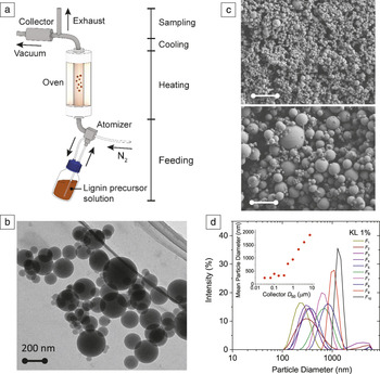
Contrary to previous cases where lignin particles were obtained in colloidal dispersions, we recently reported the facile fabrication of dry spherical lignin particles by using the aerosol flow method. This resulted in particle sizes ranging from the nano- to the micrometer scale ( Figure 3 ). Additionally, in situ separation in fractions of different size ranges was possible. In this process, particle formation is speculated to occur by a sequential process that first involves evaporation of solvent from the droplet surface. Simultaneously, a macromolecular shell forms on the droplet, while solvent remaining in the droplet interior gradually diffuses radially. Consequently, the solute condenses as the droplets evolve into dried, solid particles. Reference Ago, Huan, Borghei, Raula, Kauppinen and Rojas52 Although the number of reports that use drying is quite limited, this method is promising because of the high yield that can be achieved; in addition, it is a scalable nanomanufacturing approach that affords size control and high production capacity. Reference Ago, Huan, Borghei, Raula, Kauppinen and Rojas52
(a) Simplified schematic of an aerosol flow reactor used for the synthesis of lignin particles. (b) Transmission electron micrograph of kraft lignin (KL) solid particles from fraction F5, which corresponds to particle sizes <200 nm. The image indicates a homogeneous interior structure after isotropic shrinking from drying and perfectly spherical shapes. Scale bar = 200 nm. (c) Scanning electron micrographs are compared for solid lignin spheres obtained from two different particle fractions synthesized from organosolv lignin: top, F 5 (<200 nm) and bottom, F8 (average size: 1 μm). Scale bar = 2 μm. Reference Ago, Huan, Borghei, Raula, Kauppinen and Rojas52 (d) Distribution and mean diameter (inset) of particles obtained from 1% KL precursor solution and separated in situ into different fractions by using a collector (see [a]) with different orifices (F n or collector number). The size distribution for the different populations are displayed together with a plot of the relationship between collector orifice size (cutoff diameter, D 50) and mean particle diameter. The mean diameter of the lignin particles ranged from ∼200 to ∼1900 nm with increasing collector orifice size (fraction number). The particle size distribution became broader as the mean particle size decreased. Reference Ago, Huan, Borghei, Raula, Kauppinen and Rojas52

Use of lignin particles in organic–inorganic hybrids and multiple component systems
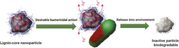
The complexation of lignin with organic or inorganic compounds provides powerful control over the structure and properties of the formed particles. For example, silver ions infused in lignin nanoparticles coated with cationic polyelectrolytes (for increased adhesiveness onto negatively charged bacteria) resulted in excellent antimicrobial effects ( Figure 4 ). Reference Richter, Brown, Bharti, Wang, Gangwal, Houck, Hubal, Paunov, Stoyanov and Velev63 The Ag-lignin hybrid particles were capable of neutralizing both gram-negative (e.g., Escherichia coli) and gram-positive (Staphylococcus epidermidis) human pathogens as well as strong strains, for example, Ralstonia-solanacearum. This was achieved while using 10× less silver than that used in conventional aqueous solutions. In addition, Ag-lignin hybrids with antimicrobial capacity afforded filters with high activity against multidrug resistant strains. Reference Rak, Friscic and Moores64,Reference Zhong, Xu and Qin65
Schematic illustration showing the antimicrobial mechanism of silver ion-infused lignin-core nanoparticles. The cationic polyelectrolyte coating promotes the attachment of the nanoparticle onto negatively charged cell walls (green). Adapted with permission from Reference Reference Richter, Brown, Bharti, Wang, Gangwal, Houck, Hubal, Paunov, Stoyanov and Velev63. © 2015 Macmillan Publishers Ltd.

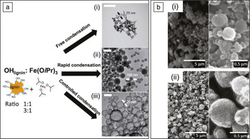
Hybrids of lignin with other metals such as iron have been reported as well. Reference Gutiérrez-Hernández, Escalante, Murillo-Vázquez, Delgado, González and Toríz66–Reference Qin, Kang, Wang, Liu, Ni, Huang, Li and Li68 In the latter case, morphology control was achieved, from hollow or solid nanospheres ( Figure 5a). Reference Lintinen, Latikka, Sipponen, Ras, Österberg and Kostiainen69 Such metal-lignin hybrids can be used as magnetic, antibacterial, catalytic, photonic, and gas- and moisture-barrier materials, depending on the components.
(a) Schematic illustration of the synthesis of different nanoparticle morphologies with iron and respective transmission electron microscope (TEM) images of the various structures obtained: (i) When a Fe(OiPr)3 solution was added to a lignin solution at OHlignin-to-Fe(OiPr)3 ratio of 1:1, a precipitate formed immediately and hydrolysis yielded Fe(OiPr)-coated lignin nanoparticles forming fishnet-type open network structures. (ii) When less Fe(OiPr)3 is used, for OHlignin-to-Fe(OiPr)3 ratio of 3:1, full condensation of OH groups with Fe(OiPr) yielded large structures, and the lignin clusters were partially covered with Fe(OiPr). The partially covered clusters fused together to form larger (ca. 200 nm) particles. (iii) Under conditions of controlled condensation (slow solution addition), the fusing of Fe:lignin nanoparticles created hollow spheres. Scale bars in all TEM images = 200 nm. Adapted with permission from Reference Reference Lintinen, Latikka, Sipponen, Ras, Österberg and Kostiainen69. © 2016 Royal Society of Chemistry. (b) Scanning electron microscope images for aggregates obtained with (i) alkaline lignin, hydroxyethyl cellulose, and sodium dodecylbenzenesulfonate as well as (ii) alkaline lignin, hydroxyethyl cellulose, NaOH, and sodium dodecylbenzenesulfonate, at (left) low and (right) high magnifications. The solvent was a mixture of water and CHCl3. Reproduced with permission from Reference Reference Shimada, Hata and Kijima78. © 2015 American Chemical Society.

Silica composites, in particular, are of high interest since this mineral enhances thermal and chemical stability, bioactivity, and biocompatibility. Reported procedures for preparing hybrid silica-lignin particles typically involve a sol-gel process. Tetraethoxysilane has been used as a silica precursor to prepare silica-lignin hybrid particles in the presence of sulfuric acid as a catalyst, Reference Hasegawa, Fujii, Yamada, Kariya and Takayama70 and mesoporous hybrids have been synthesized by using the same process. Reference Qu, Tian, Zou, Zhang, Zheng, Wang, Li, Rong and Wang71 Some features of the hybrid particles, such as pore size and surface area, were found to strongly depend on the pH and the mass ratio of lignin to silica. The resultant spherical, hybrid particles (ca. 60-nm dia.) were reported to have surface areas of ∼472 m2/g when prepared in optimized conditions (2:3 lignin:silica ratio at 90°C and pH 3 for 6 h). Coprecipitation processes were also introduced in the preparation of hybrid silica-lignin particles. Reference Xiong, Yang, Zhong, Li, Zhou and Qiu72 The obtained hybrid particles (300–400-nm dia.) underwent an undesired reduction in surface area, 386–55 m2/g with increasing lignin loading. In this aspect, black liquors obtained from rice straw pulping are of interest since they contain sodium silicates. A facile and “green” approach was proposed to coprecipitate lignin-silica particles (50–100-nm dia.) by adjusting the pH via CO2 bubbling. Reference Zhang, Zhao, Ran, Liu, Liu, Zhou and Wang73 The use of hybrid lignin particles with silica is expected to impact diagnostic and therapeutic efforts as well as the development of energy-storage materials, among others.
In the case of organic composites, functional lignin particles have been obtained via covalent conjugation. Lignin is composed of a large number of hydroxyl functional groups that can be covalently modified to develop new functionalities. For example, azo-modified (azo-lignin) particles have been synthesized with this concept in mind. Reference Deng, Zhao, Qian, Lü, Wang and Qiu45,Reference Wang, Wu, Li, Sinawang, Wang and He74 Azo-lignin with photoprocessing capability was achieved by coupling alkali lignin with diazonium salts of 4-aminobenzoic acid. The nanoparticles showed self-assembly and photo-induced morphological response. After 15-min exposure under a linearly polarized laser beam (488 nm), the colloidal spheres were deformed along the polarization direction of the light. More recently, an azo-lignin polymer was obtained by modification of lignin with aniline (C6H5NH2). Hollow lignin colloidal spheres were found to be capable of self-assembling and encapsulating photosensitive avermectins (naturally occurring macrocyclic lactone derivatives with anthelmintic and insecticidal properties). Reference Burg, Miller, Baker, Birnbaum, Currie, Hartman, Kong, Monaghan, Olson, Putter, Tunac, Wallick, Stapley, Oiwa and Ōmura75 Lignin facilitated the controlled-release behavior of the drug and also acted as a photoprotectant to avoid the photodegradation of photosensitive pesticides.
One of the important challenges underlying the formation of composite materials with lignin is the compatibility of lignin with polymers that engage in hydrophilic–hydrophobic interactions at the interface. Composite polymer-lignin particles formed thus far mainly use biopolymers such as pectin, cellulose (Figure 5b), alginate (polysaccharide), and silk sericin (protein). Reference Kwak, Shin, Yun and Lee76–Reference Borisenkov, Karmanov, Kocheva, Markov, Istomina, Bakutova, Litvinets, Martinson, Durnev, Vityazev and Popov79 For example, macroporous Ca-alginate-lignin (MCAL) beads (4.1 ± 0.4 mm) were obtained with an immobilized white rot fungus, Phanerochaete chrysosporium (P. chrysosporium), endowing the system with a capacity to degrade aromatic hydrocarbon (phenanthrene). Reference Zhang, Xu, Hua, Han, Wang, Wang, Liu and Liu80 The presence of lignin in the beads led to a ∼29-fold increase in the phenanthrene adsorption capacity, and its four-day degradation was increased to ∼42% compared to that of macroporous Ca-alginate beads. The enhanced growth and degradation indicated the role of lignin in MCAL-immobilized P. chrysosporium as an effective microbial model for intensified biodegradation of phenanthrene. Other examples of porous composite particles include those obtained after cross-linking lignin with sodium alginate via epichlorohydrin (epoxy resin with a high reactivity to phenols), which exhibited excellent adsorption efficiency for Pb ions and were used for their removal from aqueous media. Reference Li, Ge and Wan81
The formation of structured assemblies such as nano- and microparticles from lignin opens the pathway to many applications traditionally dominated by synthetic, petrochemically derived, or inorganic particles, but with the added benefits inherent to lignin such as low toxicity, biodegradability, low cost, carbon footprint reduction, and abundance.
The presence of aromatic, phenolic, and amphiphilic groups in lignin makes it an ideal candidate for the formation of material for the adsorption of metal ions and chemicals. Lignin contains high amounts of methoxy, aldehyde, keto, hydroxyl, and phenolic functional groups, providing a large range of interactions that can be exploited for specific adsorption of small molecules or for metal chelation. Reference Demirbas82–Reference Šćiban, Klašnja and Antov84 Compared to commercially available amorphous lignin powders, porous lignin particles have a larger surface area, bear more active sites, and display better efficiency for metal-ion sorption. Lignin spheres used in packed-bed columns efficiently extracted metal ions from solution and were identified as candidates for wastewater treatment or ore extraction; Reference Lintinen, Latikka, Sipponen, Ras, Österberg and Kostiainen69,Reference Kwak, Shin, Yun and Lee76,Reference Li, Ge and Wan81,Reference Krachler, von der Kammer, Jirsa, Süphandag, Krachler, Plessl, Vogt, Keppler and Hofmann85,Reference Huang, Fu, Huang and Cheng86 mesoporous TiO2-containing nanoparticles prepared via calcination of Ti-lignin systems showed excellent photocatalytic activity, which can be useful in similar applications. Reference Chen, Kuo, Lu, Hou and Kuo67
Overall, the prospects of lignin particles stem not only from their inexpensive, renewable sourcing and the structure they form, but also from the inherent macromolecular properties of lignin. This includes antioxidant behavior and high UV stability, both of which are of interest in fields such as nutritional and pharmaceutical formulations, and cosmetics. Reference Deng, Zhao, Qian, Lü, Wang and Qiu45,Reference Saidane, Barbe, Birot and Deleuze55,Reference Yearla and Padmasree58,Reference Myint, Lee, Seo, Son, Yoon, Yoon, Park, Yu, Yoon and Lee59 Addition of lignin in cellulose hydrogel beads improved the activity and stability of immobilized enzymes (lipase), suggesting potential applications for biocatalysis, as well as in biomedical and bioelectronic applications. Reference Thakur and Thakur11 Finally, other applications for lignin particles have been introduced, including templates for catalysts and supercapacitors, Reference Chen, Kuo, Lu, Hou and Kuo67,Reference Qin, Kang, Wang, Liu, Ni, Huang, Li and Li68,Reference Shimada, Hata and Kijima78 bioprotective coatings, Reference Gîlcă and Popa43 and flame retardants. Reference Shikinaka, Fujii, Egashira, Murakami, Nakamura, Otsuka, Ohara and Shigehara50
Outlook
The global market for inorganic and polymeric micro- and nanoparticles is considerable and, within this sector, lignin could play an important role because of its many advantages compared to synthetic polymers. In comparison to other polymers, lignin utilization is in its infancy. Developments that take advantage of the aromatic structure of lignin, its use along the biomass cycle as a whole, may have positive effects in the carbon dioxide targets of the global bioeconomy. Lignins are produced in biorefineries and bioproduct mills, and its properties (including surface energy and reactivity) can be tuned depending on the source of the fiber material and processing conditions. Beyond its current use in energy cogeneration as well as its potential to synthesize (carbon) fibers, lignin can be a platform for developing micro- and nanoparticles. The next generation of applications will require development of isolation processes to realize lignin streams that yield sufficient levels of purity and reactivity. Other aspects to be explored include the precise control over particle size and morphology as well as the possibility to endow new functional properties, by physicochemical transformation or by combination with other active substances.
Acknowledgments
We are grateful for the support of the Academy of Finland through funding of the SIRAF Project, and funding support from NordForsk under the Project “High-Value Products from Lignin.”

Mariko Ago is a research fellow in the Department of Bioproducts and Biosystems, School of Chemical Engineering, at Aalto University, Finland. Previously, she was with the Department of Nanomaterials and Bio-Engineering, Tokushima Bunri University, Japan. Her research focuses on new functional materials from lignocellulose. She received the 2015 Young Investigator Award from the Cellulose Society of Japan. Ago can be reached by phone at +358 50 3841759 or by email at mariko.ago@aalto.fi.

Blaise L. Tardy is a postdoctoral researcher in the Department of Bioproducts and Biosystems, School of Chemical Engineering, at Aalto University, Finland. He obtained his MSc degree in bioengineering from the Swiss Federal Institute of Science and Technology, Switzerland, in 2009, and his PhD degree in chemical and biomolecular engineering from The University of Melbourne, Australia, in 2015. His current research interests include the formation of structured materials from fiber-cell-wall components. Tardy can be reached by phone at +358 50 5979156 or by email at blaise.tardy@aalto.fi.

Ling Wang is a doctoral candidate in the Department of Bioproducts and Biosystems, School of Chemical Engineering, at Aalto University, Finland. She received her MS degree in bioproduct technology from Aalto University. Her research interests include cellulose-based filaments, lignin-based composites, and advanced biomaterials. Wang can be reached by phone at +358 50 5907467 or by email at ling.wang@aalto.fi.

Jiaqi Guo is a doctoral candidate in the Department of Bioproducts and Biosystems, School of Chemical Engineering, at Aalto University, Finland. He received his BE degree from Qilu University of Technology, China, in 2010, and his MS degree in chemical engineering from Åbo Akademi University, Finland. His current research interests focus on nanoparticle synthesis, novel chemical modification of nanocellulose and wood-derived structures, and design and synthesis of hybrid nanomaterials. Guo can be reached by phone at +358 50 3440872 or by email at jiaqi.guo@aalto.fi.

Alexey Khakalo is a doctoral candidate in the Department of Bioproducts and Biosystems, School of Chemical Engineering, at Aalto University, Finland. He is a renewable materials engineer with interest in sustainable product development with advanced functions. His research interests include biomaterials, bio(nano)composites, surface chemistry and interactions, paper technology, packaging, and advanced materials. Khakalo can be reached by phone at +358 50 4018149 or by email at alexey.khakalo@aalto.fi.

Orlando J. Rojas is professor of bio-based materials in the Department of Bioproducts and Biosystems, School of Chemical Engineering, at Aalto University, Finland. He is an elected Fellow of the American Chemical Society (2013) and a Finland Distinguished Professor (2009–2014). He chaired the Division of Cellulose and Renewable Materials of the American Chemical Society from 2009 to 2011, and is the recipient of the 2015 Nanotechnology Division Technical Award for outstanding contributions to the nanotechnologies of renewable materials. His research focuses on the development of nanostructures from the fiber cell wall and cellulose derivatives, dynamics of enzymatic reactions, and design of stimuli-responsive materials and multiphase systems. Rojas can be reached by phone at +358 50 5124227 or by email at orlando.rojas@aalto.fi.