Introduction
In principle, cloning by nuclear transfer (NT) using somatic cells (somatic cell nuclear transfer; SCNT) is expected to produce an unlimited number of identical animals because passaged and cultured cells are used as the nuclear donor. However, the efficiency of SCNT is still low, with less than 4% of reconstructed embryos resulting in live young, regardless of species and somatic cell type (Renard et al., Reference Renard, Zhou, LeBourhis, Chavatte-Palmer, Hue, Heyman and Vignon2002; Wilmut et al., Reference Wilmut, Beaujean, de Sousa, Dinnyes, King, Paterson, Wells and Young2002). Embryonic cells (blastomeres) have also been used as the nuclear donor cells for cloning by NT (embryonic cell nuclear transfer; ECNT). Although the number of blastomeres in an embryo is limited, ECNT may help to produce normal identical calves because it has shown lower incidences of pregnancy loss and fetal/postnatal abnormalities than SCNT in cattle (Heyman et al., Reference Heyman, Chavatte-Palmer, LeBourhis, Camous, Vignon and Renard2002; Wilmut et al., Reference Wilmut, Beaujean, de Sousa, Dinnyes, King, Paterson, Wells and Young2002). Moreover, higher genetic gains for carcass traits in beef cattle were reported to be obtained in testing schemes with ECNT than in conventional progeny testing schemes (Hirooka, Reference Hirooka2000) and high similarities of meat quality (the proportion of fat to muscle and the meat's colour) were confirmed among ECNT-derived Japanese black cattle (Kuchida et al., Reference Kuchida, Ogasawara, Hidaka, Minamihashi, Sakai and Yamamoto2003). Hence, ECNT may be a more practical technology than SCNT for the genetic improvement needed to produce high quality beef.
It is important to determine the optimum developmental stage of nuclear donor embryos that can be recovered from the uterus of cattle non-surgically for efficient cloning by ECNT. Several studies have examined in vitro and in vivo development of bovine NT embryos derived from 16- and 64-cell-stage embryos (Bondioli et al., Reference Bondioli, Westhusin and Looney1990) and morulae and blastocysts (Zakhartchenko et al., Reference Zakhartchenko, Reichenbach, Riedl, Palma, Wolf and Brem1996). However, the effect of the embryonic cell cycle of the nuclear donor embryo on the efficiency of ECNT has not been studied in cattle. We therefore compared the efficiencies of ECNT using blastomeres of sixth cell cycle (32- to 63-cell)-stage and seventh cell cycle (64- to 127-cell)-stage embryos derived from Japanese black cattle, which are widely raised as a typical beef breed in Japan.
Materials and Methods
Nuclear donor embryos
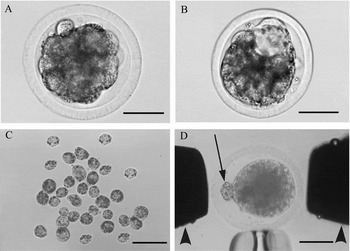
Morulae and early blastocysts were obtained from superovulated Japanese black cows (3 to 5 years old). Superovulation was induced by the injection of follicle stimulating hormone (FSH, Antorin, Kawasaki-Mitaka Pharmaceutical Co. Ltd) in declining doses twice daily for 3 days (18 mg in total). Estrus in treated cows was induced by the injection of prostaglandin F2α (40 mg, Veterinary Pronalgon-F Injection, Pharmacia Upjohn Co. Ltd) 2 days after the first injection of FSH. Cows in estrus were artificially inseminated using frozen-thawed semen from Japanese black bulls. Five or 6 days after the insemination, embryos were recovered by flushing the uterine horns non-surgically. Morphologically normal morulae and early blastocysts were incubated in 25 mM HEPES-buffered tissue culture medium 199 (TCM-199: Gibco Laboratories, Grand Island, NY, USA) supplemented with 5% (v/v) fetal calf serum (FCS, Gibco) for 5 to 6 h at 39 °C under 5% CO2, 5% O2 and 90% N2 with high humidity. Embryos at the sixth cell cycle (32- to 63-cell) and seventh cell cycle (64- to 127-cell) stages (Fig. 1) were selected as nuclear donor embryos by counting blastomere numbers as described below.
(A) 32- to 63-cell stage (6th cell cycle) embryos (number of blastomeres: 34). (B) 64- to 127-cell stage (seventh cell cycle) embryos (number of blastomeres: 86; inner cell mass and trophectoderm were observed at this stage). (C) Blastomeres of 6th cell cycle embryos (A) separated by repeated pipetting. (D) Enucleated oocyte with a blastomere (arrow) placed between needle type-electrodes (arrowheads) in Zimmerman cell fusion medium. Scale bars = 50 μm.

Preparation of blastomeres
The zona pellucida of nuclear donor embryos was removed with a microblade (Bio-cut blade, Feather Safety Razor Co. Ltd) in Dulbecco's phosphate-buffered saline (DPBS, Gibco) supplemented with 0.1% (w/v) polyvinyl alcohol (PVA: Sigma Chemical Co.) (PBS–PVA). Zona-free embryos were then exposed to Ca2+- and Mg2+-free DPBS supplemented with 0.25% (w/v) trypsin (Difco) and 0.04% (w/v) EDTA (Sigma) (trypsin EDTA solution) for 1 min. Blastomeres were separated by pipetting in HEPES-buffered Tyrode's medium (TALP–HEPES, Bavister et al., Reference Bavister, Leibfried and Lieberman1983) containing 20% (v/v) FCS (TALP–HEPES–FCS) and the blastomere numbers of each embryo were recorded.
Oocyte maturation, enucleation and activation
In vitro maturation (IVM) of oocytes was carried out according to the previously reported procedure (Saeki et al., Reference Saeki, Hoshi, Leibfried-Rutledge and First1990) with some modifications. Briefly, bovine cumulus–oocyte complexes (COCs) collected from small antral follicles of slaughterhouse-derived ovaries were cultured in a 50 μl droplet of TCM-199 supplemented with 0.5 mM sodium pyruvate (Nacalai Tesque), 1% (v/v) antibiotic–antimycotic solution (Gibco), 0.02 mg/ml of FSH, 1 μg/ml of estradiol-17β (Sigma) and 10% (v/v) FCS for 18 to 19 h at 39 °C under 5% CO2 in air with high humidity (10 COCs/droplet). Cumulus cells were removed by exposing the COCs to DPBS supplemented with 1 mg/ml of hyaluronidase (Sigma) and 0.1% PVA for 10 min and vortexing for 5 min or pipetting. The cumulus-free oocytes with a first polar body were placed into DPBS supplemented with 5 μg/ml of cytochalasin B (Sigma) and 10% FCS (PBS–FCS) for 5 to 10 min prior to enucleation. Enucleation was accomplished by aspirating a small volume of the cytoplasm adjacent to the polar body with a micropipette as described elsewhere (Yang et al., Reference Yang, Jiang, Farrell, Foote and McGrath1993). Oocytes stained with PBS–FCS containing 5 μg/ml of Hoechst 33342 (Sigma) were observed for a few seconds using a fluorescence microscope to confirm successful enucleation. Enucleated oocytes (recipient oocytes) were washed with Ca2+- and Mg2+-free TALP–HEPES [TALP–HEPES(–)] and then exposed to TALP–HEPES(–) containing 5 mM ionomycin (Calbiochem, San Diego, CA, USA) for 45 s at 22 to 24 h after the initiation of IVM to induce activation (Sims & First, Reference Sims and First1994). This was followed by another 15 min culture in TALP–HEPES–FCS. The oocytes were then further incubated in TALP–HEPES–FCS containing 10 μg/ml of cycloheximide (CHX, Sigma) for 5 h (First et al., Reference First, Leibfried-Rutledge, Northey and Nuttleman1992) prior to use for NT.
Nuclear transfer, fusion and embryo culture
Individual blastomeres were transferred into the perivitelline space of recipient oocytes. The oocyte–blastomere complexes (couplets) were fused according to the previously reported procedure (Mitani et al., Reference Mitani, Utsumi and Iritani1993) with some modifications. Briefly, couplets were individually placed in a drop of Zimmerman cell fusion medium (Wolfe & Kraemer, Reference Wolfe and Kraemer1992) in a petri dish and fixed in position using a holding pipette. With the aid of a micromanipulator, two needle-type electrodes (Unique Medical Imada) connected to an electrofusion apparatus (ECM200, BTX) were positioned as shown in Fig. 1. For fusion, a single DC pulse of 20 V/mm for 20 μs was used. After receiving the fusion pulse, couplets were transferred to synthetic oviduct fluid medium (SOF: Tervit et al., Reference Tervit, Whittingham and Rowson1972) supplemented with 20 amino acids, 3 mg/ml of bovine serum albumin (A4378, crystallized and lyophilized, Sigma) and 1% (v/v) antibiotic–antimycotic solution (m-SOF: Takahashi & First, Reference Takahashi and First1992) and maintained at 39 °C. Fusion rates were determined 30 to 60 min after applying the DC pulse.
Fused couplets were cultured in m-SOF at 39 °C under 5% CO2, 5% O2 and 90% N2 with high humidity (Nagao et al., Reference Nagao, Saeki, Hoshi and Nagai1995). Cleavage and blastocyst development of NT embryos were examined under a stereomicroscope (×60) on days 7 and 8 of culture (day 0 = day of fusion).
Embryo transfer
To investigate the subsequent viability of NT embryos, blastocysts derived from NT embryos were transferred non-surgically to the uterine horn of Holstein heifers (recipient animals) 6 to 8 days after estrus (one or two embryos/recipient animal). Pregnancy was diagnosed by ultrasonography 42 to 44 days after the embryo transfer.
Statistical analysis
The statistical significance of data was analysed with the chi-squared test (fusion and developmental rates) or Fisher's exact test (pregnancy and calving rates). p < 0.05 was considered significant.
Results and discussion
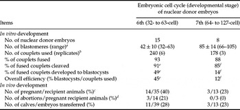
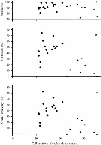
The results obtained in the present study are shown in Table 1 and Fig. 2. There was no difference in the fusion rates between blastomeres derived from sixth cell cycle (32- to 63-cell)-stage and seventh cell cycle (64- to 123-cell)-stage embryos when using a needle-type electrode, although blastomeres derived from 32-cell-stage embryos were reported to give higher fusion rates than those from 64-cell-stage embryos when using a chamber-type electrode (Ushijima et al., Reference Ushijima, Tsunoda, Eto and Imai1991). A low fusion rate was obtained in terms of a small area of contact between a small blastomere and oocyte (Ritchie & Campbell, Reference Ritchie and Campbell1995). When the needle-type electrode was used to induce fusion between a blastomere and oocyte (couplet), the couplets could be manually aligned so that the surface of contact between the blastomere and oocyte was parallel to the electrodes, leading to a high fusion rate, regardless of the size of the blastomere. In contrast, alignments with small blastomeres are difficult with a chamber-type electrode and so the fusion rates with blastomeres from 64-cell-stage embryos might be lower than those with blastomeres from 32-cell-stage embryos (Ushijima et al., Reference Ushijima, Tsunoda, Eto and Imai1991).
In vitro and in vivo development of nuclear transfer embryos reconstructed with blastomeres from 6th and 7th cell cycle stage embryos

a Number of blastomeres were expressed as mean ± SD.
b Two to three nuclear donor embryos were used for one replicate, and not all blastomeres were used for making the couplets in each replicate.
c Number of pregnant recipient animals 42 to 44 days after the transfer of NT embryos.
d Number of abortions within 90 days.
e, f Values with different superscripts in the same row are significantly different (p < 0.05).
Scatter plot showing the rate of fusion (A), blastocysts (B), and overall efficiency (C) of nuclear transfer embryos reconstructed with different cell numbers in blastomeres of in vivo-derived embryos. % were based on the number of couplets used (A and C) and on the number of couplets fused (B). (●) Derived from the sixth cell-cycle stage (32- to 63-cell stage). (▲) Derived from the seventh cell cycle stage (64- to 127-cell stage).

Cleavage, blastocyst development and the overall efficiency of obtaining NT embryos reconstructed with blastomeres from sixth cell cycle (32- to 63-cell)-stage embryos were higher than those from seventh cell cycle (64- to 127-cell)-stage embryos in the present study (p < 0.05). Coordination of the cell cycle stages of nuclear donor cells and recipient oocytes should be required for the maintenance of correct ploidy and the normal development of the NT embryo (Campbell et al., Reference Campbell, Loi, Otaegui and Wilmut1996). About 80% of the blastomeres of 32- to 63-cell-stage embryos were reported to be in S phase (Barnes et al., Reference Barnes, Collas, Powel, King, Westhusin and Shepherd1993). The cell-cycle stage of preactivated recipient oocytes, which could to be in S phase (Campbell et al., Reference Campbell, Loi, Cappai and Wilmut1994), might be synchronized with that of blastomeres of 32- to 63-cell-stage embryos, so that a high rate of cleavage and blastocyst development of NT embryos may be obtained. The reasons for the low rates of cleavage and blastocyst development of NT embryos originating from the 64- to 127-cell-stage embryos were unclear. However, it is likely that the cell-cycle stage of the recipient oocytes (preactivated oocytes) is not synchronized with that of blastomeres of 64- to 127-cell-stage embryos, although the cell-cycle stage at those embryos is not clarified. Alternatively, the embryonic differentiation at the 64- to 127-cell stage (Handyside & Johnson, Reference Handyside and Johnson1978) may affect the nuclear remodeling of NT embryos, resulting in the low rate of blastocyst development in the present study. When the preactivated oocytes were used for the recipient cytoplasm, none of the NT embryos reconstructed with cumulus cells (terminally differentiated cells) at any cell-cycle stage (G0, G1, G2 or M phase) developed beyond the 8-cell stage (Tani et al., Reference Tani, Kato and Tsunoda2001).
Nuclear donor embryos in later embryonic cell cycles tended to have a lower rate of full-term development of NT embryos than those in earlier embryonic cell cycles in mice (third versus first cell cycle: Cheong et al., Reference Cheong, Takahashi and Kanagawa1993) and sheep (eighth versus fourth cell cycle: Smith & Wilmut, Reference Smith and Wilmut1989). In the present study, however, we did not observe significant differences in the rates of pregnancy (40 versus 23%), abortion (21 versus 0%) and calving (28 versus 23%) between sixth and seventh cell cycle-stage embryos, due to the small sample sizes. The rate of abortion in the present study (21% for sixth cell cycle stage) was similar to those in previous studies (18 to 34%, Bondioli, Reference Bondioli1993; Stice & Keefer, Reference Stice and Keefer1993; Zakhartchenko et al., Reference Zakhartchenko, Reichenbach, Riedl, Palma, Wolf and Brem1996; Bourhis et al., Reference Bourhis, Chesne, Nibart, Marchal, Humblot, Renard and Heyman1998).
Calving rates after the transfer of NT embryos in the present study were lower than those for the conventional embryo transfer of non-manipulated embryos in our laboratory (504/896, 56%, unpublished data). The lower calving rates may have been due to the incomplete nuclear reprogramming of NT embryos, leading to developmental abnormalities and loss of the fetus (Wilson et al., Reference Wilson, Williams, Bondioli, Looney, Westhusin and McCalla1995; Garry et al., Reference Garry, Adams, McCann and Odde1996; Kruip & den Daas, Reference Kruip and den Daas1997, Wilmut et al., Reference Wilmut, Beaujean, de Sousa, Dinnyes, King, Paterson, Wells and Young2002). Recently, it was demonstrated that the NT embryos have undergone nuclear reprogramming by the blastocyst stage in the bovine SCNT, suggesting that low developmental efficiency of NT embryos may be caused by abnormal gene reprogramming during postimplantation fetal/placental development (Smith et al., Reference Smith, Everts, Tian, Sung, Rodriguez-Zas, Jeong, Renard, Lewin and Yang2005). Further studies are necessary to clarify the mechanism of the nuclear reprogramming during postimplantation development of ECNT embryos.
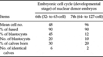
The efficiencies of ECNT in this study are summarized in Table 2. Although we did not determine the efficiency of ECNT using the fifth cell cycle (16- to 31-cell)-stage embryos (mean cell number: 24), which can be recovered from the uterus (Newcomb et al., Reference Newcomb, Rowson and Trounson1976), we could produce about 20 identical blastocysts if the rate of development of NT embryos to the blastocyst stage was about 80%. Calving rates for the fifth and sixth cell cycle (16- to 63-cell)-stage embryos were reported to be 10 to 26% (Bondioli, Reference Bondioli1993; Stice & Keefer, Reference Stice and Keefer1993; Zakhartchenko et al., Reference Zakhartchenko, Reichenbach, Riedl, Palma, Wolf and Brem1996; Bourhis et al., Reference Bourhis, Chesne, Nibart, Marchal, Humblot, Renard and Heyman1998). Thus, we could obtain about five calves at most (about 20 blastocysts × 0.26) for the fifth cell cycle (16- to 31-cell)-stage embryos, which was similar to the rate for the sixth cell cycle (32- to 63-cell)-stage embryos (Table 2). However, the rate of development to the blastocyst stage of NT embryos originating from the fifth cell cycle (16- to 31-cell)-stage embryos was reported to be lower than that of sixth cell cycle (32- to 63-cell)-stage embryos (Bondioli et al., Reference Bondioli, Westhusin and Looney1990). Blastocyst development of NT embryos reconstructed with inner cell mass cells of blastocysts was also lower than that with blastomeres of the sixth cell cycle (32- to 63-cell)-stage embryos (Zakhartchenko et al., Reference Zakhartchenko, Reichenbach, Riedl, Palma, Wolf and Brem1996). Hence, the present results indicate that sixth cell cycle (32- to 63-cell)-stage embryos yield the highest number of identical blastocysts (about 20/donor embryo) and calves (about six/donor embryo) in Japanese black cattle.
Efficiency of nuclear transfer using blastomeres of embryos at different embryonic cell cycles

Acknowledgements
We thank the Meat Inspection Offices of Hayakita and Kushiro for providing the materials used in this study.