


Congenital coronary anomalies are the second leading cause of sudden death in the young,Reference Maron, Haas, Ahluwalia, Murphy and Garberich1 with estimated prevalence varying from 0.02 to 0.7% of the population.Reference Angelini2 Anomalous aortic origin of a coronary artery from the opposite sinus of Valsalva with an intramural course is thought to be a high-risk lesion, particularly with anomalous left coronary artery.Reference Basso, Maron, Corrado and Thiene3,Reference Angelini, Velasco and Flamm4 Left main coronary artery atresia is exceedingly rare, with scattered case reports in the literature.Reference Yajima, Toda, Nishi, Yoshioka, Nakamura and Miyagawa5,Reference Sathanandam, Kumar, Boston and Facs6 We herein present a case of a patient with left coronary atresia diagnosed at 11 years of age with long-term follow-up and no exercise restrictions. This is the only patient in our cohort of 400 patients with this specific anomaly.
Case report
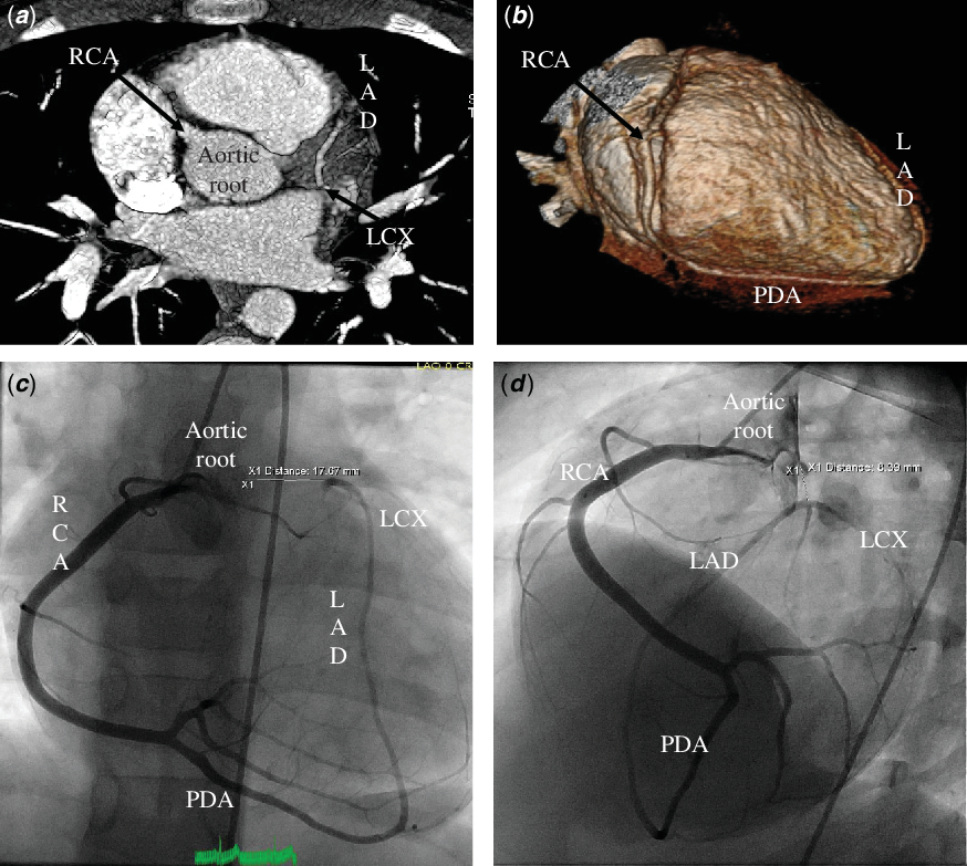
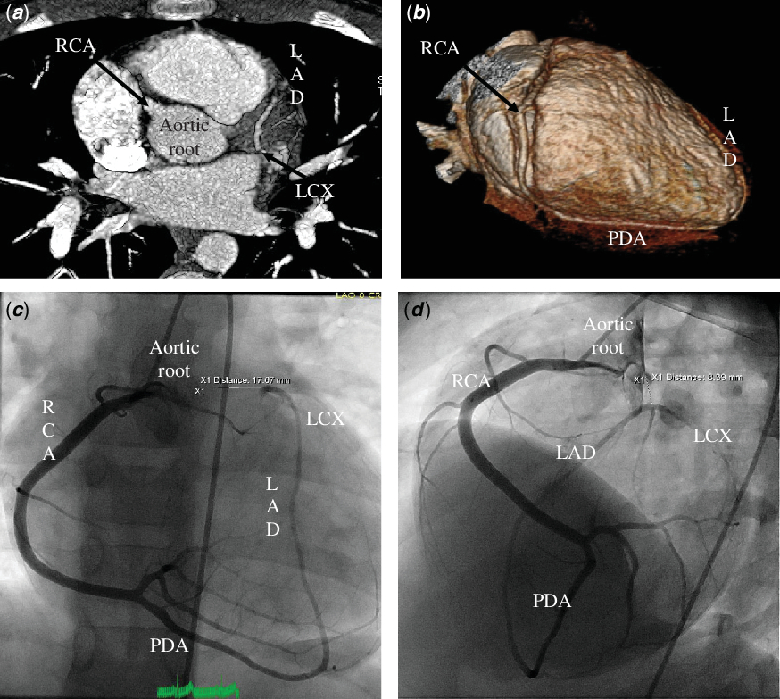
We report a 17-year old male who was diagnosed with left coronary artery atresia at the age of 11 years and has been followed by the Coronary Anomalies Program at Texas Children’s Hospital. The anomaly was detected during a workup for exertional chest pain. Upon presentation, the patient reported “stinging” chest pain with exercise only and resolving after 1–2 minutes of resting. It was not associated with dyspnoea or palpitations. No history of syncope or near syncope was reported. The patient had always been very active with excellent exercise endurance, participating in several sports, including swim team and practicing for 75 minutes 4 times a week. An echocardiogram performed failed to identify the left coronary artery. Therefore, a coronary CT scan and subsequently cardiac catheterisation with angiography confirmed the absence of the left coronary artery with retrograde filling of the left anterior descending and circumflex from the posterior descending coronary artery, which arises from the right coronary artery (Fig 1). A nuclear stress test with Sestamibi was negative for inducible myocardial ischemia.
Cardiac CT angiography shows no connection between the aortic root and left coronary artery ( a ). The right coronary artery (RCA) gives rise to the posterior descending coronary artery (PDA) with retrograde filling of the left anterior descending (LAD) ( b ). Selective angiography of the RCA shows retrograde filling of the LAD and circumflex coronary artery (LCX) from the PDA, which arises from the RCA on anteroposterior ( c ) and left anterior oblique (58o) and caudal (20o) projection ( d ).
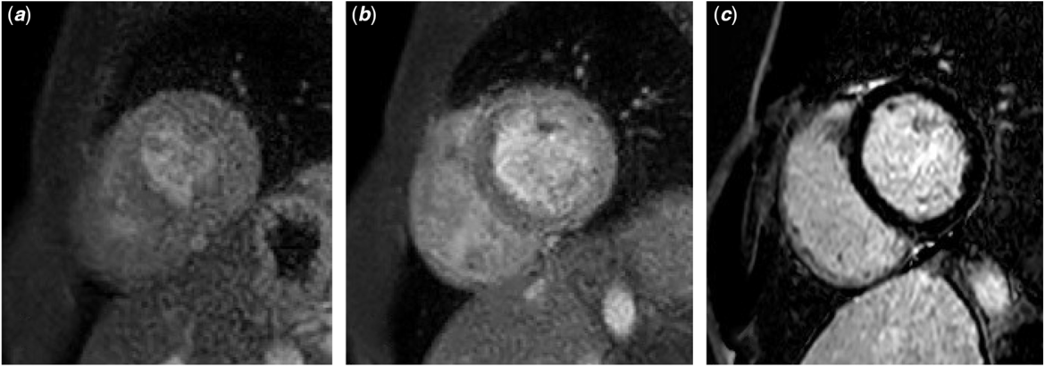
Given the lack of specific guidelines for the management of this rare entity, we reviewed all pertinent data with the multidisciplinary team in the Coronary Anomalies Program at our institution, including other experts at the national level. Chest pain was deemed to be unlikely related to myocardial ischemia. The risk of sudden cardiac arrest or death is unknown. Extensive discussion was entailed with the patient and his family regarding the lack of longitudinal data in patients with this anomaly, and shared decision-making process was exercised pertaining to physical activities, medical or surgical intervention. The shared decision was to allow him to participate fully in all physical activities and competitive sports. The family decided to obtain an automated external defibrillator and carry with them to every sport event, unbeknown to the patient initially when he was younger. Except for a vasovagal syncopal event at 15 years of age during a church service, he remained asymptomatic during exertion. On his most recent follow-up, 6 years after his initial diagnosis, he underwent repeat myocardial functional studies. A cardiopulmonary exercise stress test was normal, with the patient exercising for 18 minutes (endurance at 100th percentile for age and gender) following the Bruce protocol, work level of maximal metabolic equivalent of task 24.1, and maximum oxygen consumption of 58.4 ml/kg/minute (118% predicted). A vasodilator stress cardiac MRI using regadenoson was also performed which showed no evidence of hypoperfusion either at rest or during provocative coronary vasodilation (Fig 2).
Regadenoson stress cardiac MRI shows no evidence of myocardial hypoperfusion (a,b), and myocardial viability sequence shows no late gadolinium enhancement ( c).

Acknowledgements
We would like to thank all members of the multidisciplinary team at the Coronary Anomalies Program at Texas Children's Hospital, Houston, USA for their insights in the management of children with rare coronary artery anomalies.
Financial Support
This research received no specific grant from any funding agency, commercial or not-for-profit sectors.
Conflicts of Interest
None.
Ethical Standards
The authors assert that all testing performed on this patient were clinically indicated and comply with the ethical standards of the relevant national guidelines on human experimentation and with the Helsinki Declaration of 1975, as revised in 2008. An informed consent was not obtained for the case report.



Comment
Left main coronary artery atresia is an exceedingly rare condition with sparse case reports in the literature. Its presentation includes variable manifestations of myocardial ischemia from early infancy to late adulthood.Reference Yajima, Toda, Nishi, Yoshioka, Nakamura and Miyagawa5–Reference Pandya, Venugopal, Wildschut and Alphonso7 Cases reported include those with evident ischemia and variable approaches to surgical intervention. Best management of these patients is uncertain given lack of longitudinal follow-up and determination of the denominator to define those who are and those who are not at risk.
To our knowledge, this is the first report of a patient with left coronary artery atresia who has continued to participate fully in athletic activities, with long-term follow-up and myocardial functional assessment. Physical activity and engagement in sports are extremely beneficial not only from a cardiovascular health perspective but also importantly for peer relations during puberty and adolescence. The true risk of sudden cardiac death or arrest in these patients is unknown and, thus, it makes it difficult to impose exercise restrictions in these patients. Furthermore, revascularisation with coronary artery bypass in the youth comes with morbidity and uncertain freedom from re-intervention or ischemic events over time.Reference Yajima, Toda, Nishi, Yoshioka, Nakamura and Miyagawa5,Reference Sathanandam, Kumar, Boston and Facs6 It is critically important that all objective data and current knowledge about the child’s specific coronary artery anomaly is shared with the patient and family in a transparent manner. Currently, at 17 years of age, the patient is well aware of his condition and the decision-making of continued exercise and clinical follow-up is endorsed by him and his family.
Tomanek and Angelini recently published a comprehensive review on embryology, anatomy, and pathophysiology of coronary anomalies. They stated the term “atresia” of a coronary artery when there is incomplete connection to the sinus of Valsalva and a “cul de sac” is seen on angiography.Reference Tomanek and Angelini8 In the case we report, this finding was not evident. The left coronary artery system was fed by the right dominant coronary system, with minimal change in caliber of the connecting vessels. Intriguingly, this could also be considered as an anomalous origin of the left coronary artery supplied by the right coronary artery.
Risk stratification of this rare entity to predict myocardial ischemia remains to be established. Our patient has been free of events for 6 years with full athletic participation. His symptoms upon presentation were not deemed related to angina, and various studies performed under provocative stress to evaluate myocardial perfusion have been negative. Awareness of high-risk conditions with clear discussion of the many unknowns is essential for shared decision-making to drive management, especially if surgical intervention is entertained. The use of automatic external defibrillator is essential for secondary prevention. It is unclear, at this time, if recommendation should be given to families affected with congenital coronary anomalies to carry a defibrillator at all times or during sporting events/exercise activities and if this measure will impact the occurrence of sudden cardiac arrest and/or death.