I. INTRODUCTION
Magnesium aluminum (MgAl2O4) spinel is considered to be an important ceramic material, and it possesses attractive features such as high mechanical strength, good chemical stability, high thermal shock resistance, and low electrical losses.Reference Tokariev, Schnetter, Beck and Malzbender1–Reference Rothman, Kalabukhov, Sverdlov, Dariel and Frage3 It also has apparent optical properties, which are similar to those of glass, and it has been applied in lighting and laser technologies.Reference Morita, Kim, Yoshida and Hiraga4–Reference Ikesue and Aung6 MgAl2O4 spinel ceramic is one of the main materials used in military and space applications because of its simple synthesis, excellent mechanical properties, and transparency over a wide range of 0.2–6 μm.Reference Roy and Hastert7–Reference Tokariev, Steinbrech, Schnetter and Malzbender9 The potential applications of MgAl2O4 spinel ceramic materials are therefore vast. Ceramic powder preparation is fundamental in the synthesis and application of ceramic materials. MgAl2O4 nanoparticles with a narrow size distribution and a low degree of agglomeration are desirable in these applications.
Various approaches have been used to synthesize disperse MgAl2O4 spinel nanoparticles, such as solid-state reaction, co-precipitation, and sol–gel syntheses.Reference Ganesh, Srinivas, Johnson, Saha and Mahajan10–Reference Ye and Troczynski13 Among these, the solid-state reaction route is suitable for producing nanoparticles on a large-scale, but it usually results in particle coarsening because of high reaction temperature used. The co-precipitation route is simple, economical, and suitable for industrial production, but the as-prepared particles are agglomerated sometimes. Ultrafine particles can be obtained by the sol–gel route, but partial sintering occurs. The sintering, coarsening, and hard agglomeration of particles are unfavorable for their performance and application, especially in ceramic green body densification during sintering.Reference Goldstein14,Reference Reimanis and Kleebe15 The simple synthesis of well-dispersed MgAl2O4 spinel nanoparticles therefore still poses a challenge.
In this paper, we report a simple method to synthesize well-dispersed MgAl2O4 spinel nanoparticles. The method is based on the concept of isolation-medium-assisted calcination.Reference Du, Zhao, Liu, Li, Chen and Cui16 In the process, excess magnesium salt than its stoichiometry is introduced during precursor preparation, and a large excess of MgO is formed in the precursor mixture during calcination. A small amount of MgO acts as the reactant to synthesize target product via a solid-state reaction, while most of MgO acts as the isolation medium to separate the spinel particles as they are nucleated and to prevent their agglomeration and coarsening during calcination. The results indicate that using MgO as the isolation medium to assist calcination can prevent the agglomeration and contamination of MgAl2O4 nanoparticles. This study uses a modified sol–gel method to synthesize well-dispersed MgAl2O4 spinel nanoparticles. The changes in crystalline structure, crystallite size, specific surface area, and particle morphology were studied.
II. EXPERIMENTAL PROCEDURE
A. Sample preparation
A solution was prepared from aluminum nitrate (Al(NO3)3•9H2O) and magnesium nitrate (Mg(NO3)2•6H2O) dissolved in deionized water. The concentration of Al3+ ions was adjusted to 0.005 M and the Mg/Al atomic ratio was 20. Acrylamide (C3H5NO) and N,N′-methylenebisacrylamide (C7H11N2O2) were used as the polymer monomer and crosslinker, respectively, and added to the solution. The molar ratios of polymer monomer to Al3+ ions and monomer to crosslinker were adjusted to 40 and 25, respectively. The mixture was stirred at room temperature until the polymer network agent dissolved. Ammonium persulfate [(NH4)2S2O8] used as the initiator was introduced into the solution to promote the polymerization reaction of a network agent. The molar ratio of the polymer monomer to the initiator was adjusted to 20. The solution was stirred until an ivory polymer gel was formed. The precursor, a metal ion polymer complex, was obtained after drying the polymer gel at 80 °C. The precursors were calcined at 500–1000 °C, and the calcined products were washed once with 3 M dilute hydrochloric acid and twice with deionized water to obtain the samples. All the chemical reagents were purchased from Sinopharm Chemical Reagent Co., Ltd, China.
B. Characterization
The thermal behavior of the precursor was examined by thermogravimetric-differential thermal analysis (TG-DTA) on a Netzsch STA 449C thermal analyzer (Wald-kraiburg, Germany) at a heating rate of 10 °C/min. Crystal structures of the samples were analyzed by x-ray diffraction (XRD) on a Rigaku D/max-2400 diffractometer (Tokyo, Japan) using nickel filtered Cu Kα radiation in the 2θ range of 10°–80°. Crystallinity degree of the sample was estimated quantitatively from the relative areas of crystalline and amorphous regions from the diffraction pattern after drawing a smooth curve with the help of MDI Jade 5 x-ray data analysis program.Reference Vu, Haber, Gocmez, Swab, Halbig and Mathur17 The average crystal size (D XRD) of MgAl2O4 nanoparticles was calculated using the Scherrer formula: 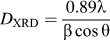 ${D_{{\rm{XRD}}}} = {{0.89\lambda } \over {\beta \cos \theta }}$, where λ is the radiation wave length (0.15406 nm), β is the full width at half maximum of the diffraction peaks, and θ is the Bragg angle. The specific surface areas of the samples were determined by N2 adsorption–desorption isotherms using an ASAP 2010 Micrometrics, and the equivalent average particle size (D BET) was calculated via the formula:
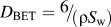 $D_{{\rm{BET}}} = {\raise0.7ex\hbox{$6$} \!\mathord{\left/ {\vphantom {6 {\left( {\rho S_{\rm{w}} } \right)}}}\right.\kern-\nulldelimiterspace}\!\lower0.7ex\hbox{${\left( {\rho S_{\rm{w}} } \right)}$}}$, where ρ = 3.55 g/cm3 is the theoretical density for MgAl2O4 spinel and S w is the specific surface area of the sample. The infrared spectra of the samples were measured by a Nicolet NEXUS 670 Fourier transform infrared spectrometer (FTIR; Thermo Scientific, Waltham, MA) in the range of 400–4000 cm−1 to identify the nature of bonding in the samples. The microstructures of the samples and the sinter were analyzed by a scanning electron microscope (SEM, Hitachi S-4800, Japan) and a transmission electron microscope (TEM, Tecnai G2 F30, Hillsboro, OR). The particle size distribution was calculated from the TEM micrograph using Gatan Digital Micrograph analysis software. The relative density of the sintered spinel pellet was evaluated using the Archimedes method.
III. RESULTS AND DISCUSSIONS
The TG-DTA curves for the as-prepared precursor are shown in Fig. 1. The DTA curve shows one endothermic and three exothermic events, and the corresponding TG curve shows three mass losses. A broad endothermic peak ranging from 60 to 240 °C is accompanied by a mass loss of 16.1%, and is attributed to the removal of free water and hydrate water.Reference Lee, Suematsu, Yano and Yatsui18 The sharp exothermic peak at 275 °C, accompanied by a mass loss of 19.4%, is attributed to the oxidation of residual nitrate ions in the precursor.Reference Naskar and Chatterjee19 It is well known that the degradation of polyacrylamide network is a multistep process and is completed at 300–600 °C under the air atmosphere.Reference Lee, Suematsu, Yano and Yatsui18,Reference Tahmasebpour, Babaluo, Shafiei and Pipelzadeh20,Reference Li, Sun, Chen, Cui and Ren21 The broad exothermic peaks centered at 376 °C, accompanied by a mass loss of 29%, are assigned to a major decomposition of polymer.Reference Lee, Suematsu, Yano and Yatsui18,Reference Tahmasebpour, Babaluo, Shafiei and Pipelzadeh20,Reference Li, Sun, Chen, Cui and Ren21 The strong exothermic peak at 608 °C, accompanied by a mass loss of 18.6%, can be attributed to the thermal overlap of the crystallization transition, in which the amorphous oxide becomes nanocrystallite and the lattice energy is released, and burning and oxidation of residual polymer.Reference Li, Sun, Chen, Cui and Ren21,Reference Pati and Pramanik22 A weak endothermic peak at 650–800 °C with no obvious mass losses is due to the solid-state reaction of Al2O3 to MgO and the formation of MgAl2O4 grains.Reference Li, Sun, Chen, Cui and Ren21 The weak exothermic effect occurs above 800 °C, accompanied by a slight weight loss of 2.3%, is associated with the final spinel crystallization step and the oxidation of the residue carbon.Reference Lee, Suematsu, Yano and Yatsui18,Reference Montouillout, Massiot, Douy and Couturesl23
TG-DTA curves of the as-prepared spinel precursor.

Figure 2 shows XRD spectra of the samples obtained by calcining the precursors at different temperatures for 2 h before [Fig. 2(a)] and after [Fig. 2(b)] washing with dilute hydrochloric acid and water. The XRD spectra of calcined products at 500–900 °C [Fig. 2(a)] display sharp and strong diffraction peaks mostly due to cubic MgO species (space group Fm−3m, JCPDS No. 43-1022). Due to a gradual degradation of the metal ion polymer complex, some diffraction peaks related to the magnesium sulfate complex are observed at 500–800 °C. After calcination at 900 °C, both cubic MgO and MgAl2O4 spinel are detected. From Fig. 2(a), it can be seen that a large amount of cubic MgO in the calcined product is present. After washing with dilute hydrochloric acid and water, XRD spectra of the samples are shown in Fig. 2(b). The diffuse peaks of the sample prepared at 500 °C indicate the formation of an amorphous phase. The major diffraction peaks of MgAl2O4 spinel at 2θ = 31.27° for (220), 36.85° for (311), 44.83° for (400), 59.37° for (511), and 65.24° for (440) appear in the XRD spectrum of the sample prepared at 700 °C. This indicated that MgAl2O4 spinel was formed after 700 °C calcination, and it is consistent with the DTA result (the endothermic peak at 650–800 °C in Fig. 1). These MgAl2O4 diffraction peaks gradually become stronger with increasing calcination temperature up to 800 °C. After calcination at 900 °C, the diffraction spectrum of the sample shows the characteristic diffraction peaks of cubic MgAl2O4 (JCPDS No. 21-1152). No residual MgO impurity is detected, although this compound is observed in other methods.Reference Su, Du, Li and Li24
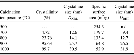
The degree of crystallinity, specific surface area, and crystalline size of the sample obtained at different temperatures are shown in Table I. The degree of crystallinity and the crystalline size increase with increasing calcination temperatures from 700 to 1000 °C. After calcining at 900 °C, an almost pure MgAl2O4 spinel phase is obtained. The crystalline size of the spinel sample increases slowly with increasing calcination temperatures from 900 to 1000 °C, and it can be confirmed by the XRD pattern [Fig. 2(b)], in which there is no obvious sharpening of diffraction peaks with the increase in calcination temperature. This can be attributed to the isolation effect of isolating medium.
Degree of crystallinity, specific surface area, and crystalline size (D XRD and D BET) of spinel samples obtained at different temperatures.
FTIR spectra of the precursor and the samples obtained at different temperatures are shown in Fig. 3. In all curves, the absorption bands at ≈3450 and 1630 cm−1 are observed. These are related to O–H stretching vibration and H2O deformation vibration, respectively, and can be attributed to the presence of a hydroxyl group in polymeric gel and adsorption water from air by nanosized samples with high surface area.Reference Silverstein and Webster25 With an increase in calcination temperature, the intensities of these absorption bands gradually weakened. This is simply attributed to the decrease in the surface area of the powder. The presence of NO3− is evidenced by the absorption bands at 1380 and 830 cm−1 (Ref. Reference Xian, Yang, Shen, Jiang, Wei and Feng26). The bands disappear with an increase in calcination temperature up to 700 °C, because of a complete oxidation of nitrate ions. The absorption bands at 1000–1300 cm−1 are consistent with stretching vibrations of C–O–C polymer groups.Reference Tahmasebpour, Babaluo, Shafiei and Pipelzadeh20 These bands disappear gradually with an increase in calcination temperature. Below 1000 cm−1, the absorption bands are related to the Al–O inorganic network. For the six-coordinated AlO6 groups, Al–O stretching and bending vibrations are expected at 500–700 and 330–450 cm−1 (Ref. Reference Kurajica, Tkalcec, Sipusic, Matijasic and Brnardic27). For the four-coordinated AlO4 groups, Al–O stretching and bending modes are expected at 700–850 and 250–320 cm−1 (Ref. Reference Kurajica, Tkalcec, Sipusic, Matijasic and Brnardic27). In curves A and B of Fig. 3, the absorption bands at 3450, 550, and 600 cm−1 are relatively broad. This can be attributed to continuous distribution of the bond length in the amorphous structure and to various distortions in the polymer-gel precursor.Reference Tahmasebpour, Babaluo, Shafiei and Pipelzadeh20,Reference Silverstein and Webster25,Reference Du, Wang, Su and Li28 In curves B and C, the two strong absorption bands at 530 and 690 cm−1 are related to the AlO6, inorganic network groups. These build up the spinel structure and therefore indicate the formation of MgAl2O4 spinel in calcined samples.Reference Tahmasebpour, Babaluo, Shafiei and Pipelzadeh20,Reference Xian, Yang, Shen, Jiang, Wei and Feng26,Reference Kurajica, Tkalcec, Sipusic, Matijasic and Brnardic27,Reference Kloprogge, Hickey and Frost29 The FTIR spectra of the products are similar in spite of different synthesized methods.
FTIR spectra of the polymeric gel precursor (a) and spinel samples obtained at different temperatures: 500 °C (b), 700 °C (c), and 1000 °C (d).
The morphology and microstructure of as-prepared MgAl2O4 spinel at 1000 °C were investigated by SEM and TEM, respectively, as shown in Fig. 4. The low-magnification and panoramic views from SEM and TEM images [Figs. 4(a) and 4(b)] show the uniformity of synthesized products, and no hard agglomerates were visible in the TEM micrograph [Fig. 4(b)]. This indicates that no undesirable agglomeration of MgAl2O4 nanoparticles occurred during the preparation process. The selected area electron diffraction (SAED) pattern of a selected area (A) in Fig. 4(b) is shown in Fig. 4(c), and it exhibits six clear and one obscure diffraction rings with d-spacings about 0.4609, 0.2881, 0.2505, 0.2075, 0.1599, 0.1457, and 0.1679 nm, which could be attributed to (111), (220), (311), (400), (511), (440), and (422) reflections of cubic MgAl2O4 spinel structure, respectively. The particle size distribution of MgAl2O4 nanoparticles [in Fig. 4(b)] is shown in Fig. 4(d). The histograms for particle size distribution and mean particle sizes in Fig. 4(d) were determined from 280 samplings of spinel particles in the sample, using Digital Micrograph version 3.4.3 distributed by Gatan.Reference Park, Choi, Kim and Ryoo30 The average particle size is ≈35 nm, which is similar to the results as calculated from XRD and BET data. The magnified TEM image in Fig. 4(e) shows that the as-prepared MgAl2O4 nanoparticles have a polyhedral morphology and are 22–52 nm in diameter. The corresponding high-resolution TEM (HRTEM) micrograph is shown in Fig. 4(f). The spacing between two lattice planes is 0.246 nm, which can be ascribed to the (311) crystal planes of MgAl2O4 spinel. It indicates the single-crystal nature of formed MgAl2O4 nanoparticles. From Fig. 4, it can be seen that well-dispersed MgAl2O4 single-crystal nanoparticles with narrow particle size distribution are obtained. The polymer-gel and isolation-medium-assisted calcination can prevent particle contact and reduce agglomeration.
Low-magnification (a) SEM and (b) TEM images for panoramic views, (c) SAED pattern, (d) particle size distribution, (e) high-magnification TEM image, and (f) HRTEM image of as-prepared MgAl2O4 nanoparticles at 1000 °C.
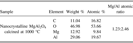
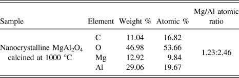
The chemical stoichiometry of the as-prepared spinel sample at 1000 °C was investigated by EDS. The result (Table II) shows that the sample consists of O, Al, and Mg. C is also present as the major component of the carbon conductive tape. The Mg/Al atomic ratio of the as-prepared sample is ≈1.23:2.46, and this result confirms the formation of a pure MgAl2O4 spinel phase.
EDS analysis data of as-prepared spinel samples at 1000 °C.
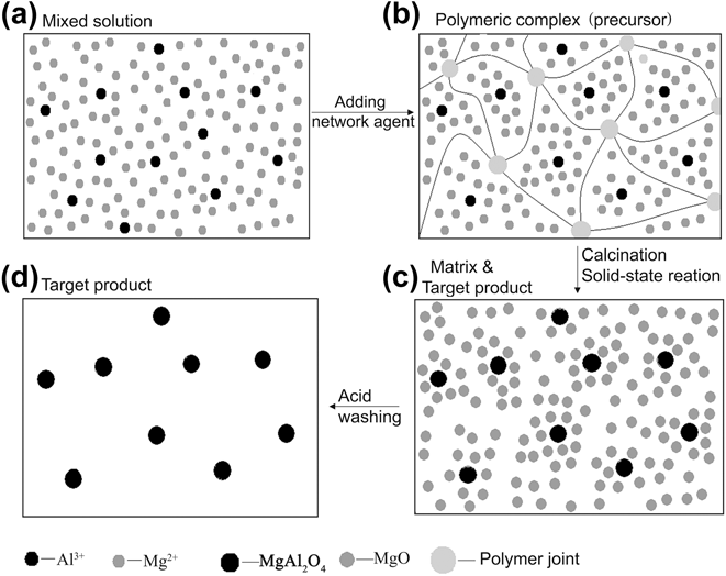
Based on the above results, the formation mechanism of well-dispersed MgAl2O4 nanoparticles can be explained as follows. A rough schematic diagram is shown in Fig. 5 to illustrate the formation of well-dispersed MgAl2O4 nanoparticles via polymer-gel and isolation-medium-assisted calcination. As shown in Fig. 5, a three-dimensional network structure formed by additional polymer network agents is used to disperse metal ions into three-dimensional grids uniformly, and the movement of Mg2+ and Al3+ ions in the mixed system is limited because of the formation of polymeric gel. It ensures that the opportunity for contacting and congregation of particles is reduced significantly during drying and the initial calcination process. During calcining, MgAl2O4 nanoparticles are fabricated in situ by a solid-state reaction of MgO and Al2O3 oxides, which are formed from precursor decomposition. Due to a large excess of magnesium nitrate introduced into the precursor, a large excess of MgO is formed after initial calcination. A large excess of MgO, 40 times the stoichiometric amount of spinel, separate the spinel particles as they are nucleated to prevent their agglomeration and coarsening during calcination. After calcining, excess MgO acting as the isolation medium is removed from calcined product by acid washing; and then well-dispersed MgAl2O4 nanoparticles are obtained. It is well known that a solid-state reaction for preparing MgAl2O4 nanoparticles always causes agglomeration because of the contact and growth of ultrafine particles.Reference Su, Du, Li and Li24 In this work, in the solid-state reaction system, excess MgO acts not only as a reactant but also as the isolation medium. It surrounds fresh MgAl2O4 grains and prevents the contact and growth of MgAl2O4 nanoparticles, which is caused by thermal aggregation of MgAl2O4 grains. After acid washing with 3 M dilute hydrochloric acid, a large excess of MgO is removed from the samples, and MgAl2O4 particles are independent. Furthermore, excess MgO as the isolation medium also prevents the contamination of MgAl2O4 nanocrystalline by introducing an isolation medium during the preparation process. High purity MgAl2O4 nanoparticles with narrow size distribution were therefore prepared.
A rough schematic diagram of a preparation process of well-dispersed MgAl2O4 nanoparticles.
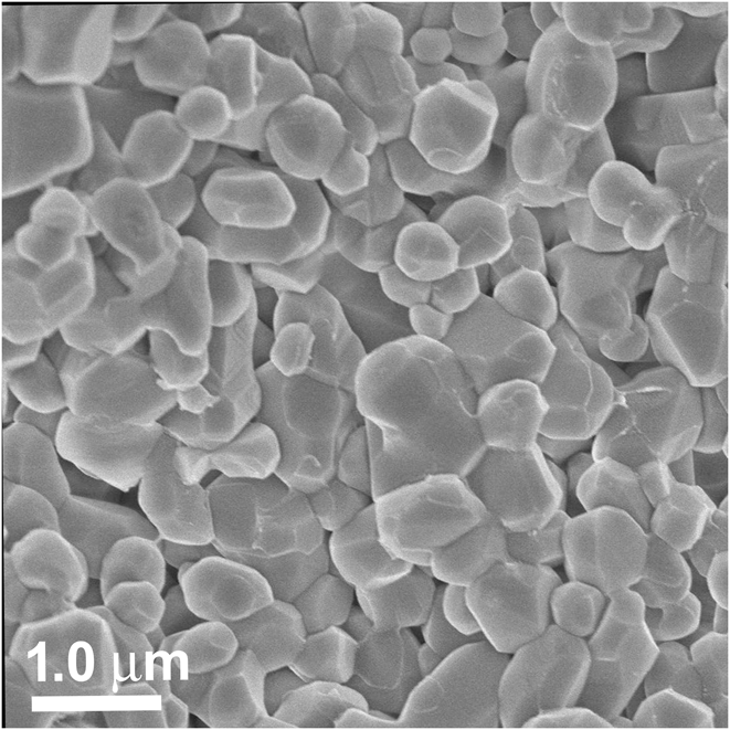
The sintering property of MgAl2O4 spinel nanoparticles with an average size of 35 nm was investigated. The sample was pressed into a disk (10 mm diameter, 1–2 mm thick) at 1100 MPa, and sintered by two-step sintering (at 1410 °C for 1 h and 1350 °C for 20 h) in a box furnace in air. The microstructure of as-sintered MgAl2O4 spinel ceramic is shown in the SEM micrograph of Fig. 6. The sintered spinel ceramic has a relative density of 95.2%, as determined by the Archimedes method. Equiaxial grains with clear grain boundaries and polyhedral morphologies are visible in the SEM micrograph. The average grain size is ≈317 nm, which is much smaller than that reported in the literature.Reference Tahmasebpour, Babaluo, Shafiei and Pipelzadeh20,Reference Li, Ikegami, Lee, Mori and Yajima31 This should be attributed to its traits such as nonagglomeration, high specific surface area, and narrow particle size distribution. The sintering schedule is the important factors in affecting the final sinter microstructure.
SEM image of MgAl2O4 spinel ceramics from starting MgAl2O4 nanopowders with an average particle size of 35 nm, pressed at 1100 MPa, and sintered at 1410 °C for 1 h then 1350 °C for 20 h.
IV. CONCLUSION
Well-dispersed MgAl2O4 spinel nanoparticles were synthesized by a polymer-gel and isolation-medium-assisted calcination method. MgAl2O4 nanoparticles are formed in situ by a solid-state reaction of MgO and Al2O3 formed from decomposition of the precursor. Using excess MgO substrate in the solid-state reaction system as an isolation medium prevents the contact and growth of fresh MgAl2O4 nanoparticles and ensures the production of a product of high purity. TEM and HRTEM results have confirmed that well-dispersed MgAl2O4 spinel nanoparticles are single crystals with an average particle size of 35 nm and a narrow particle size distribution.
ACKNOWLEDGMENTS
This work was supported by National Natural Science Foundation of China (11074160 and 221102087), Henan Provincial Natural Science Foundation (122102210486, 142102210481, and 142102210573), and Henan Provincial Educational Committee program (13A140824).