Case report
A 29-year-old primigravida woman was referred to a tertiary care centre at 18 weeks of gestation because of fetal bradycardia, and the fetus was diagnosed with complete atrioventricular block with an atrial rate of 150 bpm and a ventricular rate of 50 bpm. Although intravenous isoproterenol therapy was commenced at 21 weeks of gestation, the fetal ventricular rate did not show any increase. At 27 weeks of gestation, she was referred to our institution for pacemaker therapy.
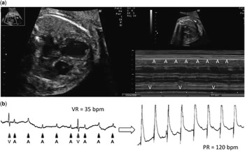
Initial fetal echocardiography at our institution revealed complete atrioventricular block with an atrial and ventricular rate of 152 and 48 bpm, respectively, cardiomegaly, a small amount of pleural effusion, and ascites. However, subcutaneous oedema was not observed. The four-chamber view showed a relatively small right ventricle with diffuse endocardial fibroelastosis and moderate hypokinesis, suggesting the association with myocarditis, and a dilated left ventricle (Fig 1a). The blood test of the mother showed elevated levels of anti-SSA/Ro antibodies (>240 U/ml; normal,<10 U/ml), anti-52 kDa SSA/Ro antibodies (165.4 index; normal,<5.1 index), and anti-60 kDa SSA/Ro antibodies (144.4 index; normal,<5.9 index). Because of the prematurity of the fetus in addition to the severity of atrioventricular block, we thought an early delivery and pacemaker therapy at this gestational age would be very risky. We thus decided to administer maternal oral dexamethasone therapy (4 mg/day) to prevent further progression of the fetal myocarditis. At 28 weeks of gestation, echocardiography revealed no sign of improvement in fetal cardiac function, and subcutaneous oedema was observed, suggesting the deterioration of the hydrops. We thought that the fetus would not be able to tolerate the haemodynamic instability any longer, and determined to deliver the baby via caesarean section and place temporary pacing leads immediately after birth.
( a ) Four-chamber view (4CV) and M-mode of the fetal echocardiograms (ECG) at 27 weeks of gestation. The 4CV shows a relatively small right ventricle with endocardial fibroelastosis, dilated left ventricle, cardiomegaly (cardiothoracic area ratio, 48%), and plural effusion. The M-mode showed congenital complete atrioventricular block with an atrial rate of 152 bpm and a ventricular rate of 48 bpm. ( b ) The postnatal ECG monitor of the patient before (left) and after (right) temporary pacing lead placement.

At 29+2 weeks of gestation, a male infant with a birth weight of 1558 g and Apgar scores of 3 (1 minute) and 4 (5 minutes) was born. Electrocardiography revealed complete atrioventricular block with a narrow QRS at 35 bpm (Fig 1b). Soon after birth, a temporary pacing lead was placed on the surface of the right ventricle, and the pacing was started at 120 bpm at 20 minutes after birth (Fig 1b). Once the right ventricular pacing was established, another pacing lead was inserted close to the apex because we were concerned about increase in the threshold of pacing owing to myocarditis and about dyssynchrony and its negative effect on the function of the left ventricle in the long term.
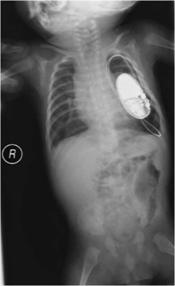
Echocardiography performed after the operation revealed no structural defect, except for a rather small right ventricle, and endocardial fibroelastosis mainly affecting the right ventricle. To prevent further damage to the myocardium due to myocarditis, prednisolone (1 mg/kg/day for 2 weeks) and intravenous immunoglobulin therapy (1 g/day for 3 days) were administered. Prednisolone was tapered gradually and discontinued at 1 month after birth. On day 8, the pacing threshold suddenly increased and significant accumulation of pericardial effusion was detected. Although the pacing threshold decreased immediately after drainage, the pacing had become unstable after this event, as a result of which, although the infant’s body weight was still 2020 g, permanent pacemaker implantation was performed on day 64. A generator (ADAPTA SR01, Medtronic INC., Minneapolis, Minnesota, USA) was implanted in the intrathoracic cavity with the epicardial ventricular lead (CapSure Epi unipolar, Medtronic INC., Minneapolis, Minnesota, USA) inserted in the apex of the left ventricle (Fig 2). The initial mode of pacemaker was VVI at a rate of 120 bpm. After pacemaker implantation, pacing failure was not detected, and the infant was discharged on day 120. Echocardiography before discharge revealed growth of the right ventricular cavity and good systolic function of both ventricles.
Chest radiography after permanent pacemaker implantation. The generator was implanted in the intrathoracic cavity with the epicardial ventricular lead inserted in the apex of the left ventricle.

Discussion
Recently, the presence of anti-SSA/Ro antibody was associated with congenital complete atrioventricular block, as well as with other cardiac manifestations such as fibrosis of the sinoatrial node and bundle of His, endocardial fibroelastosis, and valve damage.Reference Llanos, Friedman and Saxena 1 In cases of congenital complete atrioventricular block associated with hydrops, extremely low ventricular rate, and prematurity, the mortality has been reported to exceed 80%.Reference Balmer 2 – Reference Jaeggi, Hamilton, Silverman, Zamora and Hornberger 4 It has recently been reported that the presence of myocarditis, endocardial fibroelastosis, maternal diagnosis of systemic lupus erythematosus, and/or Sjogren syndrome, gestational age less than 20 weeks, ventricular rate <50 bpm, fetal hydrops, and impaired function of the left ventricle at diagnosis have been revealed as risk factors for prenatal and postnatal demise in children with cardiac neonatal lupus and fetuses with second- or third-degree atrioventricular block.Reference Eliasson, Sonesson and Sharland 5 , Reference Izmirly, Saxena and Kim 6 In the present case, although the prognosis was expected to be poor, we decided to perform planned caesarean section and temporary pacemaker implantation immediately after birth to rescue the fetus in accordance with the wishes of the parents.
In recent times, the indications for pacemaker therapy in premature and low-birth-weight infants with complete atrioventricular block have been expanded. However, treatment of premature infants, especially those born at less than 30 weeks of gestation and those who have hydrops, remains a challenge. Both staged and primary permanent pacemaker implantation for atrioventricular block has been frequently reported, but which of the two is better for high-risk patients is controversial.Reference Glatz, Gaynor and Rhodes 7 , Reference Shepard, Kochilas and Vinocur 8 Glantz et al assessed the outcome of 13 high-risk patients who required pacemaker treatment within 24 hours after birth. They concluded that the staged approach is feasible as a bridge to a permanent pacemaker implantation in high-risk patients with structurally normal hearts (survival rate, 83%).Reference Glatz, Gaynor and Rhodes 7 On the other hand, Shepard et alReference Shepard, Kochilas and Vinocur 8 reported the outcome of 10 patients with birth weight <1.5 kg who required early pacemaker implantation. In the study, they revealed that the mortality was the same as that reported by Glantz et al, without any immediate complications related to pacemaker therapy, and concluded that the staged approach was not superior to the primary permanent pacemaker implantation in terms of prognosis, but it prolonged the length of stay in the intensive care service.Reference Glatz, Gaynor and Rhodes 7 , Reference Shepard, Kochilas and Vinocur 8 Although it is difficult to conclude which approach is superior to treat high-risk patients, in our case the staged approach was successful.
In the present case, not only pacemaker therapy but also maternal dexamethasone therapy followed by postnatal high-dose immunoglobulin and prednisolone therapy was administered. Although it has been thought that there is no effective treatment for complete atrioventricular block, several reports have suggested that transplacental dexamethasone therapy may improve the prognosis of patients. In addition to pacemaker therapy, the suppression of sustained inflammation of the fetal myocardium caused by maternal autoimmune antibodies might be important to prevent the worsening of the myocardial damage.Reference Jaeggi, Fouron, Silverman, Ryan, Smallhorn and Hornberger 9 In this case, the echocardiography before discharge revealed good systolic function of both ventricles, and that might suggest the improvement in myocardial damage by postnatal combination therapy using steroid and immunoglobulin.
Acknowledgements
The authors acknowledge Dr. Tomizo Nishiguchi, Dr. Kisaburo Sakamoto, and Dr. Yasuo Ono, for their help with this report.
Financial Support
This research received no specific grant from any funding agency, commercial, or not-for-profit sectors.
Conflicts of Interest
None.
Ethical Standards
The authors assert that ethical standards of the relevant national guidelines on care were observed while evaluating the case.