INTRODUCTION
Efforts to develop an effective vaccine against malaria, which kills 2–3 million African children each year, would be greatly aided by universal recognition of the host–parasite interactions, especially the parasite immune evasion mechanisms. Their complicated and delicate evasion mechanisms include passive avoidance strategies, such as intracellular parasitism, antigenic variations and diversity, and active immune suppression (Hisaeda et al. Reference Hisaeda, Yasutomo and Himeno2005). The latter includes induction of T cell function failure (Xu et al. Reference Xu, Wipasa, Yan, Zeng, Makobongo, Finkelman, Kelso and Good2002), immunosuppressive regulatory T cells (Hisaeda et al. Reference Hisaeda, Maekawa, Iwakawa, Okada, Himeno, Kishihara, Tsukumo and Yasutomo2004), or bone marrow dysfunction (Helleberg et al. Reference Helleberg, Goka, Akanmori, Obeng-Adjei, Rodriquies and Kurtzhals2005).
Dendritic cells (DCs) are antigen-presenting cells that play a central role in both innate and adaptive immune responses. Several pathogens interfere with the host immune response by targeting different functions of DCs, including their maturation and migration (Rescigno et al. Reference Rescigno and Borrow2001). Erythrocytes infected with several species of Plasmodium activate DCs in terms of their maturation and T cell stimulation (Ing et al. Reference Ing, Segura, Thawani, Tam and Stevenson2006), but Plasmodium falciparum- and Plasmodium yoelii-infected erythrocytes also suppress DC maturation and T cell responses (Urban et al. Reference Urban, Ferguson, Pain, Willcox, Plebanski, Austyn and Roberts1999; Ocana-Mongner et al. Reference Ocana-Mongner, Mota and Rodriguez2003; Millington et al. Reference Millington, Di Lorenzo, Phillips, Garside and Brewer2006), suggesting a role for DCs in malaria-induced immune suppression.
A series of reports has shown that activated catabolism of tryptophan in DCs induces peripheral immune tolerance, such as feto-maternal tolerance (Munn et al. Reference Munn, Zhou, Attwood, Bondarev, Conway, Marshall, Brown and Mellor1998), tumour growing microenvironment (Munn et al. Reference Munn, Sharma, Hou, Baban, Lee, Antonia, Messina, Chandler, Koni and Mellor2004), and prevention of autoimmune diseases (Sakurai et al. Reference Sakurai, Zou, Tschetter, Ward and Shearer2002; Gurtner et al. Reference Gurtner, Newberry, Schloemann, McDonald and Stenson2003). Activated catabolism of tryptophan, an essential amino acid for mammals, is mediated by the inducible rate-limiting enzyme indoleamine 2,3-dioxygenase (IDO), which is also reported to play various roles in host–pathogen relationships. Activated IDO reduces tryptophan and increases its metabolites, including kynurenine, which might have an important role in antimicrobial activity, especially against microbes unable to synthesize tryptophan. On the other hand, tryptophan starvation and accumulation of toxic metabolites leads to apoptosis or anergy of host immune cells, resulting in immunosuppression (Munn et al. Reference Munn, Sharma, Hou, Baban, Lee, Antonia, Messina, Chandler, Koni and Mellor2004). It has been reported that infection with microbes, even those that synthesize tryptophan, such as malaria parasites, activates host IDO (Sanni et al. Reference Sanni, Thomas, Tattam, Moore, Chaudhri, Stocker and Hunt1998). However, the biological significance of IDO activation in these infections remains unclear. We demonstrate here that malaria infection in mice activated IDO, and inhibition of IDO during infection partially protected mice against lethal infection with malaria parasites.
MATERIALS AND METHODS
Mice and parasites
Female 8-week-old C57BL/6 mice were purchased from Kyudo, Tosu, Japan. A rodent malaria parasite, P. yoelii was used in this study. Commonly, 25 000 P. yoelii L or NL strain (PyL or PyNL)-infected red blood cells (RBCs) per mouse were intraperitoneally injected, and the percentage parasitaemia (ratio of parasitized RBCs to total RBC population) was monitored by microscopical evaluation of thin blood films stained with Giemsa solution. All experiments using mice were conducted according to the guidelines for animal experimentation of Kyushu University.
When required, parasitized RBCs were isolated using the Percoll enrichment technique, as previously described (Tosta et al. Reference Tosta, Sedegah, Henderson and Wedderburn1980). Briefly, blood of malaria-infected mice was collected with heparin, and passed through a cellulose column to remove white blood cells (WBCs). The RBC solution was added to 70% (v/v) Percoll/PBS and centrifuged, and parasitized RBCs were collected from the interface.
Tryptophan and kynurenine measurement
Tryptophan and kynurenine concentration was measured by high performance liquid chromatography (HPLC). Mouse sera were kept at −30°C until measurement. All samples were pre-treated with 10% (v/v) 2·4 m perchloric acid, mixed and kept at 4°C for 15 min. The cells were centrifuged at 10 000 g for 2 min, and the supernatants were used for analysis after passing through 0·45 μm filters. HPLC was performed as previously described, with slight modification (Pawlak et al. Reference Pawlak, Tankiewicz, Mysliwiec and Buczko2002).
In vivo IDO inhibition
1-methyl tryptophan (1mT; Aldrich), the strongest competitive inhibitor of IDO (Cady et al. Reference Cady and Sono1991) was first dissolved in 1 m hydrochloric acid, at a ratio of 1 g 1mT in 10 ml of 1 m HCl, and then dissolved in drinking water at 5 mg/ml 1mT in water. The pH was adjusted with 5 m sodium hydroxide solution. This method gave a 2×10⁻³ μm 1mT concentration in serum, as in the study of Uyttenhove et al. (Reference Uyttenhove, Pilotte, Theate, Stroobant, Colau, Parmentier, Boon and van den Eynde2003). This 1mT treatment was started 5 days before malaria infection, and continued until the end of each experiment.
In vitro P. yoelii culture with 1mT or chloroquine diphosphate
In vitro culture of P. yoelii was started at 10% haematocrit, 5% parasitized RBCs/total RBCs, in RPMI1640 supplemented with 100 IU/ml penicillin, 100 μg/ml streptomycin, 20 mmN-2-hydroxyl-ethylpiperazine- N′-2-ethanesulfonic acid (HEPES), 50 mm NaHCO3, 2 mm L-glutamine, and 10% inactivated non-infected mouse serum. Chloroquine diphosphate (Sigma), an anti-malaria agent, was used as an assay control. Incubation was carried out under mixed gas (N2 90%, O2 5%, CO2 5%) at 37°C for 20 h. 1mT was added at 2×10⁻²–10−7 m, chloroquine diphosphate was 5×10⁻⁵–10−10 m. The IC50 of chloroquine diphosphate to P. yoelii is 5×10⁻⁸ m (Fujioka et al. Reference Fujioka, Kato, Fujita, Fujimura and Nishiyama1990).
Cell purification and culture
Cell purification was performed using a magnetic cell sorting system, according to the manufacturer's instructions (MACS; Miltenyi Biotech, Bergisch Gladbach, Germany). Spleens of mice were prepared as single cell suspensions. To purify CD4+CD25− cells, the suspensions were incubated with fluorescein isothyocyanate (FITC)-anti-CD4 and phycoerythrin (PE)-anti-CD25 antibodies, then anti-PE microbeads were added, and CD25+ cells were removed. Anti-FITC microbeads were added to the flow-through and CD4+CD25− cells were obtained. For DC purification, FITC-anti-CD11c antibody was used. The purity of each cell subset usually exceeded 85%.
Purified T cells were cultured with parasitized RBCs in the presence of DCs in 200 μl of RPMI1640 supplemented with 100 IU/ml penicillin, 100 μg/ml streptomycin, 20 mm HEPES, 50 mm NaHCO3, 2 mm L-glutamine, and 10% inactivated foetal bovine serum on round-bottomed 96-well plates. ConA was added as an assay control at 2·5 μm final concentration. DCs were irradiated with 30 Gy before co-culture with other cells. The T cell population was commonly 1×10⁵ cells per well, and parasitized RBCs and DCs were added at the ratio described for each experiment. Culture was performed for 68–72 h in 37°C air supplemented with 5% CO2, including 10–16 h co-culture with 1 μCi[3H]-thymidine/well. Cells were harvested onto glass fibre filter mats, dried, and [3H]-thymidine intake was measured by liquid scintillation counter.
Interferon (IFN)-γ ELISA
IFN-γ production in vitro was measured in the supernatant of CD4+CD25− T cells, parasitized RBCs and DC co-culture. Supernatants were collected after 60 h co-culture, which was performed as described above, and kept at −80°C until measurement. IFN-γ concentration was measured by ELISA, using the Mouse IFN-γ ELISA Development Kit, DuoSet (R&D Systems, Minneapolis, MN, USA), according to the manufacturer's instructions.
Statistical analysis
We used Student's t-test for statistical analysis. P<0·05 was considered significant.
RESULTS
Infection with P. yoelii induces hyper-catabolism of tryptophan
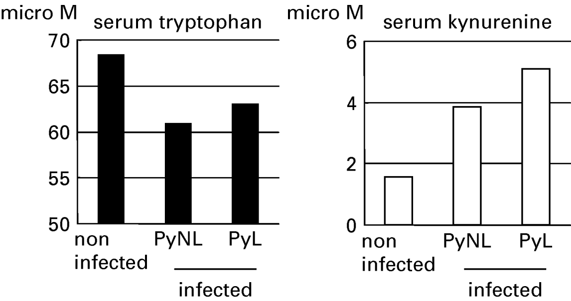
To evaluate the involvement of inducible tryptophan catabolism in the host–parasite relationship during malaria, we first measured its substrate tryptophan, and its metabolite kynurenine. P. yoelii has 2 substrains, PyL and PyNL, apparently differing in their virulence for mice. Mice were infected with either of the 2 strains, and serum samples obtained from mice 5 days after infection were analysed with HPLC (Fig. 1). Mice infected with strain Py showed decreased tryptophan and increased kynurenine levels, resulting in a lower ratio of tryptophan to kynurenine. These alterations occurred in mice infected with strains PyL or PyNL, suggesting that infection with malaria parasites induces hyper-catabolism of tryptophan regardless of parasite virulence.
Tryptophan metabolism is activated in P. yoelii-infected mice. Tryptophan (left panel) and kynurenine (right panel) were measured in sera from mice 5 days after infection with PyNL or PyL by HPLC. Data represent micromolar concentrations.

Inhibition of IDO partially protects mice from infection with strain PyL
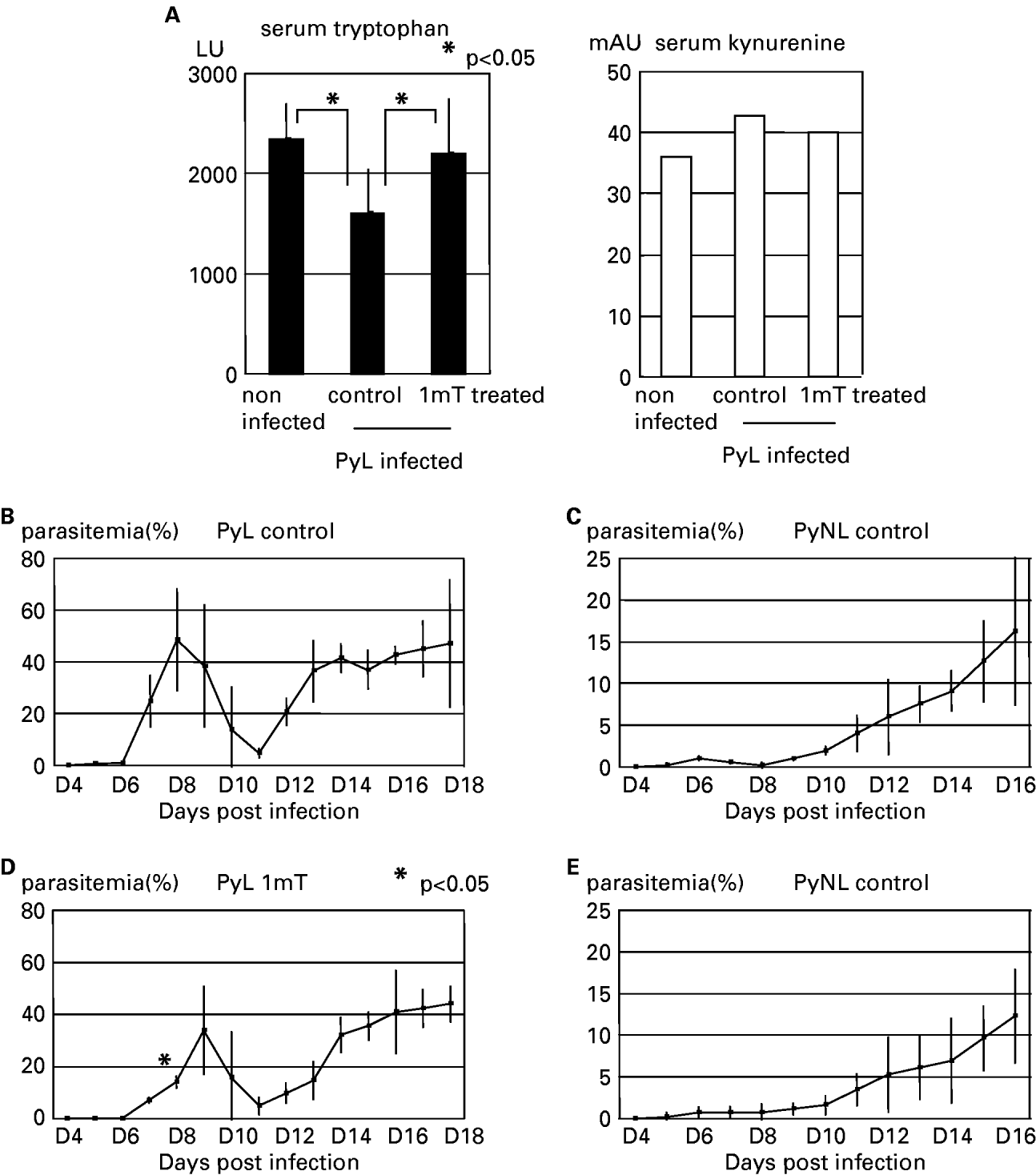
As IDO is known as an immunosuppressive enzyme, we hypothesized that IDO-mediated hyper-catabolism of tryptophan during malaria leads to less resistance to infection. To test this hypothesis, we used experimentally infected mice treated with IDO inhibitor 1mT. Prior to evaluation of the course of infection, we examined the inhibitory action of 1mT by measuring tryptophan and kynurenin in serum samples from PyL-infected mice treated with 1mT, using HPLC. As shown in Fig. 2A, the treatment suppressed the tryptophan to kynurenine ratio in PyL-infected mice, confirming that infection-induced hyper-catabolism of tryptophan was inhibited by 1mT administration. The mice treated with 1mT exhibited decreased parasite density and a delayed peak of parasitaemia during infection with strain PyL (Fig. 2B, C), although the survival rate was not significantly different among these mice (data not shown). Treatment with 1mT did not alter the course of infection with strain PyNL (Fig. 2D, E). These results demonstrate that inhibition of inducible catabolism of tryptophan by treatment with 1mT enhanced resistance to infection with strain PyL.
Effect of IDO inhibition on malaria infection. (A) Serum samples from PyL-infected mice treated with or without (control) 1mT were analysed as in Fig. 1. Data represent luminescence units for tryptophan or milli-absorbance units for kynurenine because the standards for tryptophan and kynurenine were not available in this experiment. Asterisks represent statistical significance (P<0·05) with Student's t test. (B–E) Frequency of parasitized RBCs (percentage parasitemia) in mice infected with PyL (B, D) or PyNL (C, E), and treated with (D, E) or without 1mT (B, C). Data are presented as the mean±s.d. from 5 mice per group. Asterisk on (D) represents statistical significance (P<0·05) with Student's t test between parasitaemia (%) of PyL control and of PyL 1mT at 8 days post-infection. Similar results were obtained in the 2 repeated experiments.

1mT does not have a direct parasiticidal effect on Plasmodium spp
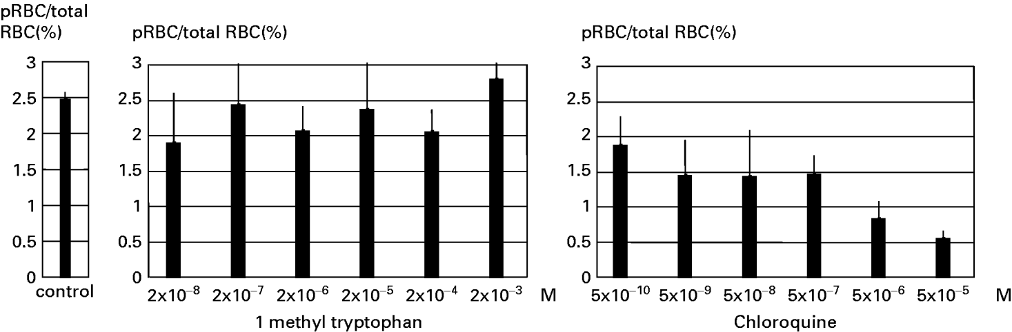
As shown in Fig. 2, 1mT treatment of mice was found to significantly enhance protection against strain PyL, probably through cancelling immunological suppression by IDO. To exclude any direct parasiticidal effect of 1mT on malaria parasites, we cultured PyL-parasitized RBCs with various concentrations of 1mT, and analysed parasite growth (Fig. 3). Chloroquine, an anti-malaria drug, reduced parasite growth in a dose-dependent manner. However, 1mT did not affect parasite growth even at concentrations 10 times more than those in the serum of mice treated with 1mT. Taken together with the finding that 1mT did not affect strain PyNL infection, it seems that 1mT does not have any direct effect on parasites.
1mT does not have direct parasiticidal effect on Plasmodium spp. PyL-infected RBCs were cultured with 1mT (centrer panel) or chloroquine (right panel) at the indicated concentrations. Parasitaemia at the culture start point was 5%, and was evaluated after 20 h. Data are presented as the mean±s.d. from triplicate cultures.

Inhibited catabolism of tryptophan suppresses malaria-specific immune responses
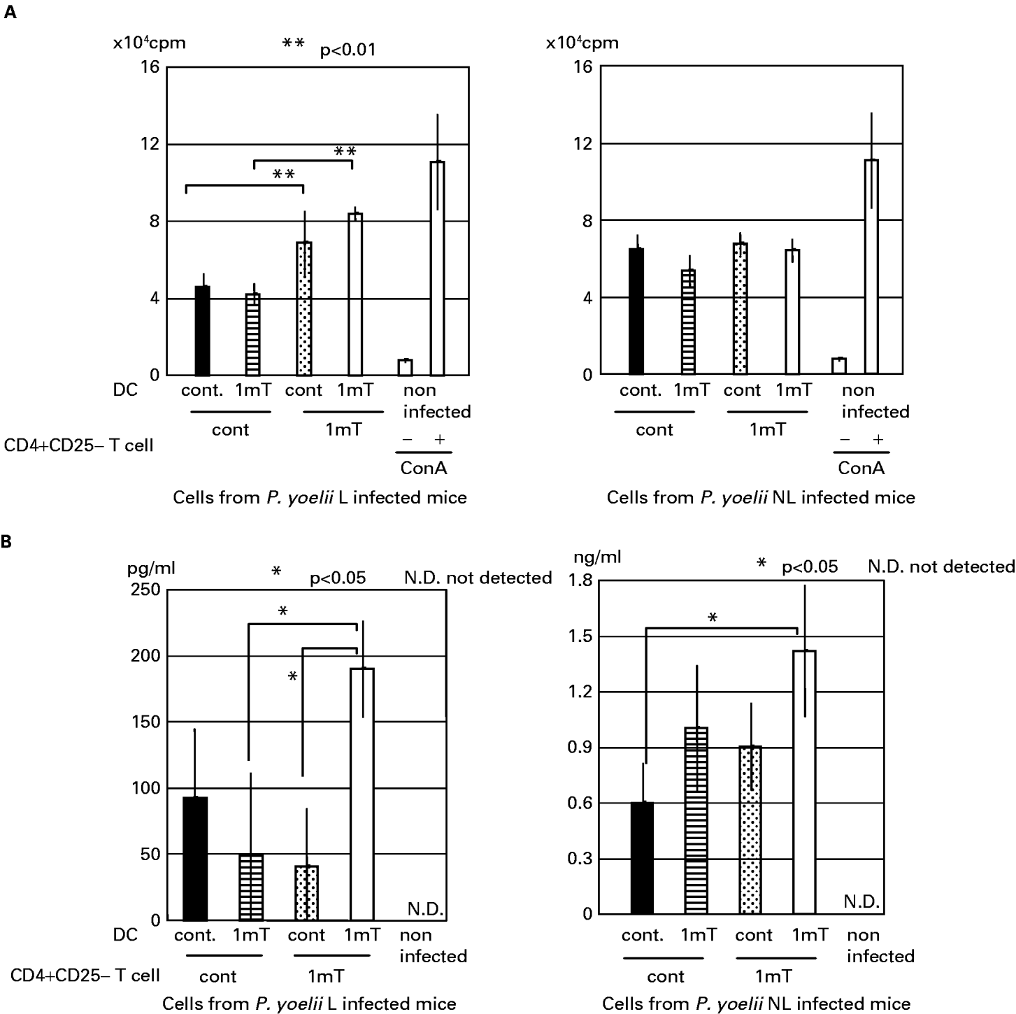
To analyse the mechanisms involved in the development of effective anti-malaria immunity under inhibition of tryptophan catabolism, we assessed the immune responses in Py-infected mice that had been treated with 1mT. Spleen CD4+ T cells isolated from infected mice were stimulated with parasitized RBCs in the presence of DCs, and were analysed for proliferation and production of IFN-γ. CD4+ T cells from PyL-infected mice treated with 1mT were found to proliferate more than those from infected mice not receiving 1mT, regardless of DC origin (Fig. 4A). In contrast, 1mT treatment of PyNL-infected mice did not promote the immune response, in accordance with our result that 1mT failed to enhance protection against PyNL infection (Fig. 2B, D).
Enhanced immune responses in PyL-infected mice treated with 1mT. (A) Proliferation of spleen CD4+CD25− T cells isolated from mice infected with PyL (left panel) or PyNL (right panel), and treated with or without (control) 1mT. T cells and DCs were obtained 5 days after infection, and T cells (1×10⁵) were cultured with PyL-parasitized RBCs (1×10⁵) in the presence of DCs (1×10⁴) from the indicated mice. One of the non-infected mouse-derived samples was cultured with ConA as an assay positive control. Results represent the mean±s.d. of triplicate cultures. Asterisks represent statistical significance (P<0·01) with Student's t-test. (B) CD4+CD25− T cells isolated from mice infected with PyL (left panel) or PyNL (right panel), and treated with or without (control) 1mT were cultured as in (A). IFN-γ in the supernatant was measured by using ELISA. Data are mean±s.d. (n=3). Asterisks indicate statistical significance (P<0·05) with Student's t-test. N.D., Means not detected.

IFN-γ secreted from CD4+ T cells is very important for protection against blood-stage malaria (Shear et al. Reference Shear, Srinivasan, Nolan and Ng1989). We quantified IFN-γ concentration in culture supernatants obtained from CD4+ T cells and DC co-culture, as above. CD4+ T cells from PyL-infected mice produced more IFN-γ in the presence of DC from the same mice than did cells from control mice (Fig. 4B). In agreement with the in vivo infection experiment, IFN-γ secretion in PyNL-infected mouse-derived cells did not show a significant difference between 1mT-treated and control mice. When these CD4+ T cells were mixed with DCs from control mice, they did not produce IFN-γ. These results indicate that when infection-induced hyper-catabolism of tryptophan was inhibited, CD4+ T cells were effectively primed for proliferation and production of IFN-γ. However, DCs with impaired IDO still played a crucial role in the recall reaction of IFN-γ production by CD4+ T cells.
DISCUSSION
Here, we provide evidence that infection with malaria parasites resulted in systemic activation of tryptophan catabolism mediated by IDO. Furthermore, inhibition of IDO in vivo partially protected mice from lethal infection, in association with enhanced lymphocyte proliferation and IFN-γ production by CD4+ T cells specific for malaria parasites. This enhancement was also observed when parasitaemia of cell-origin mice was almost the same between 1mT treated and control mice (data not shown). Interestingly, the effect of IDO inhibition on protective immunity was more obvious in infection with strain PyL than that with strain PyNL, although strain PyNL could activate IDO in a manner comparable to strain PyL. Concerning the non-lethality of the PyNL strain, we observed that in vitro IFN-γ production was suppressed by IDO, but that parasite growth was not enhanced in vivo. This suggested that even with a competent IDO mouse host, the dose of anti-malarial IFN-γ that is produced is approximately 3 times more than with IDO-incompetence with PyL strain infection. These results suggest that strain PyL evades host immunity more competitively by utilizing IDO which, in turn, could explain the higher lethality of strain PyL.
The tryptophan catabolism mediated immune suppression was found to be induced in our malaria mouse model which increased parasite load but did not shorten infected mice survival (data not shown). We did not observe any suggestive findings for reasonable interpretation on this rather contradictory result, but it is worthy to be considered that immune pathology is probably accelerated under administration of 1mT. Hyper-catabolism of tryptophan suppresses not only pathogen-reactive immunity but also host-damaging immune responses and both were activated with 1mT treatment and might have cancelled effects on survival with each other.
Although we confirmed neither protein expression nor mRNA transcription for IDO in malaria-infected mice, IDO was apparently activated in our malaria model, for the following reasons: inducible catabolism of tryptophan via the kynurenine pathway is mediated only by IDO, and IDO-specific inhibition reduces tryptophan catabolism. IDO is known to be detected in various organs and tissues such as gut, epididymis, lung and lymph node, especially antigen-presenting cells (Takikawa et al. Reference Takikawa, Yoshida, Kido and Hayaishi1986; Munn et al. Reference Munn, Sharma, Hou, Baban, Lee, Antonia, Messina, Chandler, Koni and Mellor2004). We speculate that expression of IDO in DCs is responsible for the immunomodulation observed in our model. IDO-positive DCs, especially plasmacytoid DCs (pDCs), have been reported to play the main role in IDO-mediated immunosuppression. They induce suppression of not only cellular, but also humoral immunity (Adikari et al. Reference Adikari, Lian, Link, Huang and Xiao1988). Interestingly, our data show that antigen-specific proliferation of CD4+ T cells in response to in vitro reactivation was not affected by IDO activity in DCs. These results can be interpreted as IDO-expressing DCs induce immunosuppression during T cell priming, and T cells primed by IDO+ DCs are fated to be unresponsive, with less functional plasticity than DCs.
Another important point is how the malaria parasite stimulates IDO activation. Various signals, such as IFN-α, IFN-γ, cytotoxic T lymphocyte antigen (CTLA)-4, and Toll-like receptor 9 (TLR9) ligation (CpG-oligonucleotides), are known to induce IDO in DCs (Mellor et al. Reference Mellor and Munn2004). During malaria, powerful inflammatory responses occur, and several cytokines like IFNs are secreted, which could be IDO-inducing factors. More directly, malaria parasites might stimulate, with their TLR9 ligands, the malaria pigment haemozoin (Coban et al. Reference Coban, Ishii, Kawai, Hemmi, Sato, Uematsu, Yamamoto, Takeuchi, Itagaki, Kumar, Horii and Akira2005) and some protein molecules (Pichyangkul et al. Reference Pichyangkul, Yongvanitchit, Kum-arb, Hemmi, Akira, Krieg, Heppner, Stewart, Hasegawa, Looareesuwan, Shanks and Miller2004). Our results and those of previous studies (Hisaeda et al. Reference Hisaeda, Maekawa, Iwakawa, Okada, Himeno, Kishihara, Tsukumo and Yasutomo2004) reveal that malaria parasites induce regulatory T cells, one of whose suppressive mechanisms is CTLA4 molecules. It is possible that malaria-induced regulatory T cells activate IDO in DCs via CTLA4 stimulation, and that IDO-expressing DCs might function as suppressive effector mechanisms.
In addition to immunosuppression, IDO has been reported to be involved in the host–parasite relationship in malaria. In a cerebral malaria model, IDO has been reported to be associated with pathology (Mitchell et al. Reference Mitchell, Hansen, Hee, Ball, Potter, Walker and Hunt2005). Indeed, the expression of IDO has been confirmed in the brain vascular endothelial cells in cases of cerebral malaria, using immunohistological techniques (Hansen et al. Reference Hansen, Driussi, Turner, Takikawa and Hunt2000). On the other hand, one of the metabolites of tryptophan, xanthurenic acid, has been found to be a gametocyte activating factor (Billker et al. Reference Billker, Lindo, Panico, Etienne, Paxton, Dell, Rogers, Sinden and Morris1998) which enhances the development of gametocytes into sporozoites, the infectious form of the malaria parasite, in mosquito vectors. It will be important to determine the importance of IDO in the pathology of cerebral malaria, and in transmission of malaria parasites to mosquito vectors.
In conclusion, our results demonstrate that IDO-mediated immunosuppression contributes to immune evasion by malaria parasites, providing a new insight into the host–parasite relationship. As IDO inhibition is a potent method for reducing parasite load and pathological complications, understanding the molecular mechanisms underlying IDO activation may be helpful in establishing therapeutic strategies against malaria.