Introduction
It has been estimated that about 10–15% of couples suffer from infertility, and male factor plays a role in around 20% of infertile couples worldwide (Practice Committee of American Society for Reproductive Medicine, 2012). Multiple morphological abnormalities of the sperm flagellum (MMAF) may result in male infertility, accompanied by teratozoospermia and severe asthenozoospermia. In addition, MMAF can feature a mosaic of abnormalities, including coiled, bent, irregular, short or absent flagella. Transmission electron microscopy (TEM) has been used to observe the status of central microtubules and/or dynein arms, as well as unassembled sperm fibrous sheaths (Liu et al., Reference Liu, He, Yang, Zouari, Wang, Wu, Kherraf, Liu, Coutton, Zhao, Tang, Tang, Lv, Fang, Li, Li, Zhao, Wang, Zhao, Zhang, Arnoult, Jin, Zhang, Ray, Cao and Zhang2019).
Previous studies have demonstrated that several genes are associated with MMAF, such as DNAH1, CFAP43, CFAP44, CFAP65, CFAP69 and TTC21A (Tang et al., Reference Tang, Wang, Li, Yang, Li, Liu, Li, Zhu, Wang, Wang, Zhang, Sun, Zhi, Wang, Li, Jin, Luo, Wang, Yang and Zhang2017; Liu et al., Reference Liu, He, Yang, Zouari, Wang, Wu, Kherraf, Liu, Coutton, Zhao, Tang, Tang, Lv, Fang, Li, Li, Zhao, Wang, Zhao, Zhang, Arnoult, Jin, Zhang, Ray, Cao and Zhang2019; Wang et al., Reference Wang, Tu and Tan2019). We reported previously that biallelic mutation in CFAP43 caused infertility in humans and mice. In addition there was a relationship between both CFAP43 and CFAP44 with MMAF, and biallelic mutations of these two genes may impair sperm motility and result in male infertility (Tang et al., Reference Tang, Wang, Li, Yang, Li, Liu, Li, Zhu, Wang, Wang, Zhang, Sun, Zhi, Wang, Li, Jin, Luo, Wang, Yang and Zhang2017). However, due to the absence of a specific antibody for Cillum and Flagellum Associated Protein 43 (CFAP43) the localization and specific role of the CFAP43 protein in mouse testes have still remained elusive. In Trypanosoma brucei, CFAP43 protein is located between the doublet microtubules 5 and 6 and the paraflagellar rod (Coutton et al., Reference Coutton, Vargas, Amiri-Yekta, Kherraf, Ben Mustapha, Le Tanno, Wambergue-Legrand, Karaouzène, Martinez, Crouzy, Daneshipour, Hosseini, Mitchell, Halouani, Marrakchi, Makni, Latrous, Kharouf, Deleuze, Boland, Hennebicq, Satre, Jouk, Thierry-Mieg, Conne, Dacheux, Landrein, Schmitt, Stouvenel, Lorès, El Khouri, Bottari, Fauré, Wolf, Pernet-Gallay, Escoffier, Gourabi, Robinson, Nef, Dulioust, Zouari, Bonhivers, Touré, Arnoult and Ray2018), as found in previously, however how CFAP43 can influence the formation of sperm flagella is elusive.
CFAP43 protein was firstly identified in bovine sperm centrioles (Firat-Karalar et al., Reference Firat-Karalar, Sante, Elliott and Stearns2014), and was later found to be present in human sperm (Jumeau et al., Reference Jumeau, Com, Lane, Duek, Lagarrigue, Lavigne, Guillot, Rondel, Gateau, Melaine, Guével, Sergeant, Mitchell and Pineau2015). It has been reported that cilia play a substantial role in the formation of sperm flagella (Fassad et al., Reference Fassad, Shoemark, le Borgne, Koll, Patel, Dixon, Hayward, Richardson, Frost, Jenkins, Cullup, Chung, Lemullois, Aubusson-Fleury, Hogg, Mitchell, Tassin and Mitchison2018). Transportation of some structural proteins in the sperm tail via intra-manchette transport (IMT) to the basal body at the base of the developing sperm tail has been reported by Lehti and Sironen (Reference Lehti and Sironen2016). IMT is also involved in sperm head shaping (Lehti and Sironen, Reference Lehti and Sironen2016). However, whether CFAP43 could mediate IMT and then function in the formation of sperm flagella and sperm head shaping needs to be further clarified.
In the present study, we utilized Cfap43-deficient mice to reveal the mechanism underlying multiple abnormalities of flagella. Our results demonstrated that, in mice, CFAP43-mediated IMT is essential for the formation of the sperm flagellum and sperm head shaping.
Materials and methods
Animals
Male C57BL/6 mice (age, 8 weeks old) were obtained from the Animal Center of Nanjing Medical University (Nanjing, China). To study the function of CFAP43 in spermiogenesis, we generated a Cfap43-deficient mouse model using CRISPR/Cas9 technology, as described previously (Tang et al., Reference Tang, Wang, Li, Yang, Li, Liu, Li, Zhu, Wang, Wang, Zhang, Sun, Zhi, Wang, Li, Jin, Luo, Wang, Yang and Zhang2017). The guide RNAs were designed against exon 22 of Cfap43 in accordance with the positions of pathogenic mutations as mentioned previously (Tang et al., Reference Tang, Wang, Li, Yang, Li, Liu, Li, Zhu, Wang, Wang, Zhang, Sun, Zhi, Wang, Li, Jin, Luo, Wang, Yang and Zhang2017). The frameshift mutations in Cfap43 were identified in founder mice and their offspring by polymerase chain reaction and Sanger sequencing. All animal experiments were approved by the Ethics Committee of Nanjing Medical University.
Spermatozoa counting
Spermatozoa were collected from cauda epididymides of wild-type (WT) mice or Cfap43-deficient (Cfap43 –/–) mice. Sperm were counted using a haemocytometer chamber under a light microscope (Nikon Instruments Inc., Tokyo, Japan), and the number of sperm was calculated by a standard method presented by Zhang et al. (Reference Zhang, Shen, Gude, Wilkinson, Justice, Flickinger, Herr, Eddy and Strauss2009). To assess the morphology of spermatozoa, sperm collected from cauda epididymides were spread onto slides and then fixed with 4% (w/v) paraformaldehyde (4% PFA; Servicebio Technology Co. Ltd, Wuhan, China) for 20 min. After that, they were washed three times with 1× phosphate-buffered saline (PBS) for 5 min and then stained with haematoxylin and eosin for analysis of morphology. At least 200 sperm per sample were assessed under a light microscope. Deformities were classified into two types, including abnormally shaped spermatid head and tail defects, as described previously (Soley, Reference Soley1997). Sperm abnormalities were classified based on their presumptive origin.
Periodic acid Schiff (PAS) staining
The stages in seminiferous tubule cross-sections were detected using Bouin’s solution-fixed testes from normal mice and sections were stained with PAS and haematoxylin, as described previously (Ahmed and de Rooij, Reference Ahmed and de Rooij2009). In brief, fresh mouse testicular and epididymal tissues were fixed in Bouin’s solution and 4% PFA, respectively, for over 24 h, then embedded in paraffin, and sectioned at 4-µm intervals to generate tissue slides. After deparaffinization in xylene and progressive alcohol dehydration (100%, 95%, 80% and 70%), the slides were stained with 1% PAS for 30 min at room temperature and washed under running water for 10 min. Next, slides were placed in Schiff’s reagent for 40 min and then washed with water for 10 min. Eventually, slides were washed under running water, dehydrated in alcohol, and cleared with xylene.
Transmission electron microscopy
A similar protocol was used in previous studies (Wang et al., Reference Wang, Ping, Jiang, Yu, Wang, Chen, Zhang, Xu, Wang, Li and Li2013; Liu et al., Reference Liu, He, Yang, Zouari, Wang, Wu, Kherraf, Liu, Coutton, Zhao, Tang, Tang, Lv, Fang, Li, Li, Zhao, Wang, Zhao, Zhang, Arnoult, Jin, Zhang, Ray, Cao and Zhang2019). Briefly, fresh testes tissues were fixed with 2.5% glutaraldehyde for 12 h at 48°C. Tissues were rinsed in 0.1 mol/l PBS for 1 h, then post-fixed in 1% osmic acid for 1 h at 4°C. After another rinsing in the same buffer for 30 min, testicular tissues were then treated with 2% uranyl acetate for 30 min, followed by progressive dehydration in ethanol and 100% acetone, and then embedded into Epon 812 resin. Sections were cut at 1-µm intervals for orientation of the seminiferous epithelium. Subsequently, they were stained with uranyl acetate and lead citrate for 30 min. Finally, testicular sperm ultrastructure was observed using a transmission electron microscope (TECNAI-10, 80 kV, Philips, The Netherlands) with an accelerating voltage of 80 kV.
Immunofluorescence
Bouin’s solution-fixed paraffin-embedded sections were analyzed by immunohistochemistry, as described previously (Shen et al., Reference Shen, Zhang, Yu, Guo, Gao, Liu, Zhang, Chen, Yu, Cheng, Zheng, Li, Huang, Ding and Zheng2018). In brief, endogenous peroxidase activity was quenched and sections were blocked using 5% bovine serum albumin (BSA, w/v; Sunshine, Nanjing, China), and then incubated overnight at 4°C with mouse polyclonal alpha-tubulin antibody (1:400; Sigma-Aldrich, St. Louis, MO, USA), mouse Espin polyclonal antibody (1:400; BD Biosciences, San Jose, CA, USA), rabbit polyclonal H2B antibody (1:100; Millipore, Burlington, MA, USA), rabbit polyclonal TP1 antibody (1:200; Proteintech, Rosemont, IL, USA), rabbit polyclonal P1 antibody (1:200; Santa Cruz Biotechnology Inc., Dallas, TX, USA), or rabbit polyclonal Rab27b antibody (1:400; Proteintech, Rosemont, IL, USA). Sections were washed with 1× PBS and Tween-20, and then incubated with a secondary antibody (1:2000; Alexa Fluor Plus 488 goat anti-mouse/rabbit IgG secondary antibody Invitrogen, USA) for 2 h at room temperature; the nuclei were stained with Hoechst reagent (Servicebio Technology Co. Ltd, Wuhan, China) for 5 min at room temperature. Eventually, all sections were observed under a confocal laser scanning microscope (Zeiss LSM710, Carl Zeiss, Oberkochen, Germany) and images were captured. Negative controls were used to assess the specificity of all the used antibodies (Fig. S3).
Terminal deoxynucleotidyl transferase-mediated dUTP nick end labelling (TUNEL) assay
A TUNEL FITC Apoptosis Detection Kit (Vazyme Biotech Co., Ltd, Nanjing, China) was used to detect cell death in mouse testis. Briefly, after deparaffinization and rehydration, sections of mouse testis were then incubated in proteinase K solution (20 µg/ml) at room temperature for 20 min, followed by three 5-min washes with PBS, and then incubated in TUNEL solution for 1 h at 37ºC. Next, sections were stained with 4′,6-diamidino-2-phenylindole (DAPI; 2 μg/ml) and incubated at room temperature for 10 min in the dark. Fluorescent signals were visualized using a fluorescence microscope (Nikon DS-U3, Nikon Instruments Inc., Tokyo, Japan). The total number of apoptotic cells in 50 lumens was counted, and the average number of tubules was calculated.
Statistical analysis
Data were expressed as the mean ± standard deviation. Data were analyzed using Student’s t-test and GraphPad Prism 7.0 software (GraphPad Software Inc., San Diego, CA, USA). A P-value < 0.05 was considered to be statistically significant.
Results
Effect of Cfap43 deficiency on sperm morphology, motility, count, and germ cell apoptosis
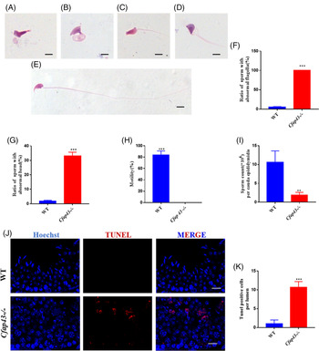
Spermatozoa collected from Cfap43-deficient mice exhibited a prominent MMAF phenotype (Fig. 1A–D) compared with WT mice (Fig. 1E). In addition to the flagellum abnormality, Cfap43-deficient mice had an abnormal sperm head (Fig. 1A–D). The ratio of sperm with abnormal flagella and heads is shown in Fig. 1(F) and Fig. 1(G), respectively. Cfap43 deficiency caused severe asthenospermia and oligozoospermia in mice (Fig. 1H, I). Using the TUNEL assay, apoptotic cells were found in Cfap43-deficient mice testes (Fig. 1J), and statistical analysis showed marked changes in apoptotic cells between WT mice and Cfap43-deficient mice (Fig. 1K).
Effect of Cfap43 deficiency on sperm morphology, motility, count, and germ cell apoptosis. (A) Amorphous head and flagellum absent. (B) Sperm with no hook and coiled flagella. (C) Amorphous head and short flagellum. (D) Amorphous head and short flagellum. (E) Wild-type (WT) spermatid. (F) Ratio of sperm with abnormal flagella. (G) Ratio of sperm with abnormal head. (H) Sperm motility. (I) Sperm count. (J) Germ cells apoptosis determination via TUNEL staining. Scale bars in (A–E) 2 μm; (J) 20 μm. TUNEL, terminal deoxynucleotidyl transferase-mediated dUTP nick end labelling. (K) Quantification of (J). **P < 0.01, ***P < 0.001, Student’s t-test, n = 3.

Effect of Cfap43 deficiency on testicular size, testicular weight and fertility
Testicular weight and testicular size were normal in Cfap43-deficient mice, and there was no significant difference in the testicular weight or size between Cfap43-deficient and WT mice (Fig. S1 A, B). Fertility tests were performed (Fig. S1 C, D). Male Cfap43-deficient mice were completely infertile, while female Cfap43-deficient mice reproduced normally.
Effect of Cfap43 deficiency on spermiogenesis
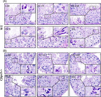
Spermiogenesis was clarified into steps based on changes in the shape of the spermatid head. Spermiogenesis began at stage I, and round spermatids could be observed from stage I to stage VIII (Fig. 2A). Concomitantly, spermatids began to take on an elongated shape as the cytoplasm was stretched out along the flagellum. At stages IX and X, sperm head shaping was obvious, and elongated spermatids (steps 9 and 10) first appeared. At stage XI, increased staining of the nuclei of elongated spermatids (step 11) could be observed. At stage XII, chromatin condensation occurred. At stages I–III, the heads of elongated spermatids (steps 13 and 14) were further shortened and appeared more dense. At stages IV–VI, the nuclei of spermatids (step 15) appeared to narrow. At stages VII and VIII, the cells (step 16) were streamlined and bore stereocilia on their luminal surface that absorbed fluid released from the testes along with sperm. In addition, spermatozoa could be observed in the lumen.
Histological changes observed by light microscopy. (A) Representative images of WT mouse testes from stage I to stage XII. Stages are marked from I to XII above the pictures. Elongated spermatids are shown in the dotted boxes. Scale bar: 5 μm. (B) Depletion of Cfap43 resulted in abnormal spermiogenesis. Elongated spermatids with abnormally shaped heads are shown in the dotted boxes. The number of spermatozoa could be observed in the seminiferous epithelium (stages VII–VIII). Scale bar: 5 μm.

Disorders of spermiogenesis were found in Cfap43-deficient mice (Fig. 2B). Normal round spermatids could be observed from stage I to stage VIII. Head shaping started at stages IX and X and morphology of elongated spermatid heads was normal compared with that of the WT. However, elongated spermatids in Cfap43-deficient mice started to show a head deformity from the beginning of head shaping (stages IX and X). Abnormally shaped spermatid heads could be observed in all stages except for stages VII and VIII. In addition, a limited number of spermatozoa could be observed in the seminiferous epithelium of Cfap43-deficient mouse (stages VII and VIII).
Effect of Cfap43 deficiency on IMT
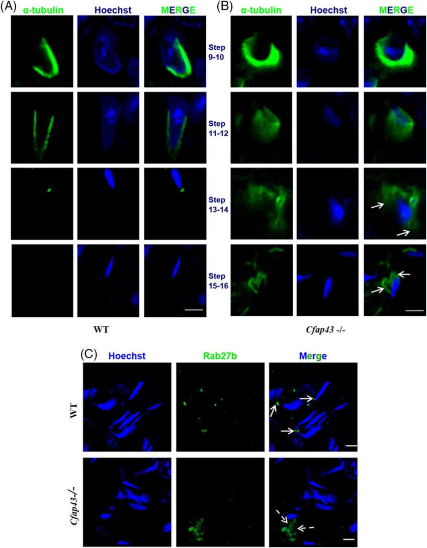
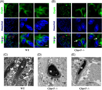
The manchette, one of the transient microtubular platforms, is essential for formation of the sperm tail and shaping of the head (Lehti and Sironen, Reference Lehti and Sironen2016). In WT mouse testes, the manchette was formed around the head by a sleeve of microtubules in step 9 and step 10 spermatids (Fig. 3A). It was gradually removed from head to neck, and became narrow in step 11 and step 12 spermatids. In steps 13–16 spermatids, the manchette became detached from the head and finally disappeared at step 16 (Fig. 3A). In the Cfap43 –/– group, the manchette was present in early elongating spermatids and girdled the caudal region of the head (Fig. 3B). With time the spermatid became elongated, however the manchette did not elongate and disengaged from the head to the tail (Fig. 3B). Following elongation (steps 15 and 16), a component of the manchette remained around the head (Fig. 3B). We also employed TEM to detect the manchette ultrastructure. In WT mice, manchette microtubules were observed to extend towards the developing flagellum, the direction of abnormal manchette microtubules observed in Cfap43-deficient testes was perpendicular to the assembly direction of the flagellum (Fig. 4C, D).
Effect of Cfap43 deficiency on intra-manchette transport (IMT) and vesicular transport. (A) Immunostaining of the manchette in wild-type (WT) testes spermatids. Scale bar: 10 µm. (B) Immunostaining of the manchette in Cfap43-deficient mouse testes spermatids. Arrows indicate markedly abnormal manchettes with some fragmentation. Scale bar: 10 µm. (C) Distribution of Rab27b in elongating spermatids. Arrows indicate that Rab27b is located in the apical region of head in step 13 spermatids of WT mouse. Dotted arrows indicate that Rab27b is in a disorganized arrangement in step 13 spermatids of Cfap43-deficient mouse. Scale bars: 5 μm.

Deficiency of Cfap43 led to spermiation failure and facilitated the abnormal assembly of manchette microtubules. (A) Immunostaining of ES in wild-type (WT) testes spermatids. (B) Immunostaining of ES in Cfap43-deficient mouse testes spermatids. Arrows show the anomalous location of the ES with some fragments. ES, ectoplasmic specializations. Scale bars: 5 μm. (C) Transmission electron microscopy (TEM) analysis of elongated spermatid in WT testes. M, manchette; N, nuclear. Scale bar: 2 μm. (D) TEM analysis of elongated spermatid in Cfap43-deficient mouse testes. Dotted arrows indicate manchette microtubules that appear to be much shorter compared with the WT. Scale bar: 2 μm. (E) TEM analysis of ES in Cfap43-deficient mouse testes. Dotted arrows indicate ES with some fragments. Scale bar: 2 μm.

Effect of Cfap43 deficiency on vesicular transport
It was found that at least some of the structural proteins of the sperm tail were transported by IMT to the basal body at the base of the developing sperm tail and by intra-flagellar transport to the assembly site in the flagellum. Rab27b is an important molecule that regulates vesicle trafficking. It controls vesicular transport by acting as a molecular switch that oscillates between the GTP-bound active form and the GDP-bound inactive form (Hayasaka et al., Reference Hayasaka, Terada, Suzuki, Murakawa, Tachibana, Sankai, Murakami, Yaegashi and Okamura2008). As illustrated in Fig. 3(C), Rab27b is located at the apical head region in WT spermatids, while its orientation is disordered in the Cfap43-deficient mouse model. Vesicular transport was disrupted in the Cfap43-deficient mouse model, therefore it can be concluded that depletion of CFAP43 protein resulted in disruption of vesicular transport and failure of flagella formation.
Effect of Cfap43 deficiency on spermiation
ES is a unique testis anchoring junction type that is typified by the presence of actin filament bundles sandwiched between the cisternae of the endoplasmic reticulum and the plasma membranes of Sertoli cells (Borg et al., Reference Borg, Wolski, Gibbs and O’Bryan2010). Espin, an ES marker, was used here to assess this structure (Fig. 4A, B). In WT mouse testes, ES resembles a cap and was localized to the apical head of elongating spermatids (Fig. 4A). However, in Cfap43 –/– mice, ES was disordered in elongating spermatids compared with WT spermatids (Fig. 4B). Using TEM, ES was observed to surround the apical head of spermatids, while it was randomly distributed in the Cfap43-deficient group (Fig. 4C, E).
Effect of Cfap43 deficiency on histone-to-protamine replacement
Using immunofluorescence analyses with antibodies specific for histone 2B (H2B), transition protein 1 (TP1) and protamine 1 (P1), we found that H2B was expressed specifically in spermatogonia, primary spermatocytes and round spermatids (Fig. S2 A) and that TP1 and P1 were expressed in elongating spermatids (Fig. S2 B, C). No significant differences were found between Cfap43-deficient and WT mice.
Discussion
In the present study, by using a gene knock-out model, we revealed that deficiency of Cfap43 in mouse caused MMAF, sperm head abnormality and oligozoospermia, and finally led to male infertility. Mechanistically, CFAP43 could regulate IMT, which promoted spermatogenesis by maintaining normal sperm head shape and flagellum formation. Moreover, we also found that CFAP43 could drive spermiation by regulating ES junction dynamics. Overall, our study provided a new insight into the pathogenesis of MMAF.
Depletion of numerous associated proteins may lead to severe abnormal elongation of the manchette and shaping of the spermatid head (Zhou et al., Reference Zhou, Du, Qin, Hu, Huang, Bao, Han, Mansouri and Xu2009; Lehti and Sironen, Reference Lehti and Sironen2016). HOOK1 is a manchette-associated proteins mainly expressed in the manchette; Hook1-KO mice displayed abnormal sperm head shapes and abnormal flagellum structures (Cole et al., Reference Cole, Meistrich, Cherry and Trostle-Weige1988; Mochida et al., Reference Mochida, Tres and Kierszenbaum1998). Elongated spermatids in Arl3-KO mice showed abnormal manchette formation, whereas spermatozoa displayed abnormal head and tail structures (Qi et al., Reference Qi, Jiang, Yuan, Bi, Zheng, Guo, Huang, Zhou and Sha2013). We have previously stated that disruption of IMT may contribute to the failure of flagellum formation and result in MMAF (Yang et al., Reference Yang, Li, Wang, Shi, Cheng, Wang, Li, Hou and Wen2015), but there was no evidence to support this hypothesis. In the present study, we used Cfap43-deficient mice to study the mechanisms underlying MMAF. The present research suggested that CFAP43 influenced sperm head shape and flagellum formation through the IMT pathway.
To date, two potential transport routes have been proposed in IMT: (i) an F-actin cytoskeletal pathway involving the myosin Va/Rab27a/b complex; and (ii) a microtubule cytoskeletal pathway involving the kinesin motor protein variant KIFC1 (Kierszenbaum, Reference Kierszenbaum2002; Hayasaka et al., Reference Hayasaka, Terada, Suzuki, Murakawa, Tachibana, Sankai, Murakami, Yaegashi and Okamura2008). Non-acrosomal vesicles can be transported to the basal body at the base of the developing sperm tail by IMT and participate in flagellum assembly. Rab proteins are monomeric GTPases of the Ras superfamily (Seabra et al., Reference Seabra, Mules and Hume2002). In human, F-actin-related proteins, e.g. MyRIP and Myosin Va and Rab27b, are localized to the manchette (Hayasaka et al., Reference Hayasaka, Terada, Suzuki, Murakawa, Tachibana, Sankai, Murakami, Yaegashi and Okamura2008). In the current research, we found that the location of Rab27b was obviously altered in Cfap43-deficient mice compared with WT mice. This suggested that CFAP43 may influence transport vesicles by regulating Rab27b, and unstable IMT may lead to disruption in flagellum formation and cause MMAF.
It has been argued that nuclear shape may be the result of a pattern of chromatin condensation (Fawcett et al., Reference Fawcett, Anderson and Phillips1971). Histone substitution by P1 and P2 plays a significant role in condensing chromatin, which is required to induce the quality of potent spermatozoa (Li et al., Reference Li, Wu, Kim, Zhao, Hearn, Zhang, Meistrich and Mills2014). Histone–protamine replacement is vital in chromatin condensation during sperm head shaping and disruption of the process may lead to abnormal sperm head shaping (Li et al., Reference Li, Wu, Kim, Zhao, Hearn, Zhang, Meistrich and Mills2014). However, the results of the present study demonstrated that the process of histone-to-protamine replacement was not disrupted in Cfap43-deficient mice, suggesting that an abnormal shaped head was not correlated with histone–protamine replacement in Cfap43-deficient mice.
In conclusion, although we did not have antibodies specific for CFAP43, our present studies demonstrated that CFAP43 was strongly associated with spermiogenesis and participated in flagella assembly through IMT transport. Consequently, Cfap43 deficiency resulted in an inability of the associated structural protein to be transported to the flagellum assembly site through the IMT and caused MMAF. In addition, abnormal sperm head morphology was linked to defects in the microtubule manchette. Furthermore, it could be implied that spermiation failure caused oligospermia in the Cfap43-deficient mice. However, we did not obtain human testes tissues with CFAP43 mutations, therefore whether CFAP43 is involved in sperm flagella via IMT in human needs to be further studied.
Supplementary material
To view supplementary material for this article, please visit https://doi.org/10.1017/S0967199420000556
Acknowledgements
We thank Li Wang from Zhejiang University for her assistance in performing TEM. We extend our thanks to Minxi Liu (State Key Laboratory of Reproductive Medicine, NJMU) and Wangjie Liu (Fudan University) for their constant advice. We extend our thanks to J. Cusick from the Medsci Group (Shanghai, China) for editing the English text of the manuscript.
Financial support
This study was supported by the Medical and Health Brand Discipline of Ningbo (PPXK2018-06), the Foundation of Maternal and Child Health Funding Project of Jiangsu Province (F201866), the Foundation of the Suzhou Key Laboratory of Male Reproduction Research (SZS201718), the Foundation of the Health Commission of Jiangsu Province (H2018050), the National Natural Science Foundation of China (81901532), and the Natural Science Foundation of Jiangsu Province (BK20190188).
Conflicts of interest
None.
Ethical standards
This study was approved by the institutional ethics review board of the Nanjing Medical University (2018-572).