Introduction
Sialadenoma papilliferum is a rare, benign, exophytic, painless, slow-growing salivary gland neoplasm which often occurs in the oral cavity, most commonly on the palate. It was first reported by Abrams and Finck in 1969.Reference Abrams and Finck1 Although more than 50 cases of sialadenoma papilliferum of the oral cavity, parotid gland, submandibular gland, nasopharynx and oesophagus have been reported, the present case is the first to be reported in the nasal cavity.Reference Argyres and Golitz2–Reference Kronenberg, Horowitz and Leventon4
Case report
A 67-year-old woman presented complaining of recurrent epistaxis from the right nasal cavity for the last two years. She had no significant previous medical history.
On examination, an exophytic, well circumscribed, painless mass with a diameter of 5 mm was identified on the anterior cartilaginous nasal septum mucosa of the right nasal cavity. The preliminary diagnosis was fibrous polyp.
The lesion was completely excised under local anaesthesia. The specimen was fixed in 10 per cent buffered formalin and referred for histopathological examination.
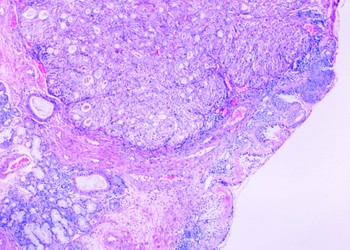
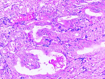
The surgical specimen consisted of an 8 × 8 × 4 mm, wedge-shaped segment of mucosa. On gross examination, a well circumscribed, nodular, 5 mm diameter lesion was observed on the mucosa. Microscopically, the lesion was characterised by broad-based, wedge-shaped but unencapsulated cellular proliferation with both exophytic papillary and submucosal components. The papillary fronds extending above the level of the adjacent nasal mucosa were covered by stratified squamous epithelium with no atypia (Figure 1). At the base of the fronds, transition from squamous epithelium to columnar ductal epithelium was seen. Towards the lower portion of the lesion, these papillary fronds were in continuity with several ducts exhibiting papillary folds of the luminal surface (Figure 2). At the base of the tumour, mucous salivary gland tissue was present. Ducts and their papillary folds were lined by a double row of cells – a basal layer of cuboidal cells and a luminal layer of columnar cells (Figure 3). A diagnosis of sialadenoma papilliferum was made based on this characteristic histological pattern.
Photomicrograph showing papillary fronds extending above the level of the adjacent nasal mucosa (H&E; ×40).

Photomicrograph showing duct-like structures in continuation with the overlying epithelium (H&E; ×100).

Photomicrograph showing ducts with papillary projections and double-cell layer lining (H&E; ×200).

The lesion was considered to have been completely removed; consequently, no supplementary treatment was required. Follow up four months after the operation showed smooth healing of the wound, with no evidence of recurrence. Subsequently, the patient remained well without complaint of epistaxis.
Discussion
Sialadenoma papilliferum is a rare, benign and slow-growing neoplasm of the salivary glands. Since it was first described by Abrams and Finck in 1969, more than 50 cases have been reported, occurring in the oral cavity, parotid gland, submandibular gland, nasopharynx and oesophagus.Reference Abrams and Finck1–Reference Kronenberg, Horowitz and Leventon4 The tumour has mostly been described in the oral cavity, commonly on the hard palate near the junction with the soft palate. The present case represents the first report of this rare tumour in the nasal cavity.
Previously reported cases show that sialadenoma papilliferum is liable to occur in middle-aged adult males, presenting as an exophytic, painless, slow-growing mass on the hard palate. In these cases, the diagnosis was usually made before the lesion grew larger than 10 mm in diameter.Reference Argyres and Golitz2 Our case occurred in a 67-year-old woman presenting with recurrent epistaxis for two years. An exophytic, well circumscribed, painless lesion was identified on the anterior cartilaginous nasal septum mucosa of the right nasal cavity. Compared with previously reported cases of sialadenoma papilliferum, our patient's symptoms and tumour localisation were unusual. However, on gross examination the lesion's macroscopic appearance was compatible with descriptions in the literature.
On physical examination, our patient's lesion was initially diagnosed as a fibrous polyp. Malignant tumours such as malignant melanoma and squamous cell carcinoma should be considered in the differential diagnosis of such a lesion. In our case, microscopic examination of the tumour enabled a diagnosis of sialadenoma papilliferum. Due to the complete resection of the tumour, no further treatment was required, and no recurrence was observed four months after excision. Although sialadenoma papilliferum is a benign neoplasm, Shimoda et al. have reported a case of malignant transformation of sialadenoma papilliferum of the palate.Reference Shimoda, Kameyama, Morinaga, Tanaka, Hashiguchi and Shimada5 Cases of recurrent sialadenoma papilliferum have also been reported.Reference Pimentel, Lopez Amado and Garcia Sarandeses6, Reference Rennie, MacDonald and Critchlow7
Conclusion
Sialadenoma papilliferum is a rare salivary gland neoplasm which occurs mostly in the oral cavity. The present case represents the first report of this entity in the nasal cavity, presenting with epistaxis. In this case, total excision enabled complete resolution.