New, multidetector CT generations – above or equal to 64 detectors – can now provide submillimetre resolution and discrimination between two points – spatial resolution – and track moving points over the cardiac cycle – temporal resolution – in the rapidly moving heart.
In comparison with conventional angiography, electrocardiographic-gated multidetector CT overcomes certain limitations such as the overlap of adjacent cardiovascular structures, difficulties in depicting systemic and pulmonary vascular systems simultaneously, and catheter-related complications,Reference Haramati, Glickstein, Issenberg, Haramati and Crooke 1 especially a prolonged exposure time and higher effective dose exposure. At present, low kilovoltage electrocardiographic-gated multidetector CT has an exposure of about 1 mSv, whereas it is 10 mSv in a diagnostic catheter with a consequent higher risk for radiation-induced malignancies.Reference Efstathopoulos, Katritsis and Kottou 2 , Reference Watson, Mah and Joseph 3
Multidetector CT allows simultaneous evaluation of cardiovascular structures and lung parenchyma, features that are important to be assessed in patients with CHD,Reference Leschka, Oechslin and Husmann 4 and offers data about ventricular wall motion and function.
Electrocardiographic-gated multidetector CT currently provides the highest spatial resolution, allowing the best post-processing of anatomical details for three-dimensional reconstructions. This is particularly important in patients with complex CHD, in whom anatomical details and relationships are of great importance for surgical and interventional catheter procedure planning.
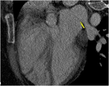
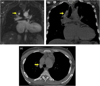
Electrocardiographic-gated multidetector CT avoids the long duration of MRI examination, which needs cooperative patients or general anaesthesia in the paediatric age group. In addition, electrocardiographic-gated multidetector CT offers higher resolution images compared with available MRI protocols for arrhythmias (Figs 1 and 2) and allows imaging of patients with pacemakers or other MRI-incompatible devices.
A 37-year-old female with cortriatriatum presented with supraventricular arrhythmia. Electrocardiographic-gated multidetector CT four-chambers view clearly identifies, with its high resolution capability, the junction between the left atrium and the pulmonary vein (yellow arrow). The figure is quoted (after permission) from the library of cardiac imaging cases, Radiology Department, Ain Shams University Hospital, Cairo, Egypt.

A 25-year-old male presented with arrhythmia and a past history of a modified Blalock–Taussig shunt, right pulmonary artery to right subclavian artery, for double right ventricle outflow tract. ( a ) Dynamic MRI with contrast hardly assessed the shunt patency, whereas electrocardiographic-gated multidetector CT without contrast ( b , c ) precisely identified the shunt and the black thrombotic material inside (yellow arrows). The figure is quoted (after permission) from the library of cardiac imaging cases, Radiology Department, Ain Shams University Hospital, Cairo, Egypt.

Advantages of electrocardiographic-gated multidetector CT
Electrocardiographic-gated multidetector CT provides excellent anatomical information with respect to the following:
The great vessels and their connections to the heart, a matter of particular importance in cases of CHD: accurate mapping of the arterial system avoids injury during electrophysiological procedures.
Coronary arteries: coronary arterial anomalies can lead to fatal cases and arrhythmias, such as a malignant interarterial course of the coronary arteries or anomalous origin of the left main coronary artery from the pulmonary artery.
Coronary venous system: electrocardiographic-gated multidetector CT provides accurate assessment of the coronary venous system, which acts as a bridge for the conduction system of the heart, transferring normal and abnormal signals – for example, the coronary sinus can transfer atrial fibrillation waves from abnormal foci in the atria to the ventricles. This bypasses the natural protective barrier of the heart, the atrioventricular node.Reference Takatsuki, Mitamura, Ieda and Ogawa 5 – Reference Volkmer, Antz, Hebe and Kuck 7 The coronary sinus can also be the origin of an arrhythmia as in the case of a persistent vein of MarshallReference Sun, Arruda and Otomo 6 , Reference Volkmer, Antz, Hebe and Kuck 7 or persistent left superior caval vein;Reference Saremi and Krishnan 8 on the other hand, cardiologists need to map and measure the dimensions and extensions of the venous system as it is the site of insertion of leads for cardiac re-synchronisation therapy.Reference Khan, Virdee and Gopalan 9
Pulmonary venous system: electrocardiographic-gated multidetector CT provides important information about the pulmonary venous system in the case of atrial fibrillation – namely, normal anatomy, anatomical variants and the number of pulmonary veins, and the ostial diameters of each vein in two orthogonal dimensions obtained in perpendicular planes to the vein – for example, coronal and axial. In addition, information can be obtained concerning the distance to the first-order branch, trunk length, as it is relevant to the choice of catheter size, the saddle distance between two adjacent pulmonary veins on one side, early branching, ostial branch, of the pulmonary vein, from the first centimetre of the trunk, and the presence of accessory or supernumerary pulmonary veins.Reference Saremi and Krishnan 8 All these variants can be the foci of arrhythmias.
The variations in the middle lobe and lingual pulmonary vein connections can be traced by using this technique, as they may be connected to the inferior pulmonary veins or directly to the left atrium, which could be a leading point of an arrhythmia.Reference Tsao, Wu and Yu 10 – Reference Sugimoto, Izumiyama, Yamashita, Baba and Hasegawa 12 This technique therefore shows congenital abnormalities of the pulmonary veins, which play an important haemodynamic role in arrhythmia generation – for example, anomalous pulmonary venous connections leading to atrial flutter or fibrillation.
It is also a useful tool for follow-up and diagnosis of complications after electrophysiological procedures – for example, the development of pulmonary venous stenosis or local thrombosis after ablation.Reference Ghaye, Szapiro and Dacher 13
Oesophagus: electrocardiographic-gated multidetector CT delineates the anatomical course of the oesophagus relative to the pulmonary veins and the posterior left atrial wall to precisely calculate the amount of energy that might be required for an ablation procedure.Reference Tsao, Wu and Chern 14
Delineation of atrial structures:
The length of the crista terminalis and its mid-point, the thickness of the crista terminalis, and the site of the sino-atrial node. This helps to decide the spectrum of energy to use and helps avoid complications.Reference Saremi and Krishnan 8
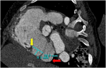
The cavo-tricuspid isthmus – paraseptal isthmus, inferolateral, and central isthmus – length, angle, muscular thickness, and depth, all of which are important for proper selection of the technique for electrophysiological manipulation (Fig 3).Reference Kajihara, Nakano and Hirai 15
A 2-year-old patient presented with mitral regurgitation and supraventricular arrhythmia. Multiplanar reconstructed electrocardiographic-gated multidetector CT clearly showing cavo-tricuspid isthmus length and depth measurement, the inferior caval vein (red arrow), and the tricuspid valve (yellow arrow). The figure is quoted (after permission) from the library of cardiac imaging cases, Radiology Department, Ain Shams University Hospital, Cairo, Egypt.

The anatomy of the sino-atrial node artery, evaluating its variable course and any pathology that may compromise the sino-atrial node blood supply.Reference Berdajs, Patonay and Turina 16 – Reference Cabrera, Ho and Climent 18
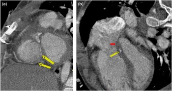
The borders of Koch’s triangle, particularly in the case of atrioventricular nodal re-entry tachycardia. This is a crucial issue, especially where there is a small triangle as in the paediatric age group to avoid the complication of heart block after the procedure. It is important to identify the location of the atrioventricular node and the distance between its arterial supply to the endocardium and the septal isthmus to avoid nodal injury (Fig 4).Reference Saremi and Krishnan 8 , Reference Cabrera and Sánchez-Quintana 19 – Reference Anderson, Ho and Becker 21
A 1-year-old male presented after repair of tetralogy of Fallot, electrocardiographic-gated multidetector CT short-axis view ( a ) shows the artery to the atrioventricular node (two yellow arrows). The four-chamber view ( b ) shows the atrioventricular septum and the artery to the atrioventricular node (yellow arrow). CT can evaluate the distance between the atrioventricular node and the Koch’s triangle (red arrow) even in postoperative CHD and avoid injury to the conduction system during electrophysiological procedures. The figure is quoted (after permission) from the library of cardiac imaging cases, Radiology Department, Ain Shams University Hospital, Cairo, Egypt.

Advances in electrocardiographic-gated multidetector CT equipment have led to the ability to predict the presence of some obstacles such as prominent tendon of Todaro and the Eustachian valve and presence of the Thebesian valve and pouch, all of which may be obstacles for catheter manipulation.Reference Saremi and Krishnan 8
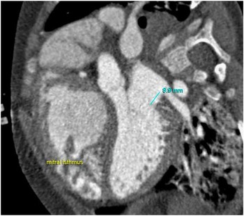
Although the majority of cases of atrial fibrillation originate from the pulmonary veins, other foci can also be detected such as the mitral isthmus, which is the area between the left inferior pulmonary vein and the leaflet of the mitral valve. Multidetector CT can provide measurements of its length, depth, and relation to vascular structures such as the left circumflex artery, the coronary sinus, and the great cardiac vein (Fig 5).Reference Chiang, Tsao and Wu 22
In the Same patient as in Figure 4, electrocardiographic-gated multidetector CT, four-chamber view, shows the mitral isthmus (area between the left inferior pulmonary vein and mitral valve attachment). The figure is quoted (after permission) from the library of cardiac imaging cases, Radiology Department, Ain Shams University Hospital, Cairo, Egypt.

Do we have ideal age-related measurements of different cardiac structures in the paediatric age group for comparison with these measurements in this patient? Do we have certain guidelines recommending certain interventional manoeuvers upon these measurements as we have in adult patients?Reference Tsao, Wu and Chern 14
A prognostic view of the risk of recurrence of supra ventricular arrhythmias before the procedure can be provided through left atrial volume analysis.
Major vessels of the heart: CT can measure the distance between the anterior left atrial wall and the aorta, which are in close proximity, and associated with a risk of perforation during the electrophysiological procedure.Reference Ho, Sanchez Quintana, Cabrera and Anderson 23
Electrocardiographic-gated multidetector CT evaluates the dimensions of the superior caval vein and the inferior caval vein, excluding any abnormality or variations in the venous drainage as well as detects areas of venous thrombosis.
The ventricular structures: an informative anatomical map of the ventricular structures can be obtained by electrocardiographic-gated multidetector CT, allowing diagnosis of ventricular congenital anomalies, non-compaction, and heart looping, all of which may have haemodynamic effects and help in creation of re-entry circuits causing arrhythmias that can progress to sudden cardiac death.Reference Walsh and Cecchin 24
Myocardium: quantification of the volume of myocardial fibrosis in cardiomyopathic patients is now possible using new software analysis of late electrocardiographic-gated multidetector CT images – a technique that has been shown to correlate well with MRI and histopathology. This can predict the development of ventricular tachyarrhythmias and sudden cardiac death, and is especially useful in patients with contraindications for MRI, such as having pacing systems.Reference Shiozaki, Senra, Edmundo Arteaga, Filho, Pita and Ávila 25
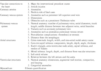
Airway anatomy is considered one of the keys for detecting the visceral situs, which helps in the prediction of anomalies and arrhythmias in certain patients – for example, patients with polysplenia and left isomerism have an increased probability of sinus nodal dysfunction and the need for cardiac pacing (Table 1).Reference Dickinson, Wilkinson, Anderson, Smith, Ho and Anderson 26 , Reference Momma, Takao and Shibata 27
Advantages of electrocardiographic-gated multidetector CT in electrophysiological procedures.

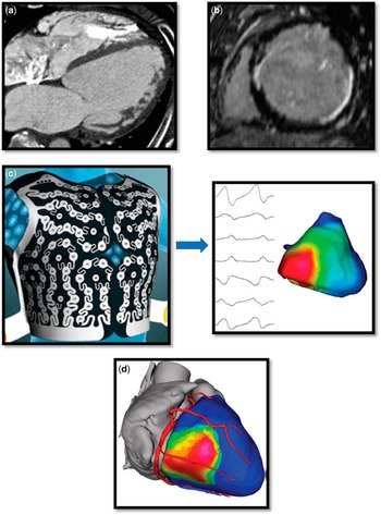
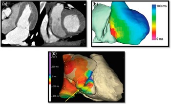
Combined anatomical and functional protocols: the real progress in the world of electrophysiology has been the ability to combine anatomical details, either normal or abnormal, with respect to congenital structural heart disease, with the information on propagation of electrical activity. Recently, cardio-radiologists have started using electrocardiographic-gated multidetector CT for accurate structural evaluation, combined with delayed gadolinium MRI images for better scar evaluation and electrical mapping of the heart, especially to record endocardiac and epicardiac activities. Non-invasive, body-surface electrodes allow determining the correlation between circuits of arrhythmias and cardiac structures. This is very important in the case of an unusual location, which may resist standard approaches to ablation. All data can be combined and compared with the three-dimensional reconstruction of catheter mapping for better localisation of arrhythmia circuits, permitting an effective ablation (Figs 6 and 7).Reference Cochet, Dubois and Sacher 28
The most recent recommended protocol before interventional procedures: ( a ) low kilovoltage electrocardiographic-gated multidetector CT for precise anatomical assessment, ( b ) delayed gadolinium enhanced image for precise calculation of fibrosis (which is the substrate of arrhythmia), and ( c ) body-surface mapping for precise assessment of cardiac electrical activity; ( d ) data are integrated in a three-dimensional model providing accurate evaluation of anatomy, myocardial fibrosis, and electrical activity (quoted fromReference Cochet, Dubois and Sacher 28 ).

A 14-year-old girl presented with Wolff–Parkinson–White syndrome, resistant to previous ablation, with history of ventricular fibrillation episodes and arrest. ( a ) Electrocardiographic-gated multidetector CT revealed a right atrium diverticulum extending towards the right ventricular free wall. ( b ) Body surface mapping precisely detects the abnormal activity. ( c ) Integrated electro-anatomical mapping showing the point of ablation (yellow arrow).

Areas for future research
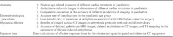
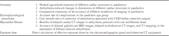
The question “Do radiologists provide adequate information for electrophysiological procedures in the pediatric age group?”, however, remains. We realise that studies are lacking on certain important aspects such as age-related measurements of different cardiac structures in the paediatric age group and whether these correspond to variations in arrhythmias (revise the question below Fig 5). Also lacking are studies comparing the degree of accuracy of measurements provided using the conventional angiography, echocardiography, MRI, and cardiac CT studies.
Other important unanswered questions include understanding the rate of complications attributable to electrophysiology procedures in the paediatric age group, the cost–benefit ratio of undertaking interventional treatment of arrhythmias associated with CHD before surgery, and the impact of such electrophysiological procedures on the severity of the disease.
There is also a need to assess the cost–benefit ratio of adding delayed cardiac images obtained with low-dose CT protocols, allowing sub milliSievert doses, to fully evaluate scars and post-ablation or postoperative fibrosis, all of which could be a structural substrate for arrhythmia. More studies are also needed to evaluate the benefit of non-invasive diagnosis of structural heart disease and associated arrhythmias through surface electrodes, electrocardiographic-gated multidetector CT, and delayed MRI images in addition to reconstructed invasive catheter mapping, in terms of dose reduction and time management in the paediatric age group.
Not all patterns of myocardial fibrosis can be evaluated with delayed gadolinium MRI images, as there are false-negative findings of fibrosis. It is argued that T1 mapping MRI technique could be a solution for assessment of other myocardial fibrosis patterns; however, the correlation between fibrosis-induced arrhythmias and T1 mapping images has not been fully assessed.
In certain types of cardiomyopathies, arrhythmias are related to hypoperfused myocardium. Further studies are needed to correlate electrocardiographic-gated multidetector CT and MRI myocardial perfusion maps with the electric activity propagation pattern.
Finally, radiologists and cardiologists have problems with the calculation of the effective doses provided by electrocardiographic-gated multidetector CT equipment, failing to provide adequate data for the assessment of long-term risk of sequelae such as lymphoid malignancies. Radiologists and CT technicians prefer to obtain effective diagnostic information and sacrifice the low-radiation dose concept. In addition, it is important to realise the shortage of well-trained technicians worldwide, and stress the importance of radiological technicians to work hand in hand with the radiologists to apply low-dose CT protocols (Table 2).
Areas for future research.
Conclusion
Electrocardiographic-gated multidetector CT supports an accurate preoperative and pre-catheter assessment of the anatomy and pathology of the heart, vessels, and extra-cardiac structures with benefits comparable with MRI and echocardiography, especially in the paediatric age group. Further comparative studies are needed to evaluate test accuracy, establish normal variations in the paediatric age group, and optimise radiation doses for ideal detailed imaging.
Acknowledgements
The poor children in Aswan Heart Center, Egypt, where I first thought how to reduce their suffering. The authors thank Claire Michael, MD, Professor of Pathology, University Hospitals, Case Western Reserve University, Cleveland, Ohio, United States of America, for her support.
Financial Support
This research received no specific grant from any funding agency or from commercial or not-for-profit sectors.
Conflicts of Interest
None.