Introductıon
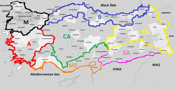
Turkey is a geographically important link between Asia, Europe and Africa and is surrounded by seas (Fig. 1). It has a population of nearly 83 million. Much of the literature on toxoplasmosis in Turkey is in Turkish. Here, we review the available literature on Toxoplasma gondii infection in humans and animals in Turkey.
Map of Turkey with seven regions and human population. Marmara region (M), Central Anatolia (CA), Aegean region (A), Mediterranean region (ME), Black Sea region (B), Eastern Anatolia (EA) and Southern anatolia (SA).

Methods for review
We consulted original manuscripts to minimize mistakes in translation. Detailed historical, serological, parasitological, clinical and genetic information on T. gondii infections in humans and other animals are summarized in tables throughout the review.
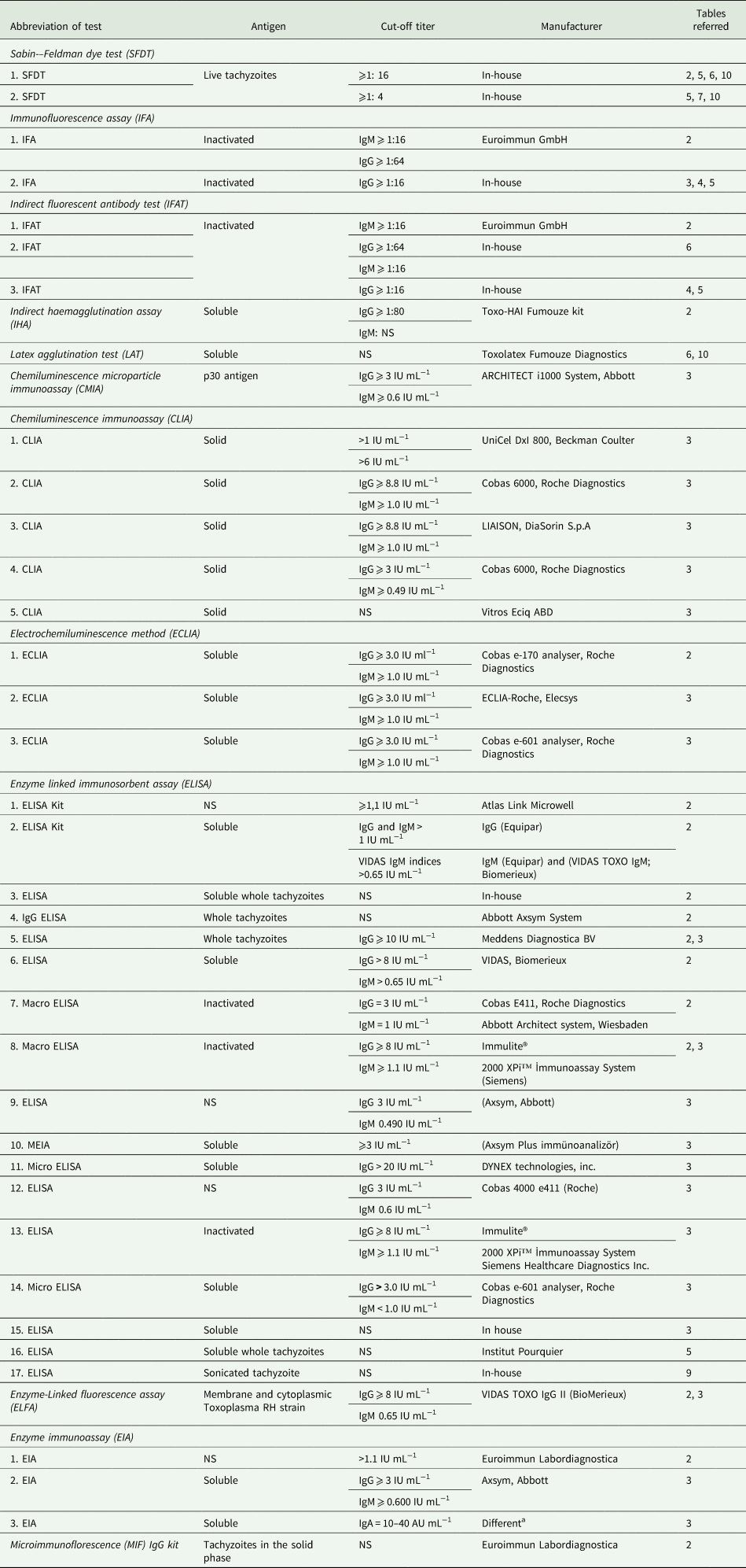
Most of the reports on toxoplasmosis relate to serological surveys with no uniformity with respect to tests used and cut-off values. In Table 1, information on serological assays used in Turkey is summarized and noted throughout the paper where applicable.
Details of serological tests used for the seropositivity of T. gondii in humans and animals in Turkey

a IgG ve IgM antibodies (Cobas Core, Roche), IgA antibodies (ETI-TOXOK-A reverse); IgG Avidity EIA Well-RADIM.
History
Although T. gondii was discovered in 1908, there was no information from Turkey until 1950 (Dubey, Reference Dubey2008). Professor Hayati Ekmen poineered research on toxoplasmosis in Turkey in 1970's. He first isolated viable T. gondii from a dog (Ekmen and Altıntaş, Reference Ekmen and Altıntaş1970) and from a child (Ekmen et al., Reference Ekmen, Altay and Altıntaş1974). The report on the baby was unfortunately published only as an abstract (Döşkaya et al., Reference Döşkaya, Caner, Ajzenberg, Değirmenci, Dardé, Can, Erdogan, Korkmaz, Üner, Güngör, Altintas and Gürüz2013). Toxoplasmosis was suspected in a child born to a mother from Ankara in 1972. The mother had been treated with anti-T. gondii therapy (sulfadiazine and pyrimethamine) during the last 2 months of pregnancy. Nothing was reported concerning the symptoms of toxoplasmosis in the newborn but the child must have had neurological signs because child's cerebrospinal fluid (CSF) was bioassayed in mice for the isolation of viable T. gondii. A virulent strain of T. gondii was recovered from the mice and designated the Ankara strain. This strain has been maintained since 1972 and used for molecular studies (Döşkaya et al., Reference Döşkaya, Caner, Ajzenberg, Değirmenci, Dardé, Can, Erdogan, Korkmaz, Üner, Güngör, Altintas and Gürüz2013).
Another noteworthy publication is an outbreak of acute toxoplasmosis in school children in Turkey (Doganci et al., Reference Doganci, Tanyuksel, Araz, Besirbellioglu, Erdem, Ozoguz, Yucel and Ciftcioglu2006). In 2002, a boarding school in Izmir, Turkey, saw 171 (9.5%) of 1797 students, aged 14–18-year, develop mild flu-like illness. All students were examined physically, including ophthalmic testing. The symptoms were typical of acquired toxoplasmosis (cervical lymph adenopathy, fever, myalgia, headache and dizziness). Antibodies to T. gondii were found in all 171 students by means of several serological techniques; all were positive for IgM antibodies and 40 of 43 randomly selected students had low-avidity T. gondii antibodies. The IgM and low-avidity antibodies are indicative of recent infection. None of the students had ocular lesions. Epidemiological investigation revealed no common source. Near the dining hall, there was a sheltering place for large numbers of stray cats. However, the school authorities removed the cats before they could be tested for T. gondii. This outbreak provided reference sera for acute toxoplasmosis for other investigations (Liang et al., Reference Liang, Döşkaya, Juarez, Caner, Jasinskas, Tan, Hajagos, Bradley, Korkmaz, Gürüz, Felgner and Davies2011).
Toxoplasmosis in humans
Toxoplasma gondii infections are prevalent worldwide but are mostly asymptomatic. However, T. gondii can cause severe illness in humans, particularly in congenitally infected children, and those with suppressed immunity, and even immunocompetent persons have died of toxoplasmosis (Robert-Gangneux and Dardè, Reference Robert-Gangneux and Dardé2012; Torgerson and Mastroiacovo, Reference Torgerson and Mastroiacovo2013; Peyron et al., Reference Peyron, Wallon, Kieffer, Graweg, Remington, Klein, Wilson, Nizet and Maldonado2016).
Serological prevalence in the general population
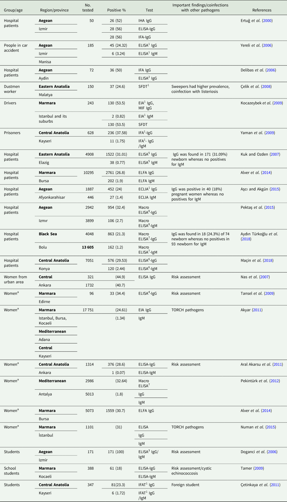
The seropositivity of T. gondii antibodies in the general population, including 15–40 years old women and those pregnant is summarized in Tables 2 and 3, respectively; seroprevalence varied between 18–100%.
Serological prevalence of T. gondii in the general human population of different regions in Turkey

a 15–50 year old.
Seroprevalence of T. gondii in pregnant women tested in hospital from different regions in Turkey

a Pregnancies with abortion (412), normal pregnancies (828).
Most of these serological studies were retrospective and based on convenience samples. There were no data on children less than 14 years of age. Notable among these surveys are reports that assayed more than 7,000 sera from different regions of Turkey. A study found T. gondii IgG antibodies in 28.8% and IgM antibodies in 1.9% of 10 295 patients from the Marmara region (Alver et al., Reference Alver, Göral and Ercan2014). Another study reported T. gondii IgG antibodies in 21.3% of 4,048 and IgM antibodies in 1.2% of 13 605 patients from Bolu province in the Black Sea region (Aydın Türkoğlu et al., Reference Aydın Türkoğlu, Karabörk, Çakmak, Orallar, Yaman and Ayaz2018). From the Central region of Turkey, IgG antibodies were found in 29.5% and IgM antibodies in 2.4% of the 7051 hospital patients (Maçin et al., Reference Maçin, Fındık, Demircan, Arslan and Türk Dağı2018). The prevalence of T. gondii antibodies in pregnant women varied a great deal (Table 3). In the largest sample of 30 863 women tested, IgG antibodies were detected in 25.5% and IgM antibodies were present in 0.3% (Çelen et al., Reference Çelen, Sargın, Çitil, Saygan, Ünlü, Danişman and Seçkin2013). Several authors emphasized the need to use multiple serological tests for diagnosis of acute infection and serological conversion during pregnancy (Tanyuksel et al., Reference Tanyuksel, Guney, Araz, Saracli and Doganci2004; Bahar et al., Reference Bahar, Karaman, Kirdar, Yilmaz, Celiloglu and Mutlu2005; Ertug et al., Reference Ertug, Okyay, Turkmen and Yuksel2005; Doğan et al., Reference Doğan, Kafkaslı, Karaman, Atambay, Karaoğlu and Colak2012; Uysal et al., Reference Uysal, Cüce, Taner, Uysal, Atalay, Göl and Köse2013; Karacan et al., Reference Karacan, Batukan, Cebi, Berberoglugil, Levent, Kir, Baksu, Ozel and Camlibel2014; Dinçgez et al., Reference Dinçgez Çakmak, Dündar, Bayram and Özgen2018).
Limited risk factor analysis data indicated that eating raw meat (Tekay and Özbek, Reference Tekay and Özbek2007; Doğan Toklu, Reference Doğan Toklu2013; Gencer et al., Reference Gencer, Cevizci, Saçar, Vural, Cakir Güngör, Uysal, Hacivelioglu, Çelik, Duru and Cosar2014), contact with soil (Doğan Toklu, Reference Doğan Toklu2013; Gencer et al., Reference Gencer, Cevizci, Saçar, Vural, Cakir Güngör, Uysal, Hacivelioglu, Çelik, Duru and Cosar2014), and consumption of raw eggs (Gencer et al., Reference Gencer, Cevizci, Saçar, Vural, Cakir Güngör, Uysal, Hacivelioglu, Çelik, Duru and Cosar2014) were the main factors associated with seroprevalence. One study found seroprevalence of T. gondii IgG in 64.6% of 84 Syrian refugees (Bakacak et al., Reference Bakacak, Serin, Aral, Ercan, Köstü, Kireçci, Bostanci and Bakacak2015).
Mother to foetus transmission of T. gondii and congenital toxoplasmosis
There are no firm data with respect to transmission of T. gondii during pregnancy. Toxoplasma gondii DNA was detected in amniotic fluid in one of 300 foetuses tested between 15th and 18th week of gestation (Gunel et al., Reference Gunel, Kalelioglu, Ermis, Has and Aydinli2012). Evidence of recently acquired infection (based on low-avidity antibodies) in one of 4651 women tested during the first trimester (Uysal et al., Reference Uysal, Cüce, Taner, Uysal, Atalay, Göl and Köse2013). The intrauterine growth of the foetus was retarded with deficient amniotic fluid; there was no follow up. Chorioretinitis was diagnosed in one of the seven babies born to mothers that had IgM antibodies.
One study followed the outcomes of pregnancy in 13 women who had seroconverted to T. gondii infection during pregnancy; all were given anti-T. gondii therapy (Samanci et al.,Reference Samanci, Ovali, Dagoğlu, Yüksel and Bengisu1995). One of the childrens had classical symptoms of congenital toxoplasmosis: chorioretinitis, intracerebral calcification and hydrocephalus at birth. Thus, the authors estimated the risk of foetal infection at 7.1% (1 of 13), based only on symptoms; there was no confirmation of congenital toxoplasmosis.
Evidence was presented that spiramycin therapy during pregnancy can reduce congenital transmission of T. gondii (Avci et al., Reference Avci, Arslan, Çiftçi, Ekiz, Tüten, Yildirim and Madazli2016). Of the 61 women who acquired T. gondii infection during pregnancy, 55 (90.2%) received spiramycin prophylaxis while six (6.6%) refused it. Obvious lesions of congenital toxoplasmosis were evident by ultrasonographic examination of foetuses of two mothers who refused spiramycin therapy. Both foetuses had intracranial calcification, enlarged ventricles and hepatomegaly. Pregnancy was terminated in foetuses from four of the six mothers who refused treatment; autopsy was not permitted. In summary, there are no estimates of the rate of congenital toxoplasmosis in Turkey.
There are only two proven cases of congenital toxoplasmosis, as documented by the isolation of viable T. gondii. The first report by Ekmen et al. (Reference Ekmen, Altay and Altıntaş1974) was discussed earlier in the history section. The second case of congenital toxoplasmosis was a child who had bilateral chorioretinitis, hepato-splenomegaly and jaundice at birth and died 2 weeks later (Döşkaya et al., Reference Döşkaya, Caner, Ajzenberg, Değirmenci, Dardé, Can, Erdogan, Korkmaz, Üner, Güngör, Altintas and Gürüz2013). The CSF collected from the baby at postmortem was inoculated intraperitoneally into mice and viable tachyzoites were found in the peritoneal fluid. This highly mouse-virulent strain was designated Ege-1 strain. Retrospectively, T. gondii IgG and IgM antibodies were found in serum samples of the child and mother; low-IgG avidity in mother serum indicating recent infection. Additionally, T. gondii DNA was detected by the polymerase chain reaction (PCR) in CSF. There was no screening for T. gondii antibodies in mother before or during pregnancy.
Clinical toxoplasmosis in adults
Ocular disease is a common sequelae of toxoplasmosis but definitive diagnosis is difficult. There are several reports of ocular toxoplasmosis in Turkey (Ozcan, Reference Ozcan1975; Küçükerdönmez et al., Reference Küçükerdönmez, Akova and Yilmaz2002; Atmaca et al., Reference Atmaca, Simsek and Batioglu2004; Tanyuksel et al., Reference Tanyuksel, Guney, Araz, Saracli and Doganci2004; Tugal-Tutkun et al., Reference Tugal-Tutkun, Corum, Otük and Urgancioglu2005; Avkan Oguz et al., Reference Avkan Oguz, Koçak, Köse, Unek, Ozbilgin, Karademir and Kaynak2012; Celebi et al., Reference Celebi, Kilavuzoglu, Altiparmak, Cosar and Ozkiris2015; Oray et al., Reference Oray, Ozdal, Cebeci, Kir and Tugal-Tutkun2015; Türkcü et al., Reference Türkcü, Şahin, Yüksel, Çınar, Cingü, Altindağ, Deveci, Özkurt and Çaça2017). However, most of these reports were based on lesions and the presence of T. gondii antibodies in the serum. One of the shortcomings of serological diagnosis is that chronically infected patients have low-antibody titers, not different than in the general population; a negative serological test, however, rules out ocular toxoplasmosis. Among the reports of ocular toxoplasmosis, two studies are discussed here. Atmaca et al. (Reference Atmaca, Simsek and Batioglu2004) reported 189 cases of ocular toxoplasmosis from 1972 to 1999; 140 (74%) were considered congenital toxoplasmosis and 49 (26%) acquired toxoplasmosis. At the first examination, 65 active lesions were detected in 65 eyes. Lesions were seen in macula of 59%. Manifestations of congenital toxoplasmosis included were: strabismus in 15%, nystagmus in 9.2%, microphthalmia in 2.6%, optic nerve atrophy in 3.1% and lens opacities in 1%. The second study (Tugal-Tutkun et al., Reference Tugal-Tutkun, Corum, Otük and Urgancioglu2005) reported on 109 consecutive patients with active ocular disease in the Department of Ophthalmology, Istambul Faculty of Medicine in the last decade. The patients had IgG but no IgM antibodies to T. gondii. Retino-choroidal scars were present in 90 (83%) of 189 patients. Bilateral lesions, suggestive of congenital toxoplasmosis, were seen in 21 patients and active lesions were detected in 55 patients. All patients were treated with anti-T. gondii drugs with favourable prognosis.
Confirmatory tests, such as the detection of T. gondii antibodies in aqueous humour or T. gondii DNA, were not used in any reports on ocular toxoplasmosis.
Lymphadenitis, dermal rash, fever, headaches, myalgia, hepatitis and chorioretinitis are some of the common symptoms of acquired toxoplasmosis. A 46-year old woman who had a history of eating undercooked meat developed maculopapular rashes, hepatomegaly and elevated liver enzymes (Atilla et al., Reference Atilla, Aydin, Demirdöven and Kiliç2015). She had both IgG and IgM T. gondii antibodies, and T. gondii-like bodies were seen in histological sections of liver biopsy.
Reactivation of chronic T. gondii infection in patients with human immunodeficiency virus (HIV) has been reported worldwide, but rarely from Turkey. We are aware of only two reports of toxoplasmosis in HIV-infected patients in Turkey. Cerebral toxoplasmosis was diagnosed in a 29-year-old woman with HIV infection who had fever, vomiting, headache, weakness of the right side and seizure (Yapar et al., Reference Yapar, Erdenizmenli, Avkan Oğuz, Çakır and Yüce2005). Cranial MRI revealed four mass lesions in the cerebellar cortex. The patient had T. gondii IgG antibodies but no IgM antibodies. She was treated with clindamycin and antiviral therapy. After 2 months, the neural lesions were resolved.
The second patient, a 37-year-old male, who had neurological signs, cranial MRI revealed multiple cerebral lesions and the diagnosis of toxoplasmosis was confirmed by brain biopsy (Midi et al., Reference Midi, Çalışkan, Toktaş, Sav and Günal2008). Despite antitoxoplasmosis, antiretroviral and antidopaminergic treatments, the patient died because of bacterial septicemia.
There is only limited information concerning toxoplasmosis in patients with other immunosuppressive disorders or transplant recipients. Possible toxoplasmosis was reported in five of 170 allogenic haematopoietic stem cell transplant patients, based on MRI, response to treatment with anti-T. gondii therapy and the PCR on CSF (Hakko et al., Reference Hakko, Ozkan, Karman and Gulbas2013). Two of these patients died of toxoplasmosis; postmortem was not performed.
Toxoplasmosis was detected in four of 40 liver transplants in a transplant service in Turkey (Caner et al., Reference Caner, Döşkaya, Karasu, Degirmenci, Guy, Kiliç, Zeytunlu, Francis, Bozoklar and Gürüz2008). Both the donors and the recipients were seropositive to T. gondii. Toxoplasmosis was suspected when symptoms of fever, headaches and nausea were noted. Diagnosis was supported by positive DNA detection in blood of patients using PCR and anti-T. gondii therapy; all four patients recovered.
A rare case of spinal cord arachnoiditis was reported by Cosan et al. (Reference Cosan, Kabukcuoglu, Arslantas, Atasoy, Dogan, Ozgunes, Kebabci and Tel2011). Toxoplasma gondii-like tachyzoites and bradyzoites were said to be detected but images are too low power to be convincing.
There are several reports of higher T. gondii serological prevalence in patients with schizophrenia (Cetinkaya et al., Reference Cetinkaya, Yazar, Gecici and Namli2007; Tamer et al., Reference Tamer, Dundar, Yalug, Caliskan, Yazar and Aker2008; Dogruman et al., Reference Dogruman, Aslan, Yalcin, Kustimur and Turk2009; Tanyüksel et al., Reference Tanyüksel, Uzun, Araz, Koru and Babür2010; Çelik et al., Reference Çelik, Kartalci, Aytaş, Aral Akarsu, Gözükara and Ünal2015; Cevizci et al., Reference Cevizci, Celik, Akcali, Oyekcin, Sahin and Bakar2015; Karabulut et al., Reference Karabulut, Bilgiç, Gürok and Karaboğa2015; Yuksel et al. Reference Yuksel, Alpay, Babur, Bayar, Saribas, Karakose, Aksoy, Aslan, Mehmetali, Kilic, Balcioglu, Hamanca, Dirican, Kucukbasmaci, Oner, Mamal Torun and Kocazeybek2010) and patients with chronic renal failure (Yazar et al., Reference Yazar, Demirtaş, Yalçin, Yaman, Tokgöz, Utas and Sahin2003; Ocak et al., Reference Ocak, Duran, Eskiocak and Aytac2005), neoplastic disorders (Yazar et al., Reference Yazar, Yaman, Eser, Altuntas, Kurnaz and Sahin2004), cirrhosis (Ustun et al., Reference Ustun, Aksoy, Dagci and Ersoz2004; Atilla et al., Reference Atilla, Aydin, Demirdöven and Kiliç2015), chronic heart failure (Yazar et al., Reference Yazar, Gur, Ozdogru, Yaman, Oguzhan and Sahin2006), reactive arthritis (Sert et al., Reference Sert, Ozbek, Paydas and Yaman2007), idiopathic Parkinson's disease (Celik et al., Reference Celik, Kamisli, Babür, Cevik, Oztuna and Altinayar2010), Alzheimer's disease (Kusbeci et al., Reference Kusbeci, Miman, Yaman, Aktepe and Yazar2011) and Multiple Sclerosis (Koskderelioglu et al., Reference Koskderelioglu, Afsar, Pektas and Gedizlioglu2017); however, the sample size and appropriate controls were lacking for cause effect relationship.
Toxoplasmosis ın anımals
Companion animals
Cats
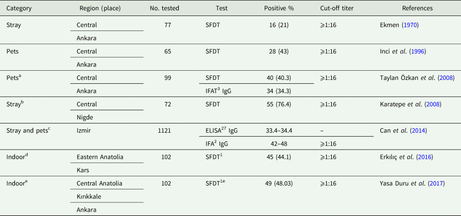
Cats are key in the epidemiology of T. gondii because they are the only hosts that can excrete environmentally resistant oocysts. Seroprevalence varied with age and life style of the cat (Table 4). Viable T. gondii was isolated from homogenates of hearts and brains in 20 of 100 cats by bioassay in mice (Can et al., Reference Can, Döşkaya, Ajzenberg, Özdemir, Caner, Iz, Döskaya, Atalay, Çetinkaya, Ürgen, Karaçali, Ün, Dardé and Gürüz2014). Additionally, T. gondii DNA was detected in tissues of 12 additional cats. These 32 T. gondii strains were used for genotyping – (see molecular epidemiology and genotyping analysis section).
Serological prevalence of T. gondii at cats in different regions of Turkey

a The seropositivity of T. gondii in cats older than 1 year (47.8%) was more about three times than cats less than a year old (13.6%). The seropositivity in indoor cats was 23.1% by IFAT and 30.8% by SFDT. The seropositivity in stray cats was 41.7% by IFAT and 52.8% by the SFDT. The presence of antibodies was significantly related to outdoor access.
b Seropositivity in male cats (48.6%) was lower than in female cats (51.4%).
c Tissues of 100 cats were used to isolate viable T. gondii (shown in the text).
d No significant differences for the seropositivity between 20 (44.4%) of 45 males and 25 (43.9%) of 57 females.
e Nested PCR was positive for four cats (8.2%). Foetal toxoplasmosis was diagnosed in one cat.
To our knowledge, there is only one report dealing with prevalence of T. gondii-like oocysts in cats from Turkey. Toxoplasma gondii-like oocysts measuring 9–14 µm in size were found in faeces of three of 36 (8.3%) cats from farms, however, results were not confirmed by bioassay or by PCR (Muz et al., Reference Muz, Altuğ and Karakavuk2013).
There are three reports of clinical toxoplasmosis in cats from Turkey. The first report concerns three cats (one, 8-month old male necropsied in 1988, one 3-month old female and one 6 month old female, both necropsied in 1984) (Haziroglu et al., Reference Haziroglu, Altınsaat, Atasever and Akın1988; Haziroglu, Reference Haziroglu1993). All three cats had disseminated toxoplasmosis and T. gondii was detected in multiple tissues of these cats histologically and by transmission electron microscopy.
Histologically confirmed disseminated toxoplasmosis was detected in a 2.5 year-old queen and her two kittens (Atmaca et al., Reference Atmaca, Dincel, Macun, Terzi, Uzunalioglu, Kalender and Kul2013). The queen was hospitalized because of dystocia. Ultrasound examination revealed that three of five kittens had died in utero. After cesarean section, the queen and the two live kittens died. The queen had pneumonia and hepatitis. The presence of T. gondii was confirmed by both immunohistochemical examination and PCR testing in the kittens and the queen, thus, confirming transplacental toxoplasmosis.
Dermal toxoplasmosis was diagnosed in 2-year old female immunocompetent Angora cat (Kul et al., Reference Kul, Atmaca, Deniz and Süer2011). The cat had an antibody titer of 1:256 using the dye test and diagnosis was confirmed immunohistochemically and by PCR testing. The dermal lesions resolved after treatment with azithromycin.
Dogs
Serologic reports are summarized in Table 5. Viable T. gondii was isolated from lungs of a dog (Ekmen and Altıntaş, Reference Ekmen and Altıntaş1970) but details of the dog's condition are missing. Clinical toxoplasmosis was diagnosed postmortem in a dog owned by an American visitor to Turkey (Akcay et al., Reference Akcay, Pamukçu and Baran1950); in retrospect it is not clear if the dog had toxoplasmosis or neosporosis (Dubey et al., Reference Dubey, Hemphill, Calero-Bernal and Schares2017).
Serological prevalence of T. gondii in dogs from different regions of Turkey

Toxoplasmosis in livestock
Sheep and goats
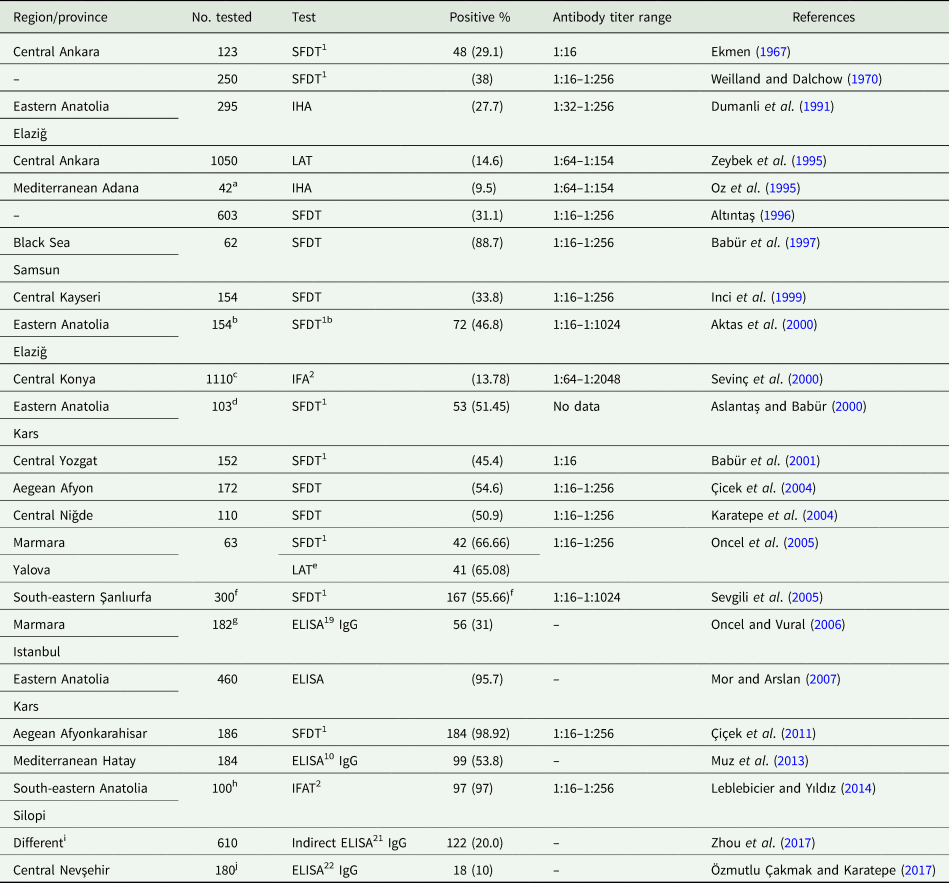
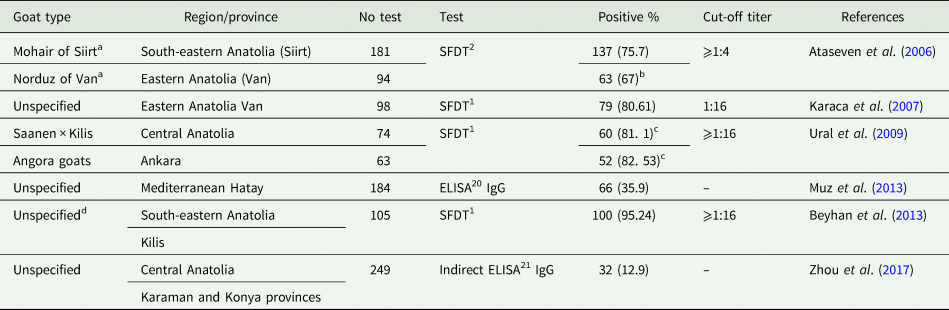
Serological surveys indicate widespread exposure to T. gondii in sheep and goats in Turkey (Tables 6 and 7). Although toxoplasmosis is a major cause of abortion in sheep and goats worldwide (Dubey, Reference Dubey2010), there is little information from Turkey. Toxoplasma gondii-associated abortion cannot be diagnosed alone by serological testing of ewes; a negative serology rules out toxoplasmosis but positive result does not establish aetiology because seroprevalence is high in general population and antibodies can remain elevated in the next pregnancy (Dubey, Reference Dubey2010). To our knowledge, there is no confirmed report of T. gondii-associated abortion in sheep or goats from Turkey. Although T. gondii DNA was found in five of 20 sheep tissues (Ergin et al., Reference Ergin, Ciftcioglu, Midilli, Issa and Gargili2009), there is no report of isolation of viable T. gondii from sheep and goats in Turkey.
Serological prevalence of T. gondii in sheep in different regions of Turkey

a 25.5% seropositivity of 259 aborted sheep with titers of 1:64–1:256.
b Pregnant 56, aborted 57 in the previous year, aborted 41 within the period of the study. Inactivating temperature for the ovine complement was 56 °C. Ovine sera should be heated to 60°C to inactivate complement (Dubey, Reference Dubey2010).
c Healthy ewes 827, ewes aborted 283 (10.16%). No significant difference between the two groups for the presence of antibody titers.
d 5 of 10 aborted sheep were seropositive for T. gondii.
e 63 sheep older than one year of age were tested. SFDT was accepted as a reference test. The specificity and sensitivity of LAT were 61.90 and 78.57%, respectively. The correlation between two tests was 73.01%.
f The seropositivity of 0–1 year old was 58.34% and >1 year age was 47.23%, respectively. The seropositivity of male (47.45%) and female (57.67%) and the seropositivity of Akkaraman breed (54.8%), Ivesi (55.71%) and Morkaraman (63.63%). No significant correlation between serum titers of age, sex and breed.
g No dissimilarity between female (31.4%) and male ewes (30). The significant difference between 0.6–1 year age (12.5%) and >1 year age (41%),
h Seropositivity of aborted 25 sheep was 96%. No significant difference between 2–4 year (96%) and 5–10 year (100%) sheep.
i Karaman, Konya provinces from Central region and Zonguldak province from Black Sea. Identify specific antibodies to T. gondii (rTgSAG2-ELISA).
j The seropositivity 1 to 2 years sheep (11.53%) and 5 to 7 years (8.51%). No statistically significant differences between the two age groups. Toxoplasma gondii antibodies were detected in 18 (11.1%) out of 162 ewes, while there was no seropositivity in the 18 rams tested.
Serological prevalence of T. gondii in goats from different regions of Turkey

a Mohair goats used for the production of mohair and blankets and Norduz are dairy breed.
b 42.85% seropositive at 1:16, 27.55% at 1:64, 7.17% at 1:256 and 3.06% at 1:1024.
c 38.33% seropositivity of 60 Saanen × Kilis goats at 1:16, 50% at 1:64, 6.67% at 1:256 and 5% at 1:1024. 36.5% seropositivity of 52 Angora goats at 1:16, 44.2% at 1:64, 11.53% at 1:256 and 7.7% at 1:1024.
d 100% of the Shami goats were seropositive with titers of 1:16 in 40 of 53, 12 at 1:64 and 1 at 1:256. 90.38% of Kilis goats were seropositive with titers of 1:16 in 36 of 52, 10 at 1:64 and 1 at 1:256.
Horses
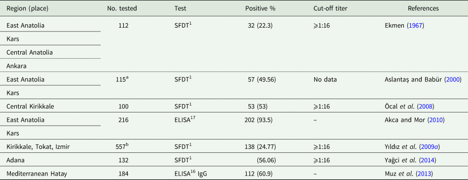
Serologic data are summarized in Table 8. There is no report of clinical toxoplasmosis in horses.
Serological prevalence of T. gondii in horses in different regions of Turkey

Cattle
There is limited information concerning toxoplasmosis in cattle in Turkey (Table 9). Additionally, the dye test used in several surveys is nonspecific and gives erratic results with cattle sera (Dubey and Beattie, Reference Dubey and Beattie1988). Finding of T. gondii DNA in five of 10 beef samples (Ergin et al., Reference Ergin, Ciftcioglu, Midilli, Issa and Gargili2009) needs confirmation.
Serological prevalence of T. gondii in cattle in different regions of Turkey

a 13 of 30 aborted cattle were seropositive for T. gondii.
b Aborted 234, pregnant cows 323.
Pigs
Toxoplasma gondii infection is not a significant direct risk for human in Turkey because pork is not eaten in Turkey due to religious restrictions, 99.8% of population is muslim ((Library of Congress. Federal Research Division website: https://en.wikipedia.org/wiki/Islam_in_Turkey#cite_note-11 (accessed 29 August 2018)).
Various other animals
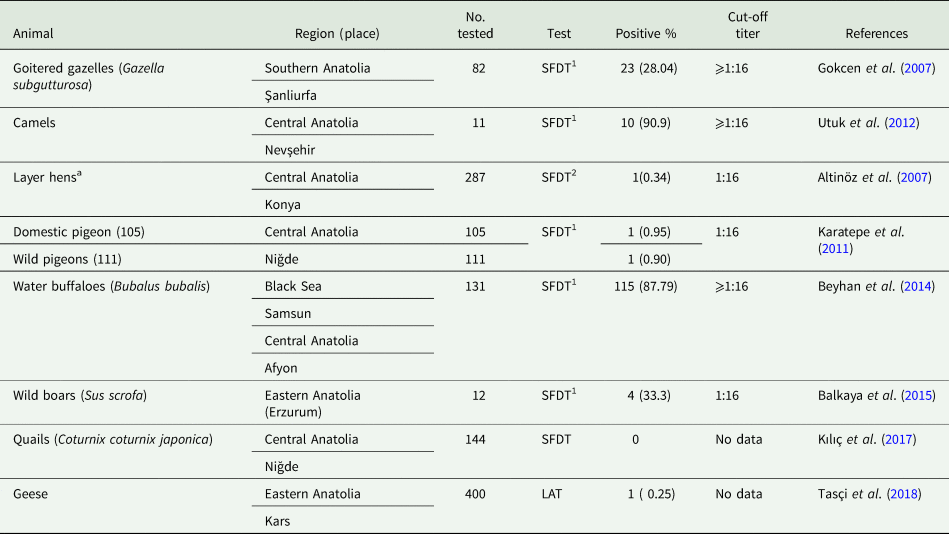
Serologic prevalence of T. gondii in miscellaneous animals is shown in Table 10. There is no confirmed report of clinical toxoplasmosis in animals, except in a zoo animal. Fatal toxoplasmosis was diagnosed in a captive kangaroo (Macropus sp.) from a zoo in Ankara (Kabak et al., Reference Kabak, Guvenc, Kul, Deniz and Gulbahar2011). Toxoplasma gondii DNA was demonstrated in the brain of a badger (Meles meles); indicating contamination of local waters by T. gondii (Karakavuk et al., Reference Karakavuk, Aldemir, Şahar, Can, Özdemir, Döşkaya, Gürüz and Döşkaya2018a).
Serological prevalence of T. gondii in miscellaneous animals in different regions of Turkey

a The SFDT does not work with chicken sera (Dubey, Reference Dubey2010).
Meat as source of T. gondii infectıon
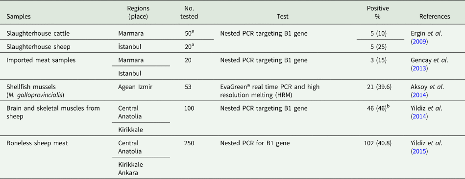
Humans acquire T. gondii infection postnatally by eating undercooked meat containing tissue cysts or ingesting food and water contaminated with oocysts (Dubey, Reference Dubey2010). There are reports of very high prevalence of T. gondii tissue cysts in sheep in Turkey destined for human consumption (Yildiz et al., Reference Yildiz, Kul, Gökpinar, Atmaca, Gençay, Gazyağci, Babür and Gürcan2014, Reference Yıldız, Pişkin, Ütük and Gökpınar2015). In the first of these two reports, antibodies to T. gondii were found in 88 of 100 sheep; tissue cysts were detected in 46, in 36 by using the Percoll concentration technique in 5 g of ovine brain. Additionally, by using immunohistochemical staining with T. gondii antibodies, tissue cysts were found in 17% of tissue sections. However, the parasites assumed to be Toxoplasma depicted in Fig. 2 of their paper are not T. gondii and are most likely Sarcocystis spp. (J. P. Dubey, own opinion). Polyclonal T. gondii antibodies can cross react with Sarcocystis (Dubey, Reference Dubey2010). In the second report by these authors, tissue cysts were detected in 21.2% of 250 sheep meat samples by the Percoll method (Yildiz et al., Reference Yıldız, Pişkin, Ütük and Gökpınar2015).
There is a similar report of high prevalence of T. gondii tissue cysts in buffalo meat imported from India into Turkey (Gencay et al., Reference Gencay, Yıldız, Gokpınar and Leblebicier2013). Tissue cysts were found in meat in three of 20 (15%) buffaloes tested by the Percoll method. The diagnosis was reported to be confirmed by PCR in both the buffalo and sheep-derived tissue cysts. However, the parasites assumed to be T. gondii depicted in Fig. 1 of their paper (Gencay et al., Reference Gencay, Yıldız, Gokpınar and Leblebicier2013) are most likely pollen grains and not T. gondii tissue cysts (J. P. Dubey, own opinion). Additionally, water buffaloes are considered resistant hosts of T. gondii, and T. gondii has not been isolated from buffalo meat in any country (Dubey, Reference Dubey2010).
These studies need confirmation because the density of T. gondii cysts in adult sheep is one tissue cyst in more than 100 g sheep tissue (Dubey, Reference Dubey2010). Reports of the presence of DNA in tissues of food animals are summarized in Table 11. However, detection of DNA does not relate to the presence of live organisms.
Environmental contamination by T. gondii oocysts
Cats can excrete millions of T. gondii oocysts and oocysts that can survive harsh conditions in the environment (Dubey, Reference Dubey2010). Toxoplasma oocyst DNA has been detected in water samples from different regions in Turkey but not in drinking water (Table 12). Overall, sedimentation and filtration procedures are efficient in trapping T. gondii oocysts, water used for irrigation and farm animals may not be filtered.
Detection of T. gondii from tissues of food animals in different regions of Turkey

a Toxoplasma gondii was found in 2% of 50 bovine brains, 6% of 50 bovine muscles, 4.17% of 120 ovine brains, 20% of 20 ovine muscles and 19% of 100 fermented sausage samples.
b Tissue cysts were at 78.2% of 36 brain and at 69.5% of 32 of skeletal muscles (masseter, tongue, diaphragm, intercostal and leg).
Detection of Toxoplasma gondii from water samples in different region of Turkey

Additionally, T. gondii oocysts can be concentrated by molluscs and fish. Toxoplasma gondii DNA was detected in 39.6% of edible shellfish (Mytilus galloprovincialis) in Izmir (Aksoy et al., Reference Aksoy, Marangi, Papini, Ozkoc, Bayram Delibas and Giangaspero2014).
Molecular characterization
Although T. gondii infections are prevalent worldwide, there is a geographic distribution of T. gondii genetic types (Shwab et al., Reference Shwab, Zhu, Majumdar, Pena, Gennari, Dubey and Su2014). Except for minor variations, T. gondii strains are broadly divided into four types: I, II, III and atypical. In general, Type I stains are rare worldwide (Dubey, Reference Dubey2010). Type II and Type III strains are prevalent worldwide, except Brazil where they are rare. The T. gondii strains in Europe are similar to those in North America. An unusual strain, designated Africa 1 genotype, has been identified in humans from sub-Saharan Africa (Döşkaya et al., Reference Döşkaya, Caner, Ajzenberg, Değirmenci, Dardé, Can, Erdogan, Korkmaz, Üner, Güngör, Altintas and Gürüz2013), and more recently in Denmark (Jokelainen et al., Reference Jokelainen, Murat and Nielsen2018).
As stated in the Introduction section, Turkey is geographically unique, a bridge between Asia, Africa and Europe therefore, providing an avenue for cross mixing of T. gondii strains.
For genotyping of T. gondii, it is important to have good quality T. gondii DNA with minimal contamination of host tissue. For this, it is often necessary to extract DNA from viable strains. This has been achieved in three studies from Turkey. Döşkaya et al. (Reference Döşkaya, Caner, Ajzenberg, Değirmenci, Dardé, Can, Erdogan, Korkmaz, Üner, Güngör, Altintas and Gürüz2013) found that both the T. gondii isolates obtained from CSF of diseased children were Africa 1 genotype. This Africa 1 genotype has been found in a cat but not yet identified in other hosts in Turkey. Of the 22 isolates of T. gondii genotyped from domestic cats in Turkey, 19 were classical Type II, two were Type III, and one was Africa 1 genotype (Can et al., Reference Can, Döşkaya, Ajzenberg, Özdemir, Caner, Iz, Döskaya, Atalay, Çetinkaya, Ürgen, Karaçali, Ün, Dardé and Gürüz2014). Of the five viable isolates of T. gondii genotyped from wild birds in Turkey, four were Type II and one was Type III (Karakavuk et al., Reference Karakavuk, Aldemir, Mercier, Atalay Şahar, Can, Murat, Döndüren, Can, Özdemir, Değirmenci Döşkaya, Pektaş, Dardé, Gürüz and Döşkaya2018b). Thus, the predominant distribution of genetic types of T. gondii in Turkey was like Europe.
Conclusıon
It is evident from this review that there are many gaps in our knowledge of toxoplasmosis in humans and animals, particular data on clinical infections are lacking. There is no centralized facility for advice and confirmation of diagnosis. Little is known about the rate of congenital toxoplasmosis, and follow up of subclinically infected children for clinical toxoplasmosis. No information is available concerning modes of transmission. Little information is available concerning the presence of viable T. gondii in edible meats, and T. gondii oocysts in the environment. Educational programs are needed to prevent T. gondii infection in humans and animals.
Acknowledgements
We thank Oliver Kwok, Meghan Sadler, Camila Cezar and Fernando Murata for their help with the paper.
Fınancıal support
We would like to thank The Scientific and Technological Research Council of Turkey (1059B191800417) for fellowship to Zeynep Kolören.
Conflıcts of ınterest
None.
Ethıcal standards
Not applicable.