INTRODUCTION
Schistosomiasis is a life-threatening parasitic disease in tropical and subtropical developing countries. According to the latest data, although effective treatment has been given, there are still 800 million people, at least, under threat around the world, and nearly 80 countries and over 200 million people have been infected (McManus, Reference McManus2012). Currently the treatment of schistosomiasis is recognized as Praziquantel, which can effectively kill the matured parasites, but it has no effect on the eggs and schistosomula.
The eggs and their secondary pathological impairment are considered as the main aetiology in schistosomiasis. The eggs arrested in liver continue to cause pathological reactions, formation of egg granulomas and secondary hepatic fibrosis, involving in the formation of some egg antigens. Another disadvantage for Praziquantel treatment is that drug-resistant strains of Schistosoma japonicum have been isolated from infected patients (Wang et al. Reference Wang, Wang and Liang2012, Reference Wang, Li, Qu, Xing, Yang, Dai and Liang2014). Therefore, killing the adult parasites is evidently insufficient in the treatment of S. japonicum. How to alleviate subsequent liver fibrosis caused by egg granulomas is the key point in reducing mortality and morbidity of schistosomiasis (Smith et al. Reference Smith, Shainheit, Bazzone, Rutitzky, Poltorak and Stadecker2009).
Recent studies showed that interleukin (IL)-13 plays a pivotal role in the process of fibrosis in many chronic infectious diseases, especially in schistosomiasis-associated liver fibrosis (Sugawara et al. Reference Sugawara, Azuma, Onodera, Tsunoka and Morimoto2010; Gray et al. Reference Gray, Ross, Li and McManus2011; Liu et al. Reference Liu, Munker, Mullenbach and Weng2012). In scahistosomiasis, IL-13 signals transmit starting from binding the IL4R/IL-13Rα1 complex receptor, then leading phosphorylation of JAK1/IRS/STAT6, and resulting in liver fibrosis ultimately (Burke et al. Reference Burke, Jones, Gobert, Li, Ellis and McManus2009; Seki et al. Reference Seki, Kumagai, Kwansa-Bentum, Furushima-Shimogawara, Anyan, Miyazawa, Iwakura and Ohta2012). The IL-13/JAK1/IRS/STAT6 pathway is the main signal pathway in schistosomiasis-associated liver fibrosis. Thereby interfering with the IL-13 and downstream signal transduction may become a new strategy to control schistosomiasis-associated hepatic fibrosis.
Corilagin (C27H22O18) is identified in several plants including Phyllanthus (Hau et al. Reference Hau, Zhu, Leung, Wong, Cheng, Lai, Tong, Lau, Chan, Wong, Lam, Cheng, Cheung, Chui, Gambari and Fong2010). Our previous studies showed corilagin is effective in hepatic protection, anti-inflammation, anti-oxidant, anti-HSV-1 encephalitis and anti-fibrosis in schistosomiasis (Zhao et al. Reference Zhao, Zhang, Tao, Pang, Jin, Guo, Dong, Ye, Zhao and Zheng2008; Jin et al. Reference Jin, Cheng, Tao, Zhang, Pang, Guo, Ye, Dong and Zhao2010; Guo et al. Reference Guo, Zhao, Li, Mei, Zhang, Tao, Zhou and Dong2010, Reference Guo, Luo, Wu, Liu, Li, Mei, Zhang, Tao, Dong, Fang and Zhao2015a ; Huang et al. Reference Huang, Zhang, Jin, Cheng, Zhou, Li, Tang, Xue, Cai, Dong and Zhao2013). It is deserved to explore which signal pathway corilagin interferes with in the anti-fibrosis activation.
Thereby, in this study we set up the cellular model with hepatic stellate cells (HSC)-T6 cells stimulated by rIL-13 and established schistosomiasis animal model and administered with corilagin to treat those models, hoping to find the mechanisms of corilagin in ameliorating schistosomiasis-associated hepatic fibrosis.
MATERIALS AND METHODS
Chemicals and reagents
Cell counting kit-8 (CCK-8) was purchased from Dojindo Company. The rIL-13 was offered from PeproTech. The Trizol reagent, RNAios and real-time polymerase chain reaction (PCR) kit were provided from TaKaRa Company. Anti-JAK1 antibody was provided by Proteinteck. Anti-IL13Rα1 antibody was offered from Abcam Biotechnology. The Immunohistochemistry (IHC) kits and HRP-labelled secondary anti-bodies were obtained from BosterBiotechnology, Wuhan, China. Praziquantel was provided from Schistosomiasis Control Institute of Hubei Province. The S. japonicum cercariae were given by Parasite Department of Tongji Medical College, Huazhong University of Science and Technology.
Cell administration
HSC-T6 cells were bought from Xiangya Central Laboratory. The cells were cultured in DMEM medium containing 10% fetal bovine serum in the incubator at 37 °C, 5% CO2 and saturated humidity. HSC-T6 cells were divided into three groups. The corilagin group was stimulated with rIL-13 (100 ng mL−1) for 2 h before corilagin (0·25 mg mL−1) was added. The model group was incubated with rIL-13 (100 ng mL−1) only. The normal group without any treatment was as negative control. After 24 h the cells were harvested for total RNA isolation and proteins extraction.
Corilagin cytotoxicity assays
The HSC-T6 cells were plated at a density of 1 × 10⁴ cells mL−1 in 96-well culture plates and different concentrations of corilagin (2, 1, 0·5, 0·25, 0·125, 0·0625 mg mL−1) were added. After 24 h, 10 µL of CCK-8 solution were added to each well followed by 2 h incubation before measuring the absorbance at 450 nm using a microplate reader. Five wells in each group were measured.
Animals and treatment
Forty male Balb/c mice, weighting 18–22 g, 6–8 weeks old, were purchased from Hubei Provincial Center for Disease and Prevention. The mice were feed in a 22–25 °C temperature and 50–60% humidity for 1 week. Then the animals were randomly divided into 6 groups, model group, normal group, praziquantel group, corilagin (10 mg kg−1) group,corilagin (20 mg kg−1) group and corilagin (40 mg kg−1) group. All the groups were infected with 20 ± 2 S. japonicum cercariae transcutaneously, except the normal group (Zhou et al. Reference Zhou, Cheng, Zhang, Li, Tang, Xue, Cai, Dong and Zhao2013). After 4 weeks, each infected mouse was given Praziquantel (500 mg kg−1 d−1) for 2 days orally to kill the schistosoma. Subsequently, corilagin with different dosage was administrated to the corilagin groups, and the other three groups were administrated with equivalent volume of saline for 4 weeks before all the mice were sacrificed. Parts of the liver tissues were fixed in 4% formalin for tissue examination and the rest were stored in a refrigerator of −80 °C for total RNA extraction and Western-blot testing.
Liver histological examination
The procedure was followed by our previous studies (Jin et al. Reference Jin, Zhang, Feng, Yuan, Zhang, Han, Li, Yu, Liu, Kong, Zhang and Zhao2015). The liver tissues that fixed in 4% formalin were embedded in paraffin. Four micrometers serial sections were obtained for haematoxylin and eosin (H&E) staining to observe the liver pathological changes. The size of egg-granuloma was observed under light microscope. Expression of Col (collagen) and Col III protein was examined by IHC following the instruction of IHC kit. For each liver section, five images were captured randomly under the microscope. Then the images were analysed by Image-Pro Plus5·0·1 software. The relative quantitative expression of Col I and Col III was estimated by mean optical density. The egg granulomas change in liver was observed by Masson staining following the instruction of manufacture.
Total RNA isolation and quantitative real-time PCR
The procedure abided by our past studies (Guo et al. Reference Guo, Luo, Wu, Mei, Peng, Liu, Li, Zhang, Dong, Fang and Zhao2015b ). Total RNA was isolated by using the Trizol reagent, and reverse transcription was performed following the protocol of kit. Amplification steps were followed with the instruction of kit and the reaction conditions were 95 °C 30 s; 95 °C 5 s, 60 °C 30 s, 40cycles; 95 °C 15 s, 60 °C 1 min, 95 °C 15 s. The results were analyzed by using 2−△△CT method. GAPDH was used as the internal control. The primers for quantitative real-time PCR were as follows:
IL-13-forward, 5′-ATGAGGAGAGACCATCCCTG-3′,
IL-13-reverse, 5′-CATCTCCCTTCCTCCTCAAC-3′,
Col I-forward, 5′-CAGCCATCGACTAGGACAGA-3′,
Col I-reverser, 5′-AGAACAGCATTGCGTACCTG-3′,
Col III-forward, 5′-GCCCGGAAGAATACG-3′,
Col III-reverse, 5′-ACATCTGGGAAGCAAA-3,
JAK-1-forward, 5′-GCCTGGAGTGGAGGTGACTTTCT-3′,
JAK-1-reverse, 5′-TTCGTTGTCATTGGTTCCGTGCC-3′,
IL13Rα1-forward, 5′-TGCTCCAGAAACTCATCGTAAA-3′,
IL13Rα1-reverse, 5′-AAGGGCTAGGCTTCTCACTTTC-3′.
GAPDH-forward, 5′-TGTGTCCGTCGTGGATCTGA-3′,
GAPDH-reverse, 5′-CCTGCTTCACCACCTTCTTGA-3′,
Western-blot analysis
The expression of JAK-1 and IL13Rα1 proteins were detected by Western blot. The cells or liver tissue proteins from each sample were separated on SDS-PAGE, and then transferred to nitrocellulose filter membranes, which were blocked overnight with 5% non-fat milk in TBST. Blots were probed over night at 4 °C with rabbit polyclonal anti-JAK1 or anti-IL13Rα1 (diluted as 1:500) and then were incubated with HRP-labelled secondary antibody for 2 h before being washed 5–6 times in TBST. After washed with TBST, ECL was added to identify the immunoreactive bands. The densitometry analysis of the immunoreactive bands was performed using the Fuji ultrasonic-doppler velocity profile system and Image J program.
Ethical considerations
All study protocols were approved by the Institutional Animal Care and Use Committee at Tongji Medical College, Huazhong University of Science and Technology, 2012 IACUC No. 307. All the surgeries were performed under 6% chloral hydrate anaesthesia by intraperitoneal injection, and all efforts were made to minimize suffering.
Statistical analysis
The data were expressed as the means ± s.d. Comparisons between groups were performed with one-way ANOVA test and Student's t-test. All data were analyzed by using SPSS12·0 statistic software (Ding et al. Reference Ding, Li, Xiong, Yin, Liu, Liu, Wang, Xia, Zhang and Zhao2015). P < 0·05 was regard as statistically significant difference.
RESULTS
Cytotoxicity of corilagin on HSC-T6 cells
It was showed that 0·25 mg mL−1 corilagin was no harm on HSC-T6 cells (data were not shown). We chose the concentration for the following treatment.
Effect of corilagin on expression of JAK-1 and IL-13Rα1 on cellular model
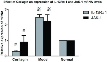
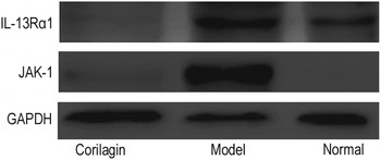
In model group the mRNA and protein levels of JAK-1 and IL-13Rα1 were significantly increased when compared with that in normal group (P < 0·01). With corilagin intervention, JAK-1 and IL-13Rα1 gene expression were significantly reduced when compared with that in model group (P < 0·01) (Fig. 1). The protein levels were also greatly decreased in the corilagin groups when compared with that in model group (Fig. 2).
Effect of corilagin on gene expression of JAK-1 and IL-13Rα1 on cellular model. ※P<0·01 compared with the normal group. #P < 0·01 compared with the model group.

Effect of corilagin on protein expression of IL-13Rα1 and JAK-1. The protein levels were greatly decreased in the corilagin groups when compared with that in model group.

Effect of corilagin on liver histopathology
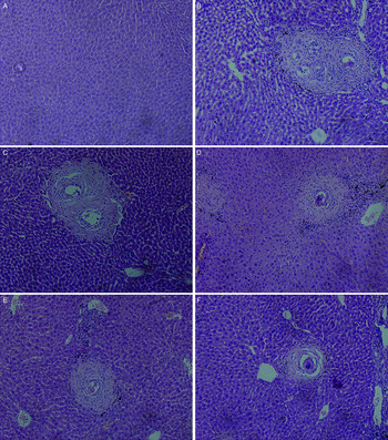
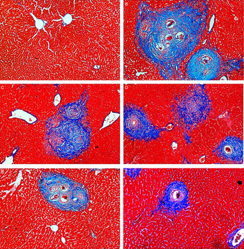
The hepatic lobules were intact and the cells were arranged regularly in normal group. H&E staining and Masson staining showed enormous egg granulomas surrounded by collagen and inflammatory cells in model group. The same pathological changes were also found in praziquantel group. The size of granulomas decreased and less inflammatory cells infiltration happened in the corilagin treated groups. (Figs 3 and 4)
Effect of corilagin on liver histopathology by H&E staining. (A) normal group, (B) model group, (C) praziquantel group, (D) corilagin 10 mg kg−1 group, (E) corilagin 20 mg kg−1 group, (F) corilagin 40 mg kg−1 group. H&E staining showed enormous egg granulomas surrounded by collagen and inflammatory cells in the model group. The same pathological changes were also found in praziquantel group. The size of granulomas decreased and less inflammatory cells infiltrated in the corilagin groups.

Effect of corilagin on liver histopathology by Masson staining. (A) normal group, (B) model group, (C) praziquantel group, (D) corilagin 10 mg kg−1 group, (E) corilagin 20 mg kg−1 group, (F) corilagin 40 mg kg−1 group. Masson staining showed the liver fibrosis. Deposition of collagen was found surrounding egg granulomas in the model group. The same pathological changes were also found in praziquantel group. The size of granulomas decreased and less collagen deposition happened in the corilagin groups.

Effect of corilagin on expression of Col I and Col III on animal model
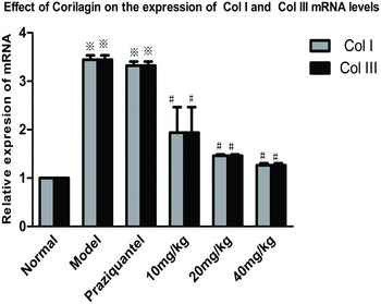
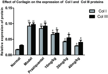
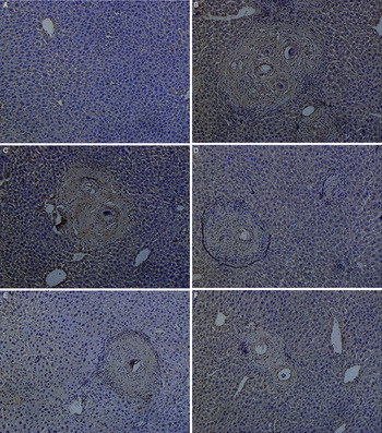
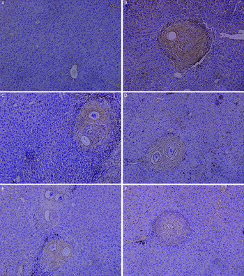
The mRNA (Fig. 5) and protein (Fig. 6) levels of Col I and Col III were increased in model group and praziquantel group when compared with that in normal group (P < 0·01), while they were significantly decreased in the corilagin groups when compared with that in the model group and praziquantel group (P < 0·01). Col I (Fig. 7) and Col III (Fig. 8) proteins were detected with IHC. Collagens were observed around the portal area, granulomas and fibrotic areas both in model and praziquantel groups. Less collagens were found in the corilagin treated groups.
Effect of corilagin on expression of ColI and ColIII mRNA levels on animal model. ※P < 0·01 compared with the normal group; #P < 0·01 compared with the model group.

Effect of corilagin on expression of Col I and Col III protein levels on animal model. ※P < 0·01 compared with the normal group; #P < 0·01 compared with the model group.

Expression Col I protein on animal model. (A) normal group, (B) model group, (C) praziquantel group, (D) corilagin 10 mg kg−1 group, (E) corilagin 20 mg kg−1 group, (F) corilagin 40 mg kg−1 group. IHC showed the Col I protein levels were increased in model group and praziquantel group compared with normal group, while the levels were significantly decreased in corilagin groups compared with that in the model group and praziquantel group.

Expression of Col III protein on animal model. (A) normal group, (B) model group, (C) praziquantel group, (D) corilagin 10 mg kg−1 group, (E) corilagin 20 mg kg−1 group, (F) corilagin 40 mg kg−1 group. IHC showed the Col III protein levels were increased in model group and praziquantel group compared with normal group, while the levels were significantly decreased in corilagin groups compared with that in the model group and praziquantel group.

Effect of corilagin on mRNA expression of IL-13 on animal model
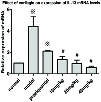
The mRNA levels of IL-13 were detected with quantitative real-time PCR. In model group and praziquantel group the mRNA level of IL-13 increased when compared with that in normal group (P < 0·01). In the corilagin groups the mRNA of IL-13 were significantly decreased when compared with that in model group and praziquantel group (P < 0·01) (Fig. 9).
Effect of corilagin on mRNA expression of IL-13 on animal model. ※P < 0·01compared with the normal group, #P < 0·01 compared with the model group.

Effect of corilagin on expression of JAK-1 on animal model
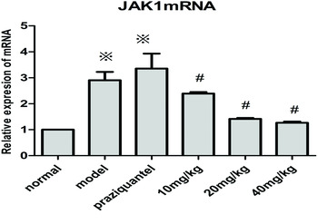
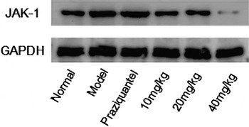
In model group and praziquantel group JAK-1 gene expression were significantly increased when compared with that in normal group (P < 0·01). In the corilagin groups JAK-1 gene expression were significantly decreased when compared with that in model group and praziquantel group (P < 0·01) (Fig. 10). Furthermore, Western blotting was employed to test changes of JAK-1 protein in the liver (Fig. 11). JAK-1 protein expressions were greatly increased in model group and praziquantel group when compared with that in normal group, while in the corilagin groups JAK-1 protein were decreased when compared with that in model group.
Effect of corilagin on JAK-1 gene expression on animal model. ※P < 0·01 compared with model group. JAK-1 gene expression was significantly increased in model group and praziquantel group. #P < 0·01 compared with the model group. JAK-1 gene expression was significantly decreased in corilagin groups.

Effect of corilagin on JAK-1 protein expression on animal model. Western-blot assay showed the JAK-1 protein expressions were greatly increased in model group and praziquantel group compared with the normal group, while in the corilagin groups JAK-1 protein were decreased compared with that in the model group.

DISCUSSION
In this study, we set up the cellular and animal model to explore the mechanisms of corilagin to control liver fibrosis caused by schistosoma. Compared with model group and praziquantel group, less inflammatory changes were found in all corilagin treated groups, which showed corilagin could ameliorate pathological changes of liver fibrosis. Furthermore, the levels of Col I, Col III were significantly decreased, and the expression of IL-13, JAK-1 and IL13Rα1 was downregulated, implying that mechanisms of corilagin to control schistosomiasis-caused fibrosis might be based on regulating IL-13 signal pathway.
IL-13, secreted mainly by activated Th2 cells, participates in various inflammatory disorders. Besides schistosomiasis-induced fibrosis, IL-13 is involved in a series of fibrotic diseases, including lung fibrosis as well as fibrotic stage in patients with HCV infection and steatohepatitis (Weng et al. Reference Weng, Liu, Chen, Huang, Xu, Godoy, Hu, Zhou, Stickel, Marx, Bohle, Zimmer, Lammert, Mueller, Gigou, Samuel, Mertens, Singer, Seitz and Dooley2009). Furthermore, elevated IL-13 levels were detected in serum of patients with rheumatoid arthritis (Spadaro et al. Reference Spadaro, Rinaldi, Riccieri, Taccari and Valesini2002). It was also shown that increased IL-13 level was found in Still's disease (Saiki et al. Reference Saiki, Uda, Nishimoto, Miwa, Mima, Ogawara, Azuma, Katada, Sawaki, Tsutsui, Matsui, Maeda and Nakanishi2004). IL-13 is the dominant cytokine that participates in the process of schistosomiasis-associated hepatic fibrosis (Morais et al. Reference Morais, Souza, Melo, Aroucha, Miranda, Domingues, Abath, Montenegro, Abath and Montenegro2008). Accumulated evidence showed that IL-13 triggered the fibrotic responses in schistosoma infection (Chu et al. Reference Chu, Du, Hu, Wu and Shen2011). The mortality was significantly reduced in IL-13 knockout mice with S. japonicum infection (Sica and Mantovani, Reference Sica and Mantovani2012; Seki et al. Reference Seki, Kumagai, Kwansa-Bentum, Furushima-Shimogawara, Anyan, Miyazawa, Iwakura and Ohta2012). IL-4 and IL-13 act synergistically by sharing the complex receptor IL4R/IL-13Rα1 (Caldas et al. Reference Caldas, Campi-Azevedo, Oliveira, Silveira, Oliveira and Gazzinelli2008).
During schistosoma infection, the Th1 immune response occurs at the early stage. However, the Th2 immune response develops and dominates in the chronic phases (Gordon and Martinez, Reference Gordon and Martinez2010; Sugawara et al. Reference Sugawara, Azuma, Onodera, Tsunoka and Morimoto2010). Studies showed that Th2 cytokines such as IL-13 and IL-4 can stimulate types of immune cells via IL13/JAK1/IRS/STAT6 pathway to product TGF-β1, Arg-1, which can promote the synthesis of extracellular matrix (ECM) and collagen deposition (Horsnell and Brombacher, Reference Horsnell and Brombacher2010).
Hepatic fibrosis is considered being excessive deposition of ECM, and Col I and Col III are the major components of ECM. HSCs have been recognized as the major source of the ECM (Fagone et al. Reference Fagone, Mangano, Mammana, Pesce, Pesce, Caltabiano, Giorlandino, Portale, Cavalli, Lombardo, Coco, Puleo and Nicoletti2015). HSCs can be activated by the injured neighbouring cells and various inflammatory cytokines including IL-13 and TGF-β (Liu et al. Reference Liu, Meyer, Muller, Herweck, Li, Mullenbach, Mertens, Dooley and Weng2011). So we set up the model of HSC-T6 cells stimulated by rIL-13, and stepped to interfere with the model by corilagin. The results showed that JAK-1 and IL-13Rα1 expression were decreased both in mRNA levels and proteins in corilagin treated groups.
In animal model, we found that collagens were observed around the portal area, granulomas and fibrotic areas both in model and praziquantel groups. But there were less collagen that was found in the corilagin treated groups, which demonstrated that corilagin can suppress the schistosome egg-induced collagen deposition in liver.
We also tested the IL-13 mRNA in the animals. Both in model group and praziquantel group IL-13 expression increased significantly. However, corilagin could downregulated IL-13 at mRNA and protein level. In addition, the expression of JAK-1 showed the same changes with IL-13, indicating that corilagin may ameliorate the schistosomiasis-associated liver fibrosis by down-regulating IL-13 and JAK-1 in vivo.
In conclusion, corilagin exerts the ability to ameliorate schistosomiasis-associated hepatic fibrosis by down-regulating the expression of IL-13 and signal molecules in IL-13 pathway. However, it is deserved to explore that corilagin may target the IL-13 pathway at more than one level or mediate anti-fibrotic effect via mechanisms other than the IL-13 pathway. We are hoping to carry on the study to find the further mechanisms that how corilagin works in the process against schistosomiasis fibrogenesis, and find a new way to control IL-13-associated liver fibrosis.
SUPPLEMENTARY MATERIAL
The supplementary material for this article can be found at http://dx.doi.org/10.1017/S0031182016001128.
FINANCIAL SUPPORT
This study is sponsored by China National Natural Science Fund No. 81371840 and No. 30901248 (to L. Z.).