Introduction
An arteriovenous malformation is an abnormal communication between an artery and a vein and is an uncommon clinical condition. Arteriovenous malformations in the head and neck are present at birth but are rarely symptomatic in the neonatal period. Instead, they manifest during late childhood, adolescence or early adulthood, and usually expand following minimal trauma, infection or hormonal changes.Reference McGill, Mulliken, Cummings, Fredrickson, Harker, Krause and Schuller1
In the head and neck, most arteriovenous malformations are located intracranially; they also occur in the cheek and parotid, ear, nose, forehead, and neck regions.Reference Wu, Bisdorff, Gelbert, Enjolras, Burrows and Mulliken2, Reference Kohout, Hansen, Piribaz and Mulliken3 Although there are large published series of head and neck arteriovenous malformations, auricular and external meatal arteriovenous malformation has been the subject only of single case reports and small series.Reference Wu, Bisdorff, Gelbert, Enjolras, Burrows and Mulliken2, Reference Yoshida, Ishizawa, Mitsuhashi, Kondo, Ohta and Sakai4–Reference Lee, Do, Yakes, Kim, Mattasi and Hyon6 The rarity of these latter arteriovenous malformations limits systematic study, their unpredictable behaviour renders prognosis difficult, and poor treatment outcomes are common.Reference Kohout, Hansen, Piribaz and Mulliken3
In this paper, we report a case of arteriovenous malformation with unilateral involvement of the auricula, external auditory meatus, mastoid process and lateral petrous segment. Such lesions are very rarely seen.
Case report
A 33-year-old woman was admitted to our clinic with a retro-auricular swelling which had enlarged over a three-year period, with accelerated expansion during her latest pregnancy, along with pain, pulsatile tinnitus and ear discharge.
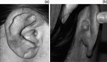
On inspection, the patient's right auricula was blue-purple in colour and hypertrophic. There was a pulsatile mass in the retro-auricular region, measuring 2 cm in diameter (Figure 1). Otomicroscopy revealed a purulent discharge in the external auditory canal. A 4 mm perforation was present in the postero-inferior quadrant of the ear drum.
Pre-operative appearance of right ear.

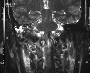
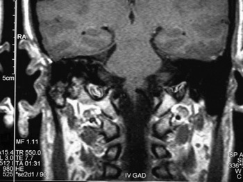
On temporal computed tomography scanning, a soft tissue density was seen to occupy the medial half of the right external auditory meatus, tympanic cavity, antrum and part of the mastoid cellules. There was also intensive contrast enhancement in the skin and subdermal regions of the external auditory canal, and the skin was thickened. These findings were initially considered to be consistent with a vascular malformation. On magnetic resonance imaging (MRI) demonstrated a lesion occupying the right auricula, external auditory canal, mastoid process of the temporal bone and lateral half of the petrous segment (Figure 2).
Coronal magnetic resonance imaging, T2-weighted turbo spin echo sequences, showing a high-flow vascular malformation occupying the external auditory canal and part of the petrous apex.

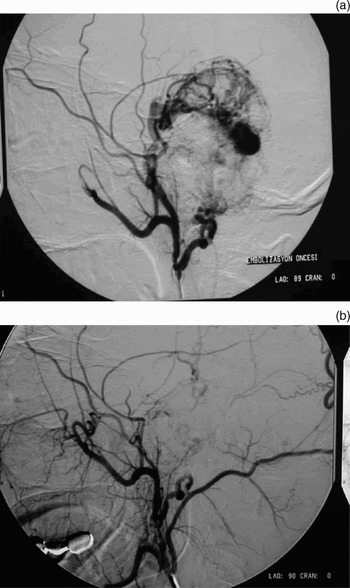
On angiography, an arteriovenous malformation was visualised at the right auricula, originating from the posterior auricular branch of the external carotid artery (Figure 3a). The lesion was embolised in the same session with polyvinyl alcohol particles. A second angiogram demonstrated almost total devascularisation of the malformation (Figure 3b).
(a) Selective external carotid angiography showing the arteriovenous malformation to be fed by the posterior auricular branch of the external carotid artery. (b) Angiogram taken after embolisation with polyvinyl alcohol particles, showing that the arteriovenous malformation has been almost completely devascularised.

Despite clinical regression in the dimensions of the mass, it persisted; therefore, a right mastoidectomy, arteriovenous malformation nidus excision and tympanoplasty were carried out. At pathological examination, the lesion was confirmed to be an arteriovenous malformation.
At three-month post-operative follow up, no recurrence was observed in the retro-auricular region, and regression in auricular size and normalisation of the colour of the covering skin were noted.
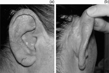
At three-year post-operative follow up, following a new pregnancy, there was complete resolution of tinnitus and continuing recovery of the dimensions and the colour of the auricula (Figure 4). An MRI taken at this stage showed no residual lesion or recurrence at the temporal bone and petrous apex, apart from a few scanty, serpiginous, vascular remnants (Figure 5).
Appearance of the right ear, three years after surgery.

Magnetic resonance imaging, Postoperative third year, showing an almost complete lack of residual or recurrent arteriovenous malformation in the temporal bone or petrous apex, apart from a few scanty, serpiginous, vascular remnants in the right auricula.

Discussion
Vascular lesions have a wide range of clinical presentations and an unpredictable course. According to the classification of Mulliken and Glowacki, vascular anomalies are divided into two main categories: haemangioma and vascular malformation. Haemangiomas are characterised by accelerated endothelial proliferation followed by regression over a five- to eight-year period. In contrast, the endothelial cells in vascular malformations have a normal rate of cellular division and do not regress. Vascular malformations are subdivided into slow-flow lesions (i.e. capillary, venous, lymphatic or combined forms) and fast-flow lesions (i.e. arteriovenous fistula and arteriovenous malformation).Reference McGill, Mulliken, Cummings, Fredrickson, Harker, Krause and Schuller1 Histologically, arteriovenous malformations may contain arteriovenous shunts with reactive, hypertrophic, thick-walled arteries and veins because of the increased blood flow.Reference Pham, Wong and Allison7
Arteriovenous malformations are thought to be present at birth but may not be clinically evident. Their increase in size is due to increased blood flow. Most vascular malformations present clinically during childhood and grow commensurately with the child. However, some malformations may remain quiescent until adolescence and in rare cases into adulthood. Enlargement of the arteriovenous malformation may be triggered by trauma, infection, puberty or other hormonal influences such as pregnancy. Our patient noted rapid enlargement of her pre-existing retro-auricular arteriovenous malformation throughout her pregnancy.
In the head and neck region, most arteriovenous malformations are located intracranially; they also occur in the cheek and parotid, ear, nose, forehead, and neck regions.Reference Wu, Bisdorff, Gelbert, Enjolras, Burrows and Mulliken2, Reference Kohout, Hansen, Piribaz and Mulliken3 In the literature, auricular and external auditory meatus arteriovenous malformations have been the subject only of single case reports and small series.Reference Wu, Bisdorff, Gelbert, Enjolras, Burrows and Mulliken2, Reference Yoshida, Ishizawa, Mitsuhashi, Kondo, Ohta and Sakai4, Reference Bapuraj, Bilwani, Khandelwal, Gupta, Nehru and Suri5, Reference Raghu, De, Higgins and Axon8 Our case demonstrated involvement of the unilateral auricula, external auditory meatus, mastoid process and the lateral regions of the petrous segment. A literature search could not identify any previously published case of such an extensive arteriovenous malformation involving the temporal area.
The clinical manifestations of arteriovenous malformations are related to abnormal blood perfusion and mass effect. An arteriovenous malformation may result in increased warmth, oedema of the affected tissues, pulsation and a bruit on auscultation. The distressing symptoms of auricular arteriovenous malformation may include throbbing pain, a buzzing sensation and pulsatile tinnitus which may prevent sleep. Furthermore, shunting may cause ischaemic injuries to part of the lesion, leading to ulceration and bleeding. In our patient, the treatment modality was determined by worsening of the pain and tinnitus to an unbearable level.
A clinical diagnosis of arteriovenous malformation is often indicated by the history and physical examination. However, MRI and angiography are essential for evaluating symptomatic malformations. Magnetic resonance imaging is the current ‘gold standard’ for assessing the extent of involvement within tissue planes and for demonstrating flow characteristics.
Angiography demonstrates the feeding vessels, as well as providing information on anastomoses with other extracranial or intracranial vessels, and on the venous drainage pattern. Angiography is usually reserved until after treatment planning is complete, and is used for ‘superselective’ embolisation before surgical extirpation. The target of embolisation must be the nidus or epicentre of the arteriovenous malformation rather than proximal feeding vessels. Embolisation may also be used for pre-operative devascularisation, in order to minimise intra-operative blood loss and to provide a dry operative field.
Clinically, it is difficult to select the optimal treatment which will maintain long-term success in arteriovenous malformation management. If the arteriovenous malformation is small and asymptomatic, treatment is unnecessary.Reference Wu, Bisdorff, Gelbert, Enjolras, Burrows and Mulliken2, Reference Pham, Wong and Allison7 For symptomatic arteriovenous malformations treatment is often difficult associated with higher risk of complications and morbidity. Optimal treatment is based on a combined approach of superselective embolisation and complete surgical excision.Reference Raghu, De, Higgins and Axon8 Aggressive control of the arteriovenous malformation nidus is essential. Kohout et al. have stated that, in terms of the extent of resection, complete excision of the nidus is critical.Reference Kohout, Hansen, Piribaz and Mulliken3 In arteriovenous malformations in any location, there is no place for proximal ligation of vessels, as this will result in re-expansion of the arteriovenous malformation.
• Arteriovenous malformations of the head and neck usually occur intracranially; occurrence outside the cranium is rare
• Although there have been single case reports and small clinical series of arteriovenous malformations involving the auricula and external auditory meatus, there has been no previous report of a case involving the temporal region so extensively
• The rarity of such arteriovenous malformations limits systematic study. Their unpredictable behaviour renders prognosis difficult, and poor treatment results are common
• Clinically, it is difficult to select treatment for such arteriovenous malformations which will maintain long-term success
• ‘Superselective’ embolisation may have a role, especially in palliation (i.e. for pain control, tinnitus or haemorrhage); it may also be used as primary therapy for surgically inaccessible arteriovenous malformations
Combined embolotherapy and sclerotherapy is a new therapeutic modality which has gained acceptance as an independent treatment. Superselective embolisation may especially have a role in palliation (i.e. for control of pain, tinnitus or haemorrhage); it may also be used as primary therapy for surgically inaccessible arteriovenous malformations.Reference Kohout, Hansen, Piribaz and Mulliken3, Reference Shinohara, Yamashita, Sugimoto, Tsuji and Omori9–Reference Lee, Do, Byun, Choo, Kim and Huh11 However, the duration of relief is usually in the order of months rather than years. Wu et al. have noted that improvement following embolisation is temporary, lasting several years at most.Reference Wu, Bisdorff, Gelbert, Enjolras, Burrows and Mulliken2 Paradoxically, some authors have reported cure with selective embolisation.Reference Vinson, Rohrer, Wilcox, Sigfred, Wheeler and Jacobs12, Reference Yakes, Rossi and Odink13 Wu et al. have stated that extra-auricular arteriovenous malformations which are limited or localised may be incorporated into auricular amputation. They also concluded that, if arteriovenous malformations are not localised, serial palliative embolisation is the only option.Reference Wu, Bisdorff, Gelbert, Enjolras, Burrows and Mulliken2
As our patient had suffered disseminated otalgia, intractable tinnitus and enlargement of the arteriovenous malformation swelling, her treatment was based on a combined approach of superselective embolisation and resection of the arteriovenous malformation nidus. A large extension to the petrous part of the temporal bone was a limiting factor, from a surgical point of view, and it was thought that embolisation would be sufficient for this extension. Moreover, when we questioned and re-examined the patient in the third post-operative year, after another pregnancy, we found no sign of recurrence or complaints due to the arteriovenous malformation itself.
Conclusion
Arteriovenous malformations involving the head and neck are rarely seen. Moreover, there has been no previously published case of such an extensive arteriovenous malformation involving the temporal region. We discuss the diagnostic difficulties and management strategies pertaining not only to this rare entity but also to other, more common arteriovenous malformations.