Introduction
The anthelmintic effects of cysteine proteinases (CPs) against nematodes in vivo have been successfully demonstrated in laboratory models (Satrija et al., Reference Satrija, Nansen, Murtini and He1995; Stepek et al., Reference Stepek, Lowe, Buttle, Duce and Behnke2006, Reference Stepek, Lowe, Buttle, Duce and Behnke2007a, Reference Stepek, Lowe, Buttle, Duce and Behnkeb), livestock (Mursof & He, Reference Mursof and He1991; Satrija et al., Reference Satrija, Nansen, Bjorn, Murtini and He1994; Buttle et al., Reference Buttle, Behnke, Bartley, Elsheikha, Bartley, Garnett, Donnan, Jackson, Lowe and Duce2011; Levecke et al., Reference Levecke, Buttle, Behnke, Duce and Vercruysse2014) and humans (Hansson et al., Reference Hansson, Veliz, Naquira, Amren, Arroyo and Arevalo1986). In comparison, data concerning the effects of CPs against cestode infections are rather inconclusive. While CPs were able to kill cestodes in vitro (He et al., Reference He, Tiuria and Retnani1992; Stepek et al., Reference Stepek, Lowe, Buttle, Duce and Behnke2007c; Mansur et al., Reference Mansur, Luoga, Buttle, Duce, Lowe and Behnke2014a), they were not able to reduce parasite burdens in mice infected with Hymenolepis nana (He et al., Reference He, Tiuria and Retnani1992; de Amorin et al., Reference de Amorin, Borba, Carauta, Lopes and Kaplan1999) and were also only minimally efficacious against Hymenolepis microstoma in vivo (Mansur et al., Reference Mansur, Luoga, Buttle, Duce, Lowe and Behnke2014b). These results, however, may be partially attributed to the use of inappropriate model systems: unlike most cestodes, H. nana does not have an absolute dependence on intermediate hosts and can re-infect its host through autoinfection, and the scolex of H. microstoma is largely protected due to its location in the bile duct, at some distance from the lumen of the intestine where most of any orally delivered, non-absorbed drug would reside during its passage through the gut (Smyth, Reference Smyth1976). Therefore, it was relevant to test the efficacy of CPs in vivo using a more typical cestode host–parasite system that does not involve autoinfection and in which the adult worms live entirely in the intestinal lumen. Hymenolepis diminuta, a natural parasite of the common brown rat Rattus norvegicus, is one such species, and a widely used laboratory model that has been investigated intensively over the years (Arai, Reference Arai1980; Andreassen, Reference Andreassen1991; Reyes et al., Reference Reyes, Leung and McKay2013).
Hymenolepis diminuta has an indirect life cycle in which arthropods such as flour beetles (e.g. Tribolium confusum) serve as intermediate hosts (Behnke, Reference Behnke, Halton, Behnke and Marshall2001). Accidental ingestion of the arthropod host releases dormant cysticercoids, which excyst in the duodenum of the vertebrate host and mature in the small intestine. Unlike H. microstoma, which possesses hooks as organs of attachment, H. diminuta is only equipped with an unarmed rostellum, so the parasite cannot embed deeply into the intestinal mucosa, but is attached only by its suckers (Smyth, Reference Smyth1976). This attachment is temporary because the worms can detach easily, enabling movement and reattachment at new sites within the intestine (Reference HopkinsHopkins, 1970a, Reference Hopkinsb; Smyth, Reference Smyth1976). The weak attachment of this species suggests that it may be a sensitive model in which to assess the host-protective effects of anthelmintics such as CPs.
If potential anthelmintic drugs are to be taken forward to clinical trials and to be developed into useful therapeutic agents, it is important to assess their efficacy in vivo in laboratory model systems that best represent the equivalent stages in parasites of medical and veterinary relevance. Having demonstrated in our earlier work that H. diminuta is susceptible to CPs in vitro (Mansur et al., Reference Mansur, Luoga, Buttle, Duce, Lowe and Behnke2014a), we present here the results of experiments that aimed to assess whether CPs from papaya also have detectable activity against this natural cestode of rats in its normal host in vivo. The hypothesis tested was that H. diminuta is susceptible to the anthelmintic activity of CPs when developing in its normal rat host.
Materials and methods
Enzyme preparation for the treatment of rats
The enzymes used in this study were cysteine proteinases (CPs) found in the supernatant of papaya latex, prepared as described previously (Buttle et al., Reference Buttle, Behnke, Bartley, Elsheikha, Bartley, Garnett, Donnan, Jackson, Lowe and Duce2011; Luoga et al., Reference Luoga, Mansur, Buttle, Duce, Garnett and Behnke2012; Mansur et al., Reference Mansur, Luoga, Buttle, Duce, Lowe and Behnke2014a, Reference Mansur, Luoga, Buttle, Duce, Lowe and Behnkeb). Papaya latex contains four cysteine proteinases: chymopapain, glycyl endopeptidase, caricain and papain (in order of relative abundance (Buttle et al., Reference Buttle, Dando, Coe, Sharp, Shepherd and Barrett1990)). The molar concentration of the active cysteine proteinase was measured by active-site titration adapted from Barrett et al. (Reference Barrett, Kembhavi and Hanada1981, Reference Barrett, Kembhavi, Brown, Kirschke, Knight, Tamai and Hanada1982) and Zucker et al. (Reference Zucker, Buttle, Nicklin and Barrett1985) with increasing concentrations of the cysteine proteinase specific inhibitor, l-trans-epoxysuccinyl-l-leucylamido-(4-guanidino) butane (E-64) (Apollo Scientific Ltd, Stockport, Cheshire, UK) with 4 mm l-cysteine as a reducing agent and N-α-benzoyl-arginine-p-nitroanilide (BAPNA) (Bachem Ltd, St. Helens, Merseyside, UK) as the substrate.
Parasites and hosts
We used the rat tapeworm H. diminuta, a widely exploited laboratory model of tapeworm infections (Arai, Reference Arai1980; McKay, Reference McKay2010). Male, 4-week-old Wistar rats (weighing 150–200 g) from Charles River (Margate, Kent, UK) were allowed 1 week to adjust to local conditions and were then infected by gavage with five H. diminuta cysticercoids each, freshly dissected on the day of administration to rats, from infected flour beetles (Tribolium confusum) (using a 1 ml syringe with a ball-tipped stainless-steel 50.8-mm, 16-gauge feeding needle (Harvard Apparatus Ltd., Sevenoaks, Kent, UK)). The infective dose of cysticercoids, and treatment with the appropriate dose of CP and water by gavage, was given in a volume not exceeding 2 ml.
The life cycle of H. diminuta has been maintained in Nottingham in a closed colony, using the intermediate beetle host T. confusum, since 1976, but for further details see Behnke (Reference Behnke, Halton, Behnke and Marshall2001) and Mansur et al. Reference Mansur, Luoga, Buttle, Duce, Lowe and Behnke(2014a). All the animals were housed in groups, at a maximum stocking density of five/cage, in standard polypropylene cages containing environmental enrichment, and provided with food and water ad libitum.
For assessment of the effectiveness of treatment, in Experiment 1 faecal samples were collected from each rat on days 21, 23, 25, 28, 30 and 32 post-infection (pi) and the numbers of eggs present (faecal egg counts = FECs) were counted using the McMaster technique modified from Behnke & Parish (Reference Behnke and Parish1979), as described by Luoga et al. (Reference Luoga, Mansur, Buttle, Duce, Garnett, Lowe and Behnke2015), and expressed as eggs per gram of faeces (EPG). For the determination of parasite burdens, rats were culled on day 32 of infection by an overdose of CO2. The small intestine was removed and worms were flushed out with Hanks’ saline using a 50-ml syringe. Individual worms were first identified by their scoleces under the light microscope before being counted and transferred into pre-weighed aluminium foil, which had been fashioned into small cups. They were weighed in these cups and the weight of empty cups was subtracted to obtain the value for the wet biomass of the parasite burdens from individual rats. In addition to the wet biomass, the dry biomass was determined by baking the worms in an oven at 60°C continuously for 5–10 h daily for two consecutive days, as opposed to the usual method of baking continuously for 24 h (Hopkins et al., Reference Hopkins, Subramanian and Stallard1972b), followed by reweighing and subtraction of the weight of the empty cups. The two periods of drying were necessary to conform to local safety regulations aimed at avoiding a fire hazard from unattended overnight use of the oven.
Experimental design
Experiment 1. Effect of papaya latex supernatant (PLS) extract on H. diminuta in vivo
Nineteen rats were used in this experiment and were divided into two groups. Since eggs usually appear in faeces 16–19 days after infection of rats (Roberts, Reference Roberts and Arai1980) and then increase for a few more days before stabilizing, we carried out the first, pre-treatment, egg count on day 21, and then every other day until day 25 pi to establish the pre-treatment baseline egg output. Each rat in treatment group 1 then received 2.4 μmol of papaya latex supernatant extract (PLS) in water, while group 2 received water. Both groups were treated for six consecutive days, starting from day 27 until day 32 pi. The effectiveness of treatment was determined through FEC on days 28 pi to 32 pi, as well as by autopsy for recovery of worms and assessment of their biomass.
Experiment 2. Effect of PLS on the viability of shed eggs
Six 22-day-old H. diminuta worms were used in this experiment, all from the rats in Experiment 1. Three worms were from the rats that had been treated for 6 days with 2.4 μmol of papaya latex and three from infected rats treated only with water. From each worm, the distal-most proglottid segments of approximately 1 cm length were cut and fed to a group of three previously starved (for 5 days) T. confusum beetles, hence nine beetles were exposed to cestode eggs from each rat treatment group. One detached proglottid segment/rat from the colon of the same rat was fed to a group of T. confusum beetles that had been starved for 5 days. After indications that the fed segments had been consumed, the usual beetle diet, consisting of a mixture of white and wholemeal flour, was added for conventional maintenance until after day 30, when all the beetles were dissected in Hanks’ saline to recover and count any developed cysticercoids.
Experiment 3. Are cysteine proteinases in PLS involved?
Twenty rats were used in this experiment and these were divided into four groups. Each rat in group 1 was treated with 2.4 μmol of PLS; group 2 with 2.4 μmol of PLS +2.8 μmol of E-64 (mixed before administration); group 3 with only 2.8 μmol of E-64; and group 4 received water. All doses were given daily and each treatment was sustained for six consecutive days, starting from day 17 until day 22 pi, earlier than in Experiment 1, because this time FECs were not carried out and treatment could begin as soon as the worms completed development to maturity. This, and the following shorter experiments during which FECs were not carried out, were also in line with the 3Rs principles of the Animals (Scientific procedures) Act (1986), UK. Thus, in Experiment 3, treatment effects were assessed only at autopsy through recovery of worms and assessment of their wet and dry biomass, as in all the experiments that follow.
Experiment 4. Scanning electron microscopical observations of surface changes to worms in PLS-treated animals
This experiment was carried out exactly as in Experiment 3, with the same four treatment groups, to observe the effects of PLS inflicted on H. diminuta in vivo. The effect of treatment was assessed through the recovery of detached proglottids found in the large intestine, which were immediately fixed in glutaraldehyde and later dehydrated in ethanol, critical-point dried and gold coated, before finally being viewed under scanning electron microscopy for any signs of damage.
Experiment 5. Dose dependency of effect of PLS on H. diminuta
An experiment was carried out to assess whether the reduction in worm burdens and biomass in PLS-treated rats, as observed in Experiment 1, was dose-dependent. Sixteen rats were used and these were arranged into four groups, and treated as follows: group 1 (n= 5) was treated with water, group 2 (n= 3) received 60 nmol of PLS, group 3 (n= 3) received 240 nmol of PLS and group 4 (n =5) received 2.4 μmol of PLS. All groups received treatment daily for 6 days starting from day 17 until day 22 pi. The outcome of treatment was assessed through worm counts and assessment of biomass at autopsy.
Experiment 6. The effect of PLS on H. diminuta 1 week after treatment
This experiment was designed to assess whether or not H. diminuta has the ability to recover in vivo after PLS treatment, since tapeworms have been shown to recover quickly and repair damage sustained by chemotherapy and the host immune response (Befus & Threadgold, Reference Befus and Threadgold1975). Nineteen rats were used for this experiment and these were divided into four groups with 4–5 rats/group. Groups 1 (n =5) and 3 (n =4) received 2.4 μmol of PLS, while groups 2 (n =5) and 4 (n =5) received only water. All groups were given treatment daily for six consecutive days from day 17 until day 22 pi. Groups 1 and 2 were culled immediately at the end of the treatment (day 22 pi) while groups 3 and 4 were left without any further treatment for another week until day 28 pi. The effectiveness of treatment was determined through autopsy for recovery of worms and assessment of their biomass on days 22 and 28 pi.
Data analysis
All data were analysed using SPSS software version 21.0 (SPSS Inc., Chicago, Illinois, USA). FEC data were log10 transformed to normalize variance and were analysed using repeated measures general linear models (rmGLM), fitting time as the within-subject factor and PLS treatment as the between-subject factor. The Huynh–Feldt adjustment to the degrees of freedom was used to interpret significance on the side of caution when the data did not meet the requirements of Mauchley's Test of Sphericity. In experiments assessing the efficacy of treatment on worm burdens, we used GLMs whenever the data met the expectations of parametric tests, and the non-parametric Mann–Whitney U and Kruskal–Wallis tests, depending on number of treatment groups being tested, when sample sizes were small and residuals did not conform to Gaussian distributions. In experiments assessing dose-dependency, data were also analysed using Spearman's correlation test.
Results
Papaya latex supernatant extract (PLS) on H. diminuta in vivo
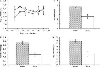
The faecal egg counts (fig. 1A) in the treatment group receiving PLS did not show any convincing reduction compared to the control group receiving water. Initially, before treatment, the group to be treated with water had higher mean log10 EPGs, but EPGs in the group to be treated with PLS caught up by day 25 pi and during treatment there was little to distinguish between the two treatment groups. Statistically there was no significant difference between the treatments (rmGLM, between-subjects test, F 1,17= 2.8, P= 0.11) and there was no evidence of a divergence between treatment groups with time during the treatment period, although the pre-treatment difference between the FEC output of the two groups generated borderline significance overall (within-subjects test, time × treatment interaction F 4.4,75.1= 2.37, P= 0.054; fig. 1A).
Effect of papaya latex supernatant extract (PLS) on H. diminuta in vivo (Experiment 1). (A) The mean log10 of eggs per gram of faeces before (days 21–25) and during (days 28–32) treatment of Wistar rats infected with H. diminuta with PLS (open squares; n= 9) or water (open circles; n= 9); (B) mean worm burden of Wistar rats infected with H. diminuta and treated with PLS 2.4 μmol or water; (C) mean wet biomass and (D) mean dry biomass. Error bars represent the standard error of the mean.

However, there were significant differences in worm burdens (fig. 1B; 44.7% reduction; Mann–Whitney U test, z= − 2.94, P= 0.003), wet biomass (fig. 1C; 53.6% reduction; Mann–Whitney U test, z= − 2.35, P= 0.011) and dry biomass (fig. 1D; 50.0% reduction; Mann–Whitney U test, z= − 2.69, P= 0.007) between the treated group and the control group.
PLS on the viability of shed eggs
Tribolium confusum beetles fed with the cut terminal proglottid segments from H. diminuta that had been treated in vivo with PLS were found to harbour intact cysticercoids. The mean number of cysticercoids recovered (9.1 ± 2.6) was not significantly different from that of the control group of beetles fed worms from rats that had been treated with water (10.7 ± 1.9; Mann–Whitney U test, P= 0.8). Beetles fed with detached proglottid segments recovered from the large intestine of treated rats also harboured intact cysticercoids (0.7 ± 0.7) and the mean cysticercoid burden was not significantly different from that in the control beetle group fed with detached proglottids from rats treated with water (1.3 ± 0.9; Mann–Whitney U test, P= 0.7).
Are cysteine proteinases in PLS involved?
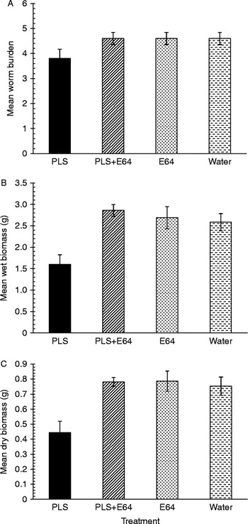
Arithmetically, there was only a minimal reduction (17%) in the mean worm burden of the PLS-treated group compared with the control group receiving water (fig. 2A). Not surprisingly, overall there was no significant effect of PLS treatment on the mean worm recovery (main effect of PLS, F 1,16= 2.0, P= 0.2) and no interaction with treatment with the CP inactivator, E-64. Post-hoc comparison of the worms from the PLS-treated group with those from the water-treated control group by the Mann–Whitney U test gave P= 0.15.
Involvement of cysteine proteinases in PLS (Experiment 2). Wistar rats were infected with H. diminuta and treated either with 2.4 μmol of PLS, 2.4 μmol of PLS pre-treated with 2.8 μmol E-64 before administration to rats, 2.8 μmol E-64 alone or water (n= 5 rats for each treatment). (A) Mean worm burden; (B) mean wet biomass; (C) mean dry biomass. Error bars represent the standard error of the mean.

However, assessments of biomass revealed more marked reductions. The mean wet biomass of the worms from the PLS-treated group was 38% lower compared to the mean of the worms from the control group (fig. 2B), and although the main effect of treatment with PLS was close to, but not significant (F 1,16= 3.8, P= 0.068), the two-way interaction with E-64 treatment was significant (F 1,16= 7.7, P= 0.014). The latter indicated that the effect of treatment with PLS was highly dependent on whether rats were also treated with E-64, and hence provided support for the conclusion that E-64 blocked the effect of PLS, since in the absence of E-64 treatment, the worms in PLS-treated animals showed reduction in wet biomass. By way of comparison, the means of the wet biomass of the worms in the group receiving PLS +E-64 and that receiving E-64 were very similar to the mean wet biomass of the worms in the water-treated control, but marginally higher, 11% and 4%, respectively. Perhaps unexpectedly, we also found a highly significant main effect of E-64 (F 1,16= 10.7, P= 0.005), arising from the higher wet biomass of worms in E-64-treated rats compared with those from rats not treated with E-64 (mean value of worms from E-64 treated rats = 2.77 ± 0.140 g, n= 10; and for worms from rats not treated with E-64 = 2.09 ± 0.217 g, n= 10).
Similarly, the dry biomass of the worms from the PLS-treated group was 41% lower compared to the worms from the group receiving water (fig. 2C). The overall effect of PLS treatment was also statistically significant (for main effect of PLS treatment on dry biomass F 1,16= 6.6, P= 0.021) and treatment diverged significantly between the E-64 treatment groups (for the two-way interaction PLS × E-64 treatment F 1,16= 6.2, P= 0.024). The group receiving PLS +E-64 and the group receiving E-64 had mean dry biomass values that were 3.9% and 4.5%, respectively, greater than that in the control group treated with water alone. Not surprisingly, there was therefore a significant effect of E-64 on dry biomass (for main effect of E-64, F 1,16= 9.3, P= 0.008; mean value of E-64 treated rats = 0.784 ± 0.035 g and for rats not treated with E-64 = 0.599 ± 0.069 g).
In conclusion, in this experiment PLS did not affect the number of worms harboured but reduced the wet and dry biomass significantly. The effect of PLS was blocked by the specific inactivator E-64, but the latter also marginally enhanced the growth of worms.
SEM observations of surface changes to worms in PLS-treated animals
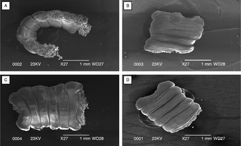
Proglottid segments of H. diminuta recovered from the rats treated with 2.4 μmol of PLS appeared to sustain treatment-related damage, which was observed to be substantial, uneven and located at the proglottid's anatomical point of attachment, exposing eggs of the parasite in the uterus (fig. 3A). The proglottids recovered from control rats treated with either 2.4 μmol PLS pre-treated with 2.8 μmol of E-64, 2.8 μmol E-64 alone or water did not show similar damage, apart from the minimal, clean and sharp tegumental breaks or cracks which are likely to be mechanical in origin (fig. 3B–D).
Scanning electron microscopical observations of surface changes to worms in PLS-treated animals (Experiment 3). SEM images of detached proglottid segments of H. diminuta recovered from the colons of infected rats treated with (A) 2.4 μmol of PLS; (B) 2.4 μmol of PLS pre-treated with 2.8 μmol E-64 before administration to rats; (C) 2.8 μmol of E-64 alone; (D) water. Note the lesions to the tegument in (A), revealing internal structures of the worm, and the otherwise smooth and intact appearance of other segments.

Dose dependence of the effects of PLS on H. diminuta
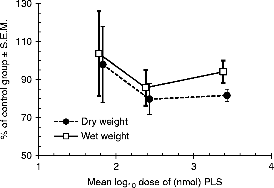
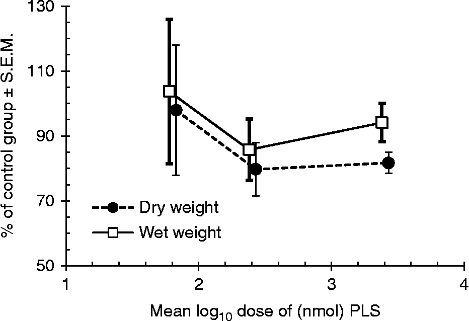
Autopsy revealed no correlation between the dose of PLS administered and worm burden. The mean worm burden of water-treated rats was identical to that of rats given the highest dose of PLS (2.4 μmol; 4.6 ± 0.25 in both cases). Similarly, there was little evidence of PLS dose-related loss of wet biomass (fig. 4), although the correlation coefficient was negative (Rs = − 0.186, n =16, P =0.49). The mean wet biomass in the water-treated group was 2.38 ± 0.135 g and that of the highest dose 2.24 ± 0.140 g. However, there was a significant negative correlation between dry worm biomass and PLS (fig. 4; Rs = − 0.531, n =16, P =0.034). The mean dry weight of the rats given 2.4 μmol PLS (0.59 ± 0.24 g) was lower than that of the control rats given just water (0.72 ± 0.37 g; post-hoc Mann–Whitney U test, P= 0.032). However, best-fit concentration–inhibition curves on data normalized to the mean of the control water-treated group and log transformed PLS dose, were not significant (for wet biomass P= 0.65; for dry biomass P= 0.12).
Log dose–response curve for PLS. Groups of rats were treated with 60, 240, 2400 nmol of PLS or water, and the data were normalized by expression as a percentage of the mean value in the control water-treated group. The SEMs for the wet weight (open squares) and dry weights (closed circles) of the control group (for means of 100% in each case) were 5.65 and 5.17, respectively. Values for dry weight have been slightly offset to avoid overlap of error bars by the addition of 0.05 to each value on the x-axis.
Duration of the effect of PLS on H. diminuta
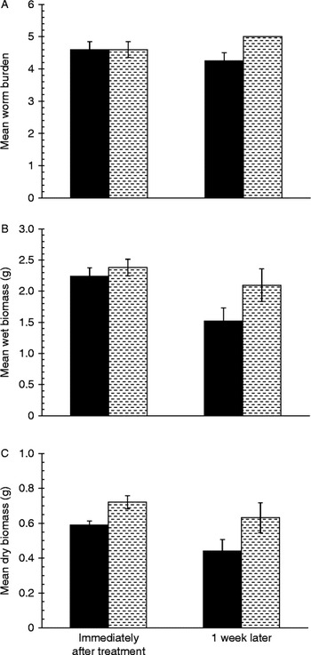
The first autopsy, immediately after completing 6 days of treatment, revealed no loss of worms as a consequence of treatment with PLS (fig. 5A) but there was a slight arithmetic reduction in the mean worm burden at the second time point, 1 week after completing treatment. However, overall there was no significant reduction in worms in this experiment (main effect of PLS, F 1,15= 3.15, P= 0.096) and no significant divergence in worm burdens between the two treatment groups over time (two-way interaction PLS × time, F 1,15= 3.15, P= 0.096). The effect of treatment with PLS on wet biomass was also close to significance (F 1,15= 3.39, P= 0.086), but the two-way interaction, PLS × time, was not significant (F 1,15= 1.25, P= 0.28), despite the apparent greater discrepancy between mean values of treated and control rats on day 28 compared with day 22. Surprisingly, in this experiment, the worms in both treatment groups lost some weight over the week following treatment (main effect of time, F 1,15= 6.66, P= 0.021), wet biomass values being lower on day 28 compared with day 22 in both groups. The effect of treatment with PLS on dry biomass was significant (main effect of PLS, F 1,15= 7.68, P= 0.014), the weight of dry biomass in PLS-treated rats being lower on both occasions (fig. 5C). However, there was no divergence over time (PLS × time, F 1,15= 0.26, P= 0.62) and, despite the generally lower mean dry weights of worms on day 28 compared with day 22, the effect of time was just the wrong side of significance (F 1,15= 4.14, P= 0.06).
The effect of PLS on H. diminuta 1 week after treatment (Experiment 5). Wistar rats were infected with H. diminuta and culled immediately after 6 days of treatment with either 2.4 μmol of PLS (black columns) or water (stippled columns) (n= 5 rats for both treatments) and 1 week after treatment was completed (n= 5 for both treatments). (A) Mean worm burden; (B) mean wet biomass; (C) mean dry biomass. Error bars represent the standard error of the mean.

Discussion
The results reported in this paper provide the first convincing evidence that entirely lumen-dwelling tapeworms, such as H. diminuta, are adversely affected by CPs in vivo, albeit moderately and not nearly as severely as nematodes in mice, sheep and pigs (Stepek et al., Reference Stepek, Lowe, Buttle, Duce and Behnke2007b; Buttle et al., Reference Buttle, Behnke, Bartley, Elsheikha, Bartley, Garnett, Donnan, Jackson, Lowe and Duce2011; Levecke et al., Reference Levecke, Buttle, Behnke, Duce and Vercruysse2014). This contrasts with previous reports based on experiments with the rodent tapeworms H. nana and H. microstoma in mice, in which fruit-derived CPs were found to be without any, or with just minor, effects on tapeworm infections (He et al., Reference He, Tiuria and Retnani1992; de Amorin et al., Reference de Amorin, Borba, Carauta, Lopes and Kaplan1999; Mansur et al., Reference Mansur, Luoga, Buttle, Duce, Lowe and Behnke2014b). Disappointingly, our results (fig. 1A) showed no reduction in the FECs of the treated group as compared to the control, in agreement with our earlier study on H. microstoma (Mansur et al., Reference Mansur, Luoga, Buttle, Duce, Lowe and Behnke2014b). However, in contrast to H. microstoma, the strobila of which is partly hidden in the bile duct from direct exposure to PLS as it passes down the length of the intestine, in this case the worms were fully exposed to the CPs, because H. diminuta is entirely lumen dwelling. Our failure to detect any significant reduction in FEC with H. diminuta, and in our earlier study with H. microstoma, clearly contrasts with the nematode systems, in which FECs fall rapidly, even a day after the commencement of treatment, and by 90% or more over the course of treatment (Stepek et al., Reference Stepek, Lowe, Buttle, Duce and Behnke2006; Luoga et al., Reference Luoga, Mansur, Buttle, Duce, Garnett, Lowe and Behnke2015). We speculated on the explanation in our earlier paper (Mansur et al., Reference Mansur, Luoga, Buttle, Duce, Lowe and Behnke2014b). Unlike in H. microstoma, in the case of H. diminuta, the scolex and the neck regions, as well as the entire strobila, would have been exposed to the CPs. In contrast to nematode infections, FECs are known to be a less accurate reflection of cestode worm burdens and, in some cases, cannot be used to estimate the intensity of infection (Proudman & Edwards, Reference Proudman and Edwards1992; Meana et al., Reference Meana, Luzon, Corchero and Gomez-Bautista1998). Tapeworms are subject to intense density-dependent regulation of their length and the weight of their strobili (Read, Reference Read1951, Reference Read2000; Roberts, Reference Roberts2000), so as worm burdens fall, the average size of the remaining parasites increases. We might have expected to see a complete loss of FECs had the worms been totally expelled but, as our data show, the only consistent effects of CPs on H. diminuta were to reduce dry weight, and never by even as much as 60%, with actual loss of worms being unpredictable and at best only a relatively weak effect. It is therefore possible that per capita egg output remained unaffected, even though the size of individual worms, as reflected in the total dry biomass, was reduced.
It was also relevant to ascertain whether the viability of the shed eggs was affected by CPs, despite no reduction in egg counts. Our results showed that the eggs of H. diminuta were not affected at all, because intact H. diminuta cysticercoids were recovered from Tribolium beetles fed with either cut terminal proglottid segments or detached proglottid segments recovered from the colon of rats infected with H. diminuta and treated with PLS, much as we had found earlier with H. microstoma (Mansur et al., Reference Mansur, Luoga, Buttle, Duce, Lowe and Behnke2014b). Unlike the benzimidazoles, which are able to adversely affect the development of H. diminuta larval stages (Evans & Novak, Reference Evans and Novak1976), PLS does not appear to have any effect on the fecundity of H. diminuta when administered to infected rats. Cestode eggshells are robust, with several layers protecting the developing hexacanth embryo within, because of the need for the eggs to survive under harsh external environmental conditions before resumption of growth and development in a suitable intermediate host (Smyth & Clegg, Reference Smyth and Clegg1959; Lethbridge, Reference Lethbridge1971). Since CPs attack specific peptide sequences in target proteins, it is equally possible that the eggshell does not contain amino acid sequences that are susceptible to digestion by papaya CPs.
In contrast to the lack of an effect on fecundity, our data revealed significant effects on the total biomass of worms harboured by treated rats; especially on the dry weight of the total biomass, which in some experiments was 46–54% lower than that in control water-treated animals. This contrasts with the findings with H. microstoma, where reductions in worm biomass were smaller. The effect on dry biomass was confirmed as being directly attributable to CP activity, because when PLS was pre-incubated with E-64, a specific and powerful inhibitor of CPs (Matsumoto et al., Reference Matsumoto, Mizoue, Kitamura, Tse, Huber and Ishida1999), the effect of PLS on both dry and wet biomass was completely blocked. Interestingly, both groups treated with E-64, whether in combination with PLS or alone, saw a moderate but significant increase in biomass (4–10%) compared to the groups not given E-64, as we had found earlier with H. microstoma (Mansur et al., Reference Mansur, Luoga, Buttle, Duce, Lowe and Behnke2014b). In the case of H. microstoma, we suggested that this slight increase in the size of worms following exposure to PLS +E-64 may be due to an exaggerated repair response following the intestinal perturbation by other components in PLS, such as the mucolytic enzymes and alkaloids that PLS is also known to contain (Mezhlumyan et al., Reference Mezhlumyan, Kasymova and Yuldashev2003). However, here, with H. diminuta, a similar increase in the biomass was found in animals treated only with E-64, so this must be a purely E-64 effect. E-64 alone might be diffusing through the tegument, causing some disturbance to the worms and subsequently an exaggerated repair response; and since tapeworms are known to possess endogenous CPs, it might suggest that a cestode-derived CP is needed in homeostatic maintenance of the cestode (White et al., Reference White, Baig and Chappell1997; Tort et al., Reference Tort, Brindley, Knox, Wolfe and Dalton1999; Sajid & McKerrow, Reference Roberts2002; Zimic et al., Reference Zimic, Pajuelo, Rueda, López, Arana, Castillo, Calderón, Rodriguez, Sheen, Vinetz, Gonzalez, Garcia and Gilman2009).
Our SEM images of detached proglottid segments from the colon of PLS-treated rats provide further evidence of PLS activity. While the control segments were either intact or possibly sustained mechanical damage (fig. 3), the segments found in the colon of the PLS-treated animals appeared to have sustained more extensive, treatment-related surface damage, which was uneven in appearance across the sampled proglottids and with occasional deeper lesions, exposing underlying structures in the subtegumental layers of the worms. However, the extent of damage was not as severe as that seen in the in vitro experiments, where the worm surface would have been exposed to higher concentrations of CPs (Mansur et al., Reference Mansur, Luoga, Buttle, Duce, Lowe and Behnke2014a). A possible explanation for the limited changes in vivo could be the remarkable ability of cestodes to repair their tegumental membranes when in their natural hosts, and to recover from surface damage (Befus & Threadgold, Reference Befus and Threadgold1975).
We also explored the dose-dependence of the effect of CPs on H. diminuta, and while some evidence of a reduction in the dry weight of biomass was detected, this experiment did not provide strong support for dose-dependence, as we might have expected with an anthelmintic drug. There was no effect on worm burden with increasing PLS dose, and wet biomass had only a weak correlation in the expected direction. The effect on dry biomass was stronger and was also reflected in a negative correlation with increasing PLS dose. Unexpectedly, in these dose–response experiments we were limited by the highest dose that could be administered to rats. No host weight loss was detected in these experiments, even in the rats given the highest dose of PLS. In pilot experiments, however, we found that some rats were more prone to gastro-oesophageal reflux at higher doses than the mice used in our earlier work, resulting in PLS finding its way into the animals’ lungs, leading to death. For humane reasons, in order to avoid these losses, we limited our treatment to 2.4 μmol.
We were also interested to see whether the effects of PLS are sustained or temporary, since cestodes are known to be able to repair damage to their surface very quickly (Befus & Threadgold, Reference Befus and Threadgold1975). Although reduction in worm burden following treatment with PLS was rare in these experiments, there remained the possibility that some loss of worms might have been attributable to destrobilated worms, whose scoleces persisted somewhere in the intestine and later regrew to the original size when the hazard from local CPs had been swept away by peristalsis. Destrobilated scoleces have been shown to regrow even when transplanted into another host (Hopkins et al., Reference Hopkins, Subramanian and Stallard1972a). However, in Experiment 6 virtually all the worms were recovered from every rat at both time points, but the surprising feature of this experiment was that biomass did not increase after the cessation of treatment, indeed mean biomass appeared to be lower a week later, with values approaching statistical significance.
Clearly, the tapeworm, H. diminuta, is affected by papaya-derived CPs administered to its rat host in vivo. It is possible that PLS was indeed responsible for some loss of H. diminuta worms in vivo, possibly through a synergistic interaction with the immune system, whereby weak and vulnerable worms, after PLS-induced damage, might not have been able to continue to evade host immunity. Even in its natural rat host, H. diminuta has been shown to be rejected immunologically in chronic infections (Andreassen & Hopkins, Reference Andreassen and Hopkins1980). However, within the dose range that we could ethically apply in our experiments, the effect on the worms was disappointing and far less than desirable for a cestocidal anthelmintic. Nevertheless, our results provide a starting point and offer the possibility of further refinement in formulation and delivery or assessment of alternative natural CPs (Behnke et al., Reference Behnke, Buttle, Stepek, Lowe and Duce2008). Since fruit-derived CPs have such potent effects on nematodes, including those of humans (Hansson et al., Reference Hansson, Veliz, Naquira, Amren, Arroyo and Arevalo1986), efforts to develop them into broad-spectrum anthelmintics should be encouraged by our data.
Acknowledgements
We thank Tim Smith for technical support and advice with the scanning electron microscopy. We dedicate this paper to Dr Wenceslaus Luoga, who, sadly, was killed in a road accident in July 2013 shortly after returning to Tanzania on the successful completion of his postgraduate studies at the University of Nottingham.
Financial support
F.M. was supported by a postgraduate studentship from the government of Malaysia, and W.L. from the Government of Tanzania.
Conflict of interest
None.
Ethical standards
The authors assert that all procedures contributing to this work comply with the ethical standards of the national guides on the care and use of laboratory animals in the UK (The Animals (Scientific Procedures) Act 1986) and were locally approved by the Animal Welfare and Ethical Review Body of the University of Nottingham. Importantly, all work was conducted within a recognized culture of care and compliance to meet the expectations of both the University and the UK Home Office Inspectorate.