Introduction
Complications of middle-ear cleft infections are currently encountered infrequently.Reference Gower and Mcguirt1 However, they are often serious and therefore need to be treated properly and as quickly as possible. As a result, the physician always needs to consider this possibility and have a thorough understanding of the pathology to prevent complications. Acute mastoiditis is the most common complication of acute otitis media. Spread of infection from the middle-ear cleft to intracranial structures is usually direct, and infection can spread either upward and into the middle cranial fossa or backwards into the posterior fossa. However, since the advent of antibiotic therapy, acute mastoiditis is less frequent, as are its secondary complications and subsequent need for surgical intervention.Reference Fliss, Leiberman and Dagan2, Reference Harley, Sdralis and Berecowitz3
A high resolution CT scan of the temporal bone is useful in the pre-operative diagnosis of otologic disease and in the investigation of patients with acute coalescent mastoiditis, a contrast enhanced CT of the brain is useful to rule out intracranial complications.Reference Graham, Goldsmith and Lee4 In this case report, we present a potential pitfall in the interpretation of the CT scans. This case demonstrates that a normal intracranial blood vessel (occipitotemporal vein) can be confused with a complication of middle-ear disease.
Case report
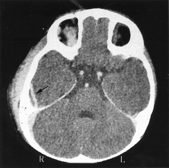
A 12-month-old male was transferred from a community hospital to a tertiary care medical centre with a diagnosis of acute mastoiditis associated with a subperiosteal abscess in the retroauricular area. He had not responded to three days of broad spectrum intravenous antibiotics. On pre-operative post-contrast CT, clouding of the mastoid air cells with a subperiosteal abscess was observed. Additionally, an epidural abscess overlaying the tegmen was suspected (Figure 1).
Pre-operative post-contrast CT demonstrating soft tissue oedema with a subperiosteal abscess adjacent to the right mastoid. The course of the vein of Labbe that can be mistaken for an epidural abscess is easily visible (arrow).

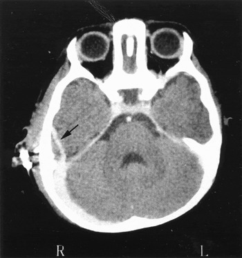
A cortical mastoidectomy was performed the same day with drainage of the subperiosteal abscess. No epidural abscess was found during exploration of the mastoid. A post-operative contrast enhanced CT showed no change (Figure 2). On revised analysis of the CT scan it was concluded that the enhancement observed in the CT scan was the occipitotemporal vein (OTV).
Post-operative contrast enhanced CT following drainage of the subperiosteal abscess. The vein of Labbe, which is still readily observed, remains unchanged in this view (arrow).

Discussion
The OTV is a superficial vessel running anterioposteriorly in the occipitotemporal sulcus, between the inferior temporal sulcus and the occipitotemporal gyri. It is a variation of the inferior anastomotic vein (vein of Labbe), connecting the superficial middle cerebral vein and the transverse sinus.Reference Sener5, Reference Williams, Warwick, Dyson and Bannister6 The OTV has been identified in 83 per cent of cadavers (52 per cent bilaterally). Post-contrast CT scans have identified the OTV in only 8 per cent of the population and all cases were seen in children.Reference Sener5 The OTV is rarely seen on CT due to the slight angulation of the vessel in comparison to the axial plane and its isodense appearance compared to brain tissue, unless a large amount of contrast material is injected and absorbed by this small vessel.Reference Sener5
The patient described in this report needed a large amount of contrast medium to rule out a sigmoid sinus thrombosis. The consequence of this was that the OTV became clearly identifiable. There are other findings on the CT (Figures 1 and 2) that further suggest a vascular structure as opposed to an epidural abscess. The soft tissue lateral to the contrast enhanced structure is isodense to brain tissue and not hypodense, as is seen with an abscess. In addition, the enhanced structure is connected to the transverse sinus which supports the diagnosis of a blood vessel. Although these considerations were taken into account prior to surgery, the surgeon's and the consultant radiologist's unfamiliarity with this vessel necessitated an exploration of the tegmen as a part of the surgical procedure. Magnetic resonance imaging (MRI) offers superior soft tissue resolution over CT, which can aid diagnosis; however, it is not readily available in emergency settings in many institutions. Therefore, the attending surgeon should be familiar with the OTV on post-contrast CT of the brain in order to avoid an erroneous diagnosis and unnecessary intracranial exploration.