Introduction
Although conventional chemotherapy has been successful to some extent, the main drawbacks of chemotherapy are its poor bioavailability, high-dose requirements, adverse side effects, low therapeutic indices, the development of multiple drug resistance, and non-specific targeting.[Reference Hood1–Reference Cho, Wang, Nie, Chen and Shin4] Nanoparticles (NPs) provide a new mode of cancer drug delivery as a carrier through fenestrations in tumor vasculature allowing direct cell access.[Reference Brigger, Dubernet and Couvreur5–Reference Yih and Al-Fandi7] The focus on nanocarrier design over the years has evolved towards core–shell-type lipid–polymer hybrid nanoparticles (LPHNPs) which have emerged as a robust and promising drug delivery platform.[Reference Malam, Loizidou and Seifalian8–Reference Sun, Zhang, Wang, Feng, Liu, Yin, Xu, Wei, Ding, Shi and Jiang10] LPHNPs are polymeric nanoparticles enveloped by lipid layers, which combine the beneficial characteristics of liposomal drug delivery—cell affinity and cell-targeting ability—with those of polymeric NPs—structural integrity, controlled release, and ease of functionalization to achieve high serum stability.[Reference Hadinoto, Sundaresan and Cheow11] The encapsulation of a drug molecule in these nanocarriers provides effective drug delivery and toxicity reduction.[Reference Ramasamy, Tran, Choi, Cho, Kim, Yong, Choi and Kim12]
The efficacy of drug delivery therapy depends on a suitable combination of drug and LPHNPs to achieve a synergistic effect on any cancer cell with minimal adverse effects.[Reference Cheow and Hadinoto13] Doxorubicin is a chemotherapeutic drug that can treat a wide variety of cancers, such as bladder, leukemia, lymphoma, gastric, head and neck, liver, kidney, and cervical and ovarian cancer.[Reference Thorn, Oshiro, Marsh, Hernandez-Boussard, McLeod, Klein and Altman14] The molecular structure of doxorubicin consists of a conjugated ring structure which gives the drug fluorescent properties. Fluorescence allows investigators to track the drug uptake into cells. Moreover, doxorubicin has already been approved for use in NPs under the trade name Doxil.[Reference Prasad15] Hence, incorporating doxorubicin in LPHNPs should improve the therapeutic index of the drug in terms of enhanced permeability, targeting ability, and active cellular intake. Accordingly, the synthesis of doxorubicin-loaded LPHNPs was attempted and presented in this paper.
Despite the extensive research on LPHNPs, none are FDA-approved and available for cancer treatment. That may be due to the two main substantial challenges in the evaluation of LPHNPs: (i) the synthesis methods employed for the LPHNP synthesis are time-consuming and not reproducible and (ii) the two-dimensional (2D) monolayer cell cultures used for their cytotoxicity evaluation yield complicated results with less apparent benefits to the patients.[Reference Bose, Ravikumar, Karuppagounder, Bennet, Rangasamy and Thandavarayan16–Reference Idrees, Chiono, Ciardelli, Shah, Viebahn, Zhang and Salbar18] In this report, doxorubicin-loaded LPHNPs have been synthesized by three-dimensional (3D)-printed herringbone-patterned multi-inlet vortex mixer (MIVM) which enables a rapid and efficient mixing of polymer and lipid solutions.[Reference Le, Bokare and Erogbogbo19] Simulation and experimental studies have also been done to elucidate the mechanism behind the rapid mixing that takes place in a 3D-printed herringbone-patterned MIVM device. Furthermore, 3D cell cultures have been used to evaluate the efficiency of doxorubicin-loaded LPHNPs and compared with the results obtained from 2D cell cultures. HeLa ATCC CCL-2 cells cultures have been used for both 2D and 3D cell cultures for the cytotoxicity evaluation of doxorubicin-loaded LPHNPs. Compared with the traditional monolayer culture system, the cells in the multicellular system are expected to closely mimic the cellular microenvironment in vivo. Results from 3D studies demonstrate that increasing the dimensionality from 2D to 3D can significantly impact cytotoxicity results. Hence, three-dimensional (3D) in vitro tumor models provide an important tool for the optimization of efficient intratumoral drug delivery.[Reference Kim, Doh and Cho20]
Materials and methods
Materials
Poly(lactic-co-glycolic acid)-ester (PLGA polymer) having molecular weight (25–35 kDa) and LA:GA (85–15) was provided by Akina Chemicals (West Lafayette, IN, USA). 1,2-Distearoyl-sn-glycero-3-phosphoethanolamine (DSPE-PEG amine) was supplied by Avanti Polar Lipids (Alabaster, AL, USA). Lecithin (soybean, refined) was supplied by Alfa Aesar. Four percent by weight aqueous ethanol, acetonitrile, plant-based sodium alginate (4–12 cPs; 1% H2O in 25 °C) (product no. A-0682), calcium chloride (product no. 449709), trypan blue, and chloroform were purchased from Sigma Aldrich (St. Louis, MO, USA). Trypsin EDTA (0.25%) was obtained from Gibco (ThermoFisher Scientific) (Waltham, MA, USA). Eagle's Minimum Essential Medium (EMEM, ATCC® 30-2003™, Manassas, VA, USA), HeLa (ATCC® CCL-2™), fetal bovine serum (FBS; ATCC® 30-2020™), and Penicillin–Streptomycin were obtained from ATCC. Doxorubicin-free base was obtained from Medakoo (Morrisville, NC, USA). A fingertip aerosol sprayer (Juvale, Amazon.com) was used to produce alginate beads used for 3D cell culture.
SolidWorks 2015, Objet30 Pro 3D printer, and COMSOL Version 5.1 were used from San Jose State University (San Jose, CA, USA). The MicroKros apparatus (part #C02-E010-10-N 10 kDa) was used from Spectrum Labs (Waltham, MA, USA).
3D-printed MIVM
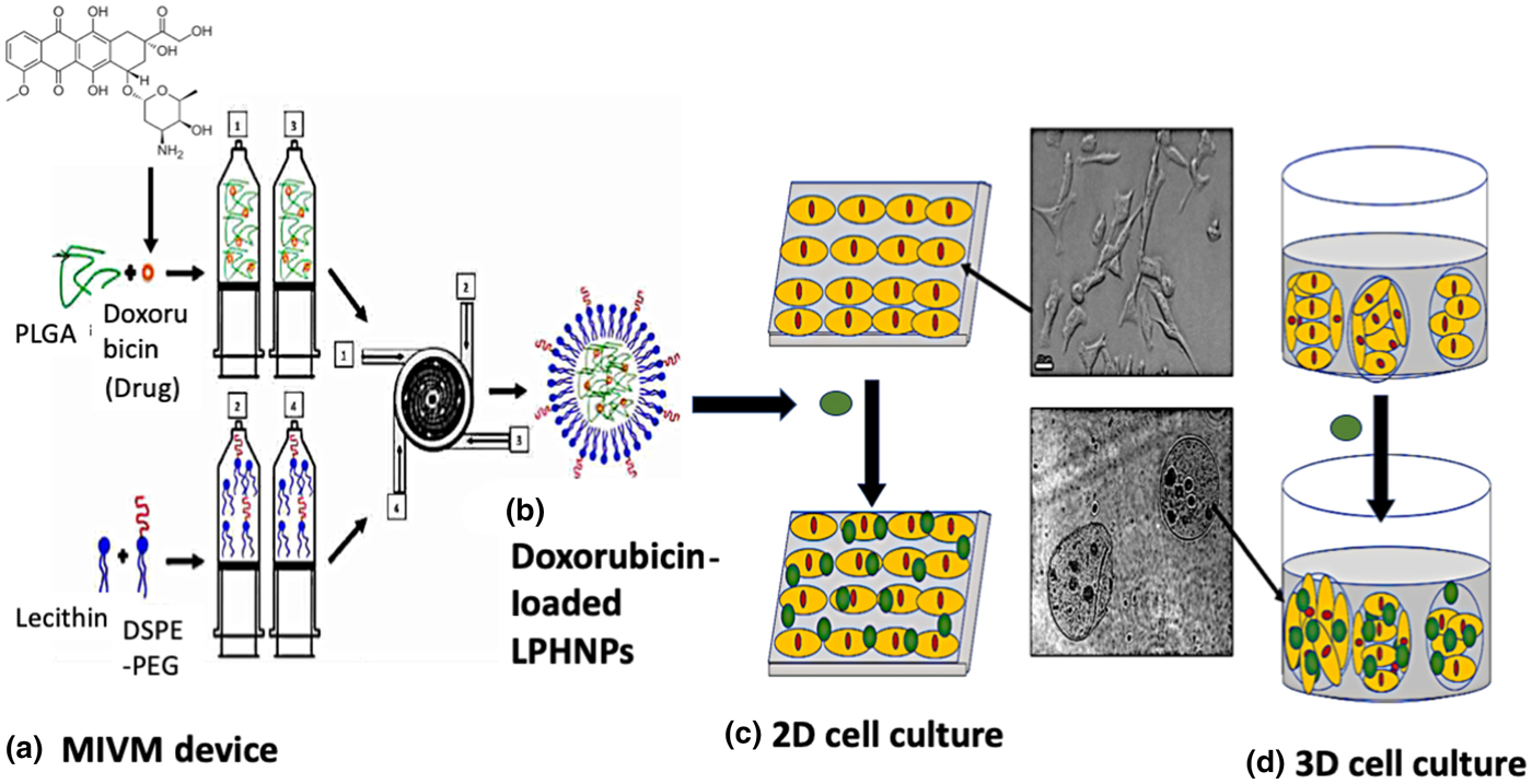
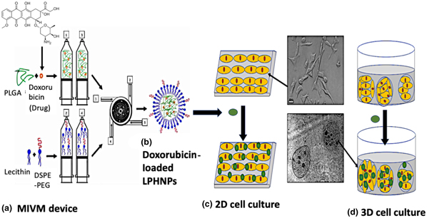
A modeling program called SolidWorks was utilized to design the MIVM device. The design was based on the design created by Liu et al. and the dimensions were approximate to the values indicated in the various literature papers.[Reference Shi, Fox and Olsen21,Reference Liu, Ramezani, Fox, Hill and Olsen22] The schematic representation of the MIVM device is shown in Fig. 1(a). It consists of four inlet channels where 1 and 3 represents the inlet for drug and PLGA solution (solvent), and 2 and 4 represents the inlet for antisolvent (DSPE-PEG) solution. An inner black chamber (as shown in Fig. 1(a)) acts as a mixing chamber as well as an outlet for flash nanoprecipitation. The multi-inlet vortex mixer is a portable nanoparticle fabrication device that does not require an external power source. This device enables fast and efficient fabrication of drug-loaded NPs. The herringbone pattern consisting of repeated patterns of grooves is fitted in each microchannel (arm) of the MIVM device. The herringbone geometries are loosely based on designs by Manda and are scaled up from the micrometer scale.[Reference Williams, Longmuir and Yager23] The model is loaded into a 3D printing software and printed using an Objet30 Pro 3D printer. Once the device is printed, it needs to be cleaned before use. The cleaning process begins by washing the device with deionized water and storing it overnight in a solution of sodium hydroxide (pH 13). Then, hydrochloric acid is added in a dropwise manner to neutralize the pH. After few hours, the device is transferred to a solution of deionized water and stored overnight.
Schematic representation of (a) MIVM device, (b) doxorubicin-loaded LPHNPs, (c) 2D cell culture, and (d) 3D cell cultures.

Fabrication of doxorubicin-loaded LPHNP
The drug-loaded nanoparticles are fabricated by combining two mixtures together in the MIVM. The first solution is the core mixture, which consists of PLGA and doxorubicin. A set mass of PLGA is weighed out into a beaker and suspended in DMSO at a concentration of 1 mg/mL. The drug loading is set to 10% or 15% the weight of PLGA, which represents 10% or 15% drug loading. The second solution is lecithin:DSPE-PEG (molar ratio as 1:2.5) lipid solution which forms the shell of the NP. DSPE-PEG:lecithin dissolved in chloroform first. After evaporating the chloroform in vacuum, the residue is dissolved in 4% ethanol and heated to a temperature of 65 °C with a hot plate. These two solutions then applied to the inlets of 3D-printed herringbone-patterned MIVM devices with 20 mL/min flow-rates. Obtained nanoparticles are then washed with distilled water using a 30 kDa pore tangential flow filtration MicroKros apparatus.
Estimation of doxorubicin in doxorubicin-loaded LPHNPs
To estimate the amounts of doxorubicin in the NP, UV–Vis spectroscopy method was used. We use subtractive inferences that allow us to estimate the amount of doxorubicin left based on the initial amount used. We measure the initial amount of doxorubicin used to make the NPs and measured the amount of left over doxorubicin found in the solution (with subsequent filtration steps).
2D monolayer cytotoxicity testing
NP cytotoxicity in a 2D monolayer is tested using HeLa (ATCC® CCL-2™). These cells are cultured as a continuous cell line. The cells require EMEM (ATCC® 30-2003™) and FBS (ATCC® 30-2020™). The cell culture follows the American Type Culture Collection culture method for HeLa ATCC CCL-2 cells. For cytotoxicity testing, HeLa cells are plated onto a 96-well plate using cells 2–3 days after a split to ensure the HeLa cells are in their exponential growth phase.[Reference Geraghty, Capes-Davis, Davis, Downward, Freshney, Knezevic, Lovel-Badge, Masters, Meredith, Stacy, Thraves and Vias24] The cells are first detached with trypsin, centrifuged, and the cells are counted using a hemocytometer with trypan blue. After an approximate measurement of the cell density, cells are spotted onto a 96-well plate at a density of 10,000 cells in 0.1 mL of media. The plate is then incubated for 24 h before cytotoxicity testing. The schematic for 2D cell culture and the corresponding bright field image are shown in Fig. 1(c).
3D cell culture and cytotoxicity
Cell preparation and culture
HeLa cells (ATCC) are grown in EMEM, supplemented with 10% FBS. Immediately prior to encapsulation into alginate beads, the cells were trypsinized and resuspended into an alginate solution at a concentration of 8–10 × 106 cells/mL.
Cell encapsulation
Cells were encapsulated into alginate beads following the work of Bigdeli et al.[Reference Bigdeli, Dettloff, Frank, Davis and Crosby25] Sodium alginate (Sigma Aldrich) was mixed into deionized water at 2% (w/v) and stirred for 4 h at room temperature. A 2% (w/v) calcium chloride solution was prepared by dissolving powder into deionized water and stirring for 1 h at room temperature. Following dissolution, the alginate solution was autoclaved, and the calcium chloride solution was sterile filtered using a vacuum filtration unit before use. A bath of approximately 300 mL of the calcium chloride solution was prepared inside a biosafety cabinet and continuously stirred, and alginate solution containing HeLa cells at a concentration of 8–10 × 106 cells/mL was sprayed into the bath using a fingertip aerosol sprayer. After a gelation period, the beads are collected from the bath and rinsed with sterile phosphate-buffered saline (pH 7.4). Subsequently, the beads are filtered through 100 µm and retained by a 70 µm cell strainer to isolate beads with a diameter between 70 and 100 µm. Beads recovered from the 70 µm cell strainer are resuspended in EMEM + 10% FBS, transferred to a six-well plate, and incubated at 37 °C and 5% CO2. Encapsulated cells were visualized on an inverted fluorescent microscope (Nikon Eclipse). Figure 1(d) shows the schematic of a 3D cell culture and the corresponding bright field image. The 3D cell culture shows cells in a spherical matrix in comparison with the 2D cell culture monolayer which shows cells adhering to a flat surface.
Viability
3-(4,5-Dimethylthiazol-2-yl)-5-(3-carboxymethoxyphenyl)-2-(4-sulfophenyl)-2H-tetrazolium, or the MTS assay, was used for the sensitive quantification of metabolically active cells by observing the conversion of the provided MTS assay compounds into a dye with a measurable absorbance at 490–500 nm by NADPH-dependent dehydrogenase enzymes. After culturing the cells for the desired time frame, the media for the cells to be analyzed were aspirated away and replenished with fresh media before the addition of the MTS reagent. About 1 µL of the MTS reagent was added for every 10 µL of media in each well. The cells were incubated for 30–45 min before the absorbance of the assay we measured at 490 nm. The obtained values were used to generate viability plots.
To visualize the viability of cells in the 3D culture environment, cells are stained with calcein-AM and ethidium homodimer using a LIVE/DEAD Viability kit. Calcein-AM has excitation at 495 nm and emission at 635 nm, which gets retained in the live cells. The live cells are detected by intracellular esterase activity turning the nonfluorescent stain into fluorescent calcein. Ethidium homodimer has high affinity for nucleic acids. Thus, cells with intact membrane do not accept this stain, while the dead cells do. The live cells appear green fluorescence and dead cells appear red fluorescence. Stained samples were visualized on an inverted fluorescent microscope (Nikon Eclipse) using fluorescence microscopy, and the survival rate is analyzed.
Results and discussion
NP synthesis by herringbone and without a herringbone pattern
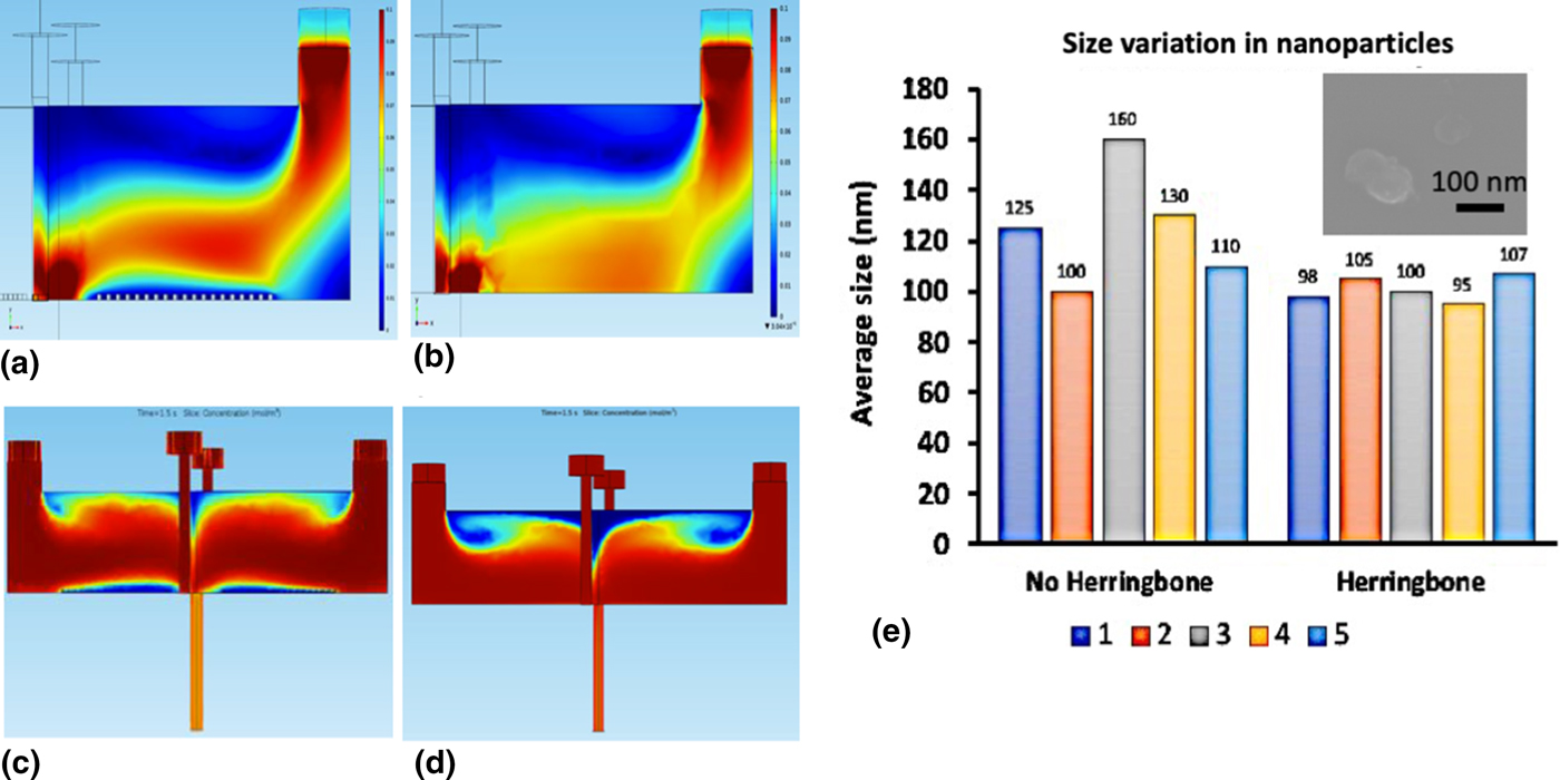
To study the effect of the herringbone pattern on the doxorubicin-loaded LPHNPs, simulations of the fluid dynamics inside the MIVM were conducted using COMSOL. Figures 2(a) and 2(b) depicts the velocity pattern through the MIVM arm with and without the herringbone pattern, respectively. The maximum velocity corresponds to the red color, whereas blue color represents minimum velocity. The grooves in the herringbone pattern creates the helical motions of the moving fluid inside the microchannel.[Reference Xu, Lu, Riordon, Sinton and Moffitt26] This leads to the twisting patterns of two liquid channels into each other at that point resulting into a higher velocity and vorticity. It is clear from Figs. 2(a) and 2(b) that the herringbone directs the flow towards the middle of the MIVM arm. This phenomenon led to higher velocity flow through the herringbone-patterned MIVM arm. The maximum velocity recorded within the MIVM arm with and without the herringbone pattern is 0.086 and 0.063, respectively. Diverting the flow towards the center of the MIVM in the herringbone pattern resulted in more favorable interactions to achieve desired NP sizes.
Velocity pattern in the MIVM arm (a) with and (b) without the herringbone pattern. Concentration gradient in the MIVM device (c) with and (d) without the herringbone pattern. (e) Size variation in NPs from five experimental runs and inset shows an SEM image agglomerated two NPs.
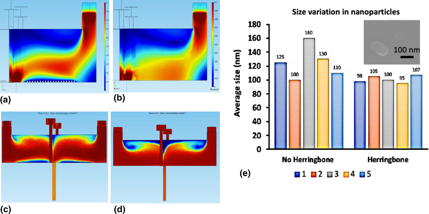
The pushing of the flow towards the center of the MIVM in the herringbone pattern may result in more favorable interactions of the shell and core solution resulting in maximum mixing efficiency at the mixing chamber and the outlet pipe. Figures 2(c) and 2(d) depict that the subtle differences in the concentration gradient develop in the MIVM pattern with and without the herringbone pattern. The maximum concentration is dark red which corresponds to 0.27 mg/mL, whereas the minimum concentration is blue which is 0 mg/mL. It is clear from Figs. 2(a) and 2(b) that the concentration of the solution inside the outlet pipe is higher in case of the herringbone-patterned MIVM device indicating maximum mixing efficiency.
Figure 2(e) represents the sizes of doxorubicin-loaded LPHNPs obtain through five experimental runs. The experiment has been conducted at different days keeping all the experimental parameters same. It is clear from Fig. 2(e) that herringbone-patterned MIVM methods yield the smallest particle size with uniform size distribution. In addition to this, it also showed more reproducibility and repeatability in the LPHNP synthesis than the MIVM device without the herringbone pattern. This can be attributed to the maximum vorticity, velocity, and mixing efficiency offered by the herringbone pattern. An SEM image of the doxorubicin-loaded LPHNPs is shown as an inset in Fig. 2(e). SEM shows a single NP of the size ~100 nm and two nanoparticles fused together.
Cytotoxicity testing by 2D culture
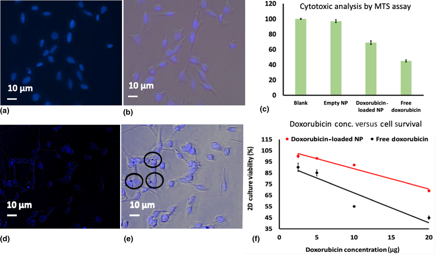
To assess the viability in bulk, HeLa cells are spotted 2 days after split onto a 96-well plate and incubated for 24 h. After the incubation, 0.1 mL of culture media are added to the control wells and 0.1 mL of NP with doxorubicin are added to the treatment wells then incubated for another 24 h. Then, the cells are stained with 300 nM of DAPI and viewed by using the EVOS Cell Imaging System. Figures 3(a) and 3(b) show the untreated HeLa cells with and without DAPI stain, respectively, whereas Figs. 3(d) and 3(e) show the HeLa cells treated with the NPs with and without DAPI stain, respectively. Figures 3(e) (encircled) clearly shows some darkened spots around the nucleus of HeLa cells which are not observed in untreated HeLa cells [Figs. 3(a) and 3(b)]. These darkened spots indicate the agglomeration and interaction of the doxorubicin-loaded LPHNPs with the nucleus of HeLa cells. This interaction exerts the toxic effect leading to cell death. Hence, the number of stained nucleus is less and unclear in case of treated cells [Fig. 3(d)] when compared with the untreated cells [Fig. 3(a)].
Untreated HeLa cells (a) with and (b) without DAPI stain; (c) cytotoxic analysis of 2D culture cells by the MTS assay; HeLa cells treated with NPs (d) with and (e) without DAPI stain; (f) cell viability in 2D culture corresponding to doxorubicin concentrations.
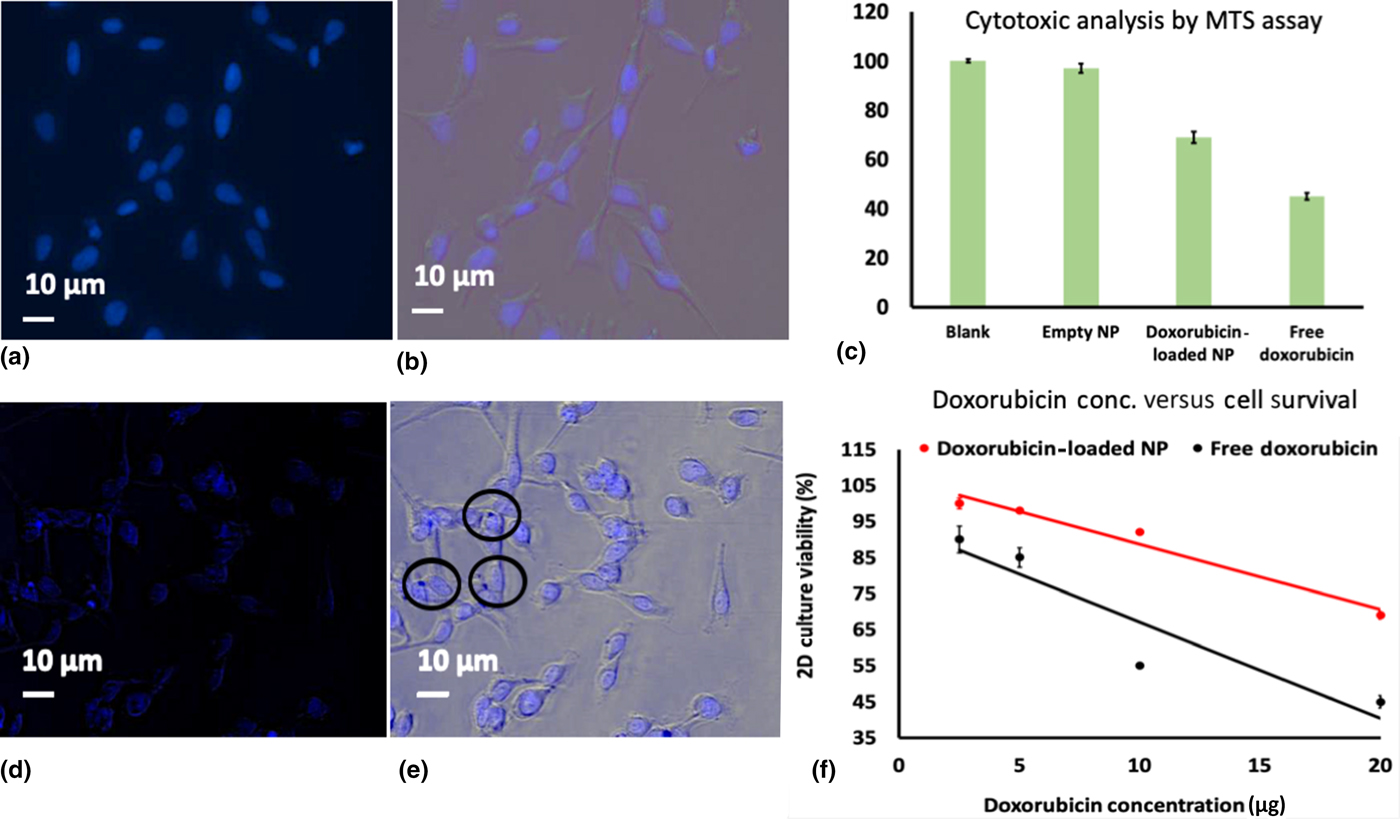
The viability of HeLa cells in 2D cell culture was further confirmed by means of the 24-h MTS assay. The amount of doxorubicin used was IC50 [DOX] for HeLa cells i.e., 0.6 mg. The cytotoxic results of blank solution, empty nanoparticle, free doxorubicin, and the doxorubicin-loaded LPHNPs are shown in Fig. 3(c). The empty NP has showed a similar cytotoxic effect as control. The free doxorubicin has a higher cytotoxicity than the samples with the doxorubicin inside the NPs. Furthermore, the viability of free doxorubicin and the doxorubicin-loaded LPHNPs against the concentration of doxorubicin are shown in Fig 3(f). It can be seen from Fig. 3(f) that at the amount of 20 µg, doxorubicin has a survival rate of 45% and the NPs with doxorubicin has a survival rate of 69%.
Cytotoxicity testing by 3D culture/spheroids
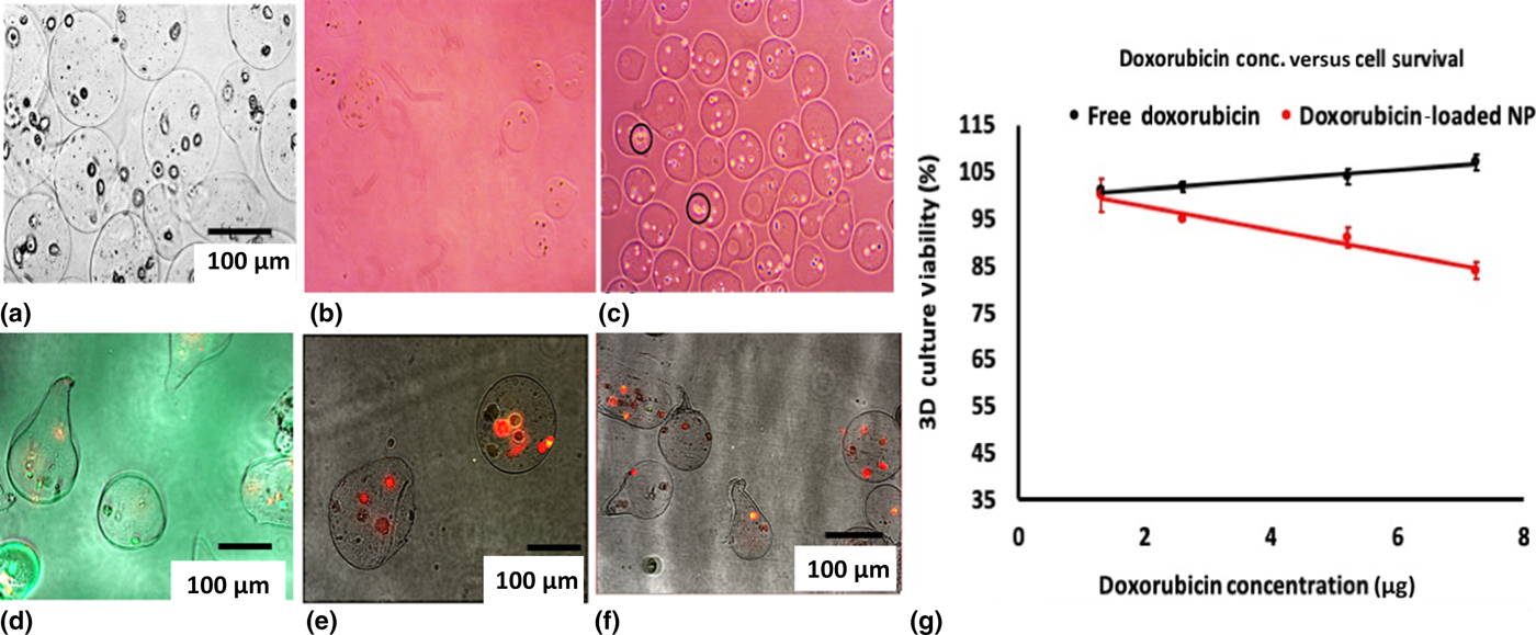
To investigate the cytotoxic effects of doxorubicin-loaded LPHNPs in 3D culture environment, HeLa cells were encapsulated in sodium alginate beads. Figure 4(a) shows the overall structure of the HeLa cells encapsulated in the sodium alginate beads (spheroids).
(a) HeLa cells encapsulated in sodium alginate bead, phase contrast microscopic images of spheroids after (b) 2 days, (c) 8 days, live/dead cells staining results, (d) control spheroids (spheroids without NPs), (e) and (f) spheroids cultured with doxorubicin-loaded LPHNPs, and (g) cell viability in 3D culture corresponding to doxorubicin concentrations.
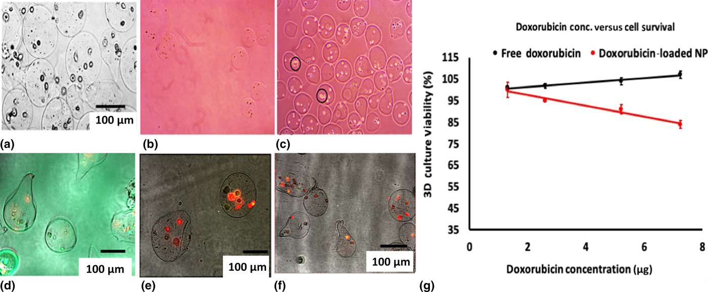
It can be noted from Fig. 4(a) that most of the spheroids have a spherical and symmetrical shape with the diameter of 70–100 µm. The phase contrast microscopic images of the spheroids at 2 and 8 days are shown in Figs. 4(b) and 4(c), respectively. After 8 days of growth period, cells are proliferated to form a multicellular aggregate which are encircled in Fig. 4(c).
To investigate the cytotoxic effect of doxorubicin-loaded LPHNPs on these spheroids, the live/dead viability assay has been carried out. In this assay, the spheroids are treated with strain containing calcein-AM and ethidium homodimer-1 and viewed under the fluorescent microscope. Bright red fluorescence of spheroids indicates dead/damaged cells, whereas intense uniform green fluorescence indicates live cells. Figure 4(d) represents the image of control (spheroids without NPs), which shows green fluorescence indicating live cells. However, along with the live cells, few dead cells can also be seen in Fig. 4(d). Figures 4(e) and 4(f) depict the images of the spheroids which are cultured with the free doxorubicin and doxorubicin-loaded LPHNPs, respectively, for 24 h. It can be noted from these images that all the cells in these spheroids show intense red color indicating dead cells. When compared with the control [Fig. 4(d)], drug-treated spheroids [Figs. 4(e) and 4(f)] show significantly more cell damage which confirms the cytotoxicity of doxorubicin-loaded LPHNPs in 3D culture. The dead cells observed in the control are may be due to the lack of nourishment. As the spheroids are not immobilized to any surface, it is not possible to provide continuous nourishment to cells over the period of 8 days, Thus, few cells which are not very healthy can form the spheroids but can not survive in long run without nourishment.[Reference Edmondson, Broglie, Adcock and Yang27]
To compare with the 2D cell cultures, the cytotoxic analyses of free doxorubicin and doxorubicin-loaded LPHNPs have been tested by the 24-h MTS assay. The concentration of doxorubicin used was IC50 [DOX] for HeLa cells in 3D cultures. The results obtained are shown in Fig. 4(g). As can be seen from Fig. 4(g), in the presence of free doxorubicin, 3D culture shows temporal growth, and doxorubicin-loaded LPHNPs show some cytotoxic effect. Unlike 2D cultures, the spheroids are resistant to the free doxorubicin drug. This resistance can be due to the differences in cell cycle distribution, inability of the drug to penetrate the spheroid, or the presence of hypoxic cells. Furthermore, it is also reported that the size of the spheroids also plays very important role in cytotoxicity.[Reference Lovitt, Shelper and Avery28] In general, cell toxicity is observed to be lower for smaller sized spheroids. In our case, the spheroids are 70–150 µm in diameter which is less than spheroid normally reported in the literature (350–800 µm), which shows higher toxicity.[Reference Gong, Lin, Cheng, Su, Zhao, Liu, Wen and Zhao29] The close 3D contact in spheroids has been implicated in this type of cytotoxicity. Proposed mechanisms for this “contact effect” include gap junctional “reciprocity”, cell shape-mediated changes in (repair-related) gene expression, and alterations in chromatin packaging which influence DNA repair. The consequences of the contact effect are especially important for multifraction exposures. Another form of resistance can be demonstrated during repetitive treatments; “regrowth resistance” reflects the capacity of spheroid cells to proliferate more efficiently to compensate for cell killing.[Reference Olive and Durand30]
Interestingly, doxorubicin-loaded LPHNPs shows a similar cytotoxic effect in 3D as in case of 2D cell culture. The exact mechanism behind is not very clear at this stage. But it may be due to different modes of penetration, selectivity, and specific targeting offered by the doxorubicin-loaded LPHNPs than the free doxorubicin drug.
Conclusions
Doxorubicin-loaded LPHNPs have been successfully synthesized by 3D-printed herringbone-patterned MIVM. The herringbone pattern observed to offer enhanced mixing efficiency and vorticity which leads to the uniform and reproducible LPHNP production. The cytotoxicity of these nanoparticles has been tested by using 2D and 3D microenvironment of HeLa cells. For 3D microenvironment, HeLa cells were successfully encapsulated inside sodium alginate beads forming spheroids of about 70–150 µm in diameter. Free doxorubicin and doxorubicin-loaded LPHNPs both showed cytotoxicity in 2D microenvironment. Whereas, in case of 3D microenvironment, HeLa cells observed to be resistant to the free doxorubicin and showed significant cytotoxicity for doxorubicin-loaded LPHNPs. This different cytotoxic behavior HeLa cells in 2D versus 3D cell culture models highlight the importance of 3D culture models in preclinical studies of anticancer drugs.