Introduction
Treacher Collins syndrome, also known as Treacher Collins Franceschetti syndrome and mandibulofacial dysostosis, is an autosomal dominant disorder of craniofacial morphogenesis which affects 1 in 25 000 to 50 000 newborns.Reference Gorlin, Cohen and Hennekam1, Reference Marsella, Scorpecci, Pacifico and Tieri2 At least 30–50 per cent of children with this disease present with bilateral, severe, conductive hearing loss due to bilateral aural stenosis or atresia and middle-ear malformation.Reference Marres3–Reference Jahrsdoerfer and Jacobson5 The results of reconstructive surgery to improve hearing are usually disappointing, because of the severity of middle-ear malformation and the presence of combined anomalies.Reference Jahrsdoerfer, Aguilar, Yeakley and Cole6
Active middle-ear implants are a new development in aural atresia surgery. The Vibrant Soundbridge implant (Med-El, Medical Electronics, Innsbruck, Austria) offers new possibilities for satisfactory audiological rehabilitation following atresia surgery.Reference Frenzel, Hanke, Beltrame, Steffen, Schonweiler and Wollenberg7, Reference Colletti, Carner, Mandala, Veronese and Colletti8 Since the new approach of coupling the floating mass transducer to the round window membrane was introduced and clinically proven, the use of Vibrant Soundbridge technology for patients with aural atresia has expanded the range of its application substancially.Reference Colletti, Soli, Carner and Colletti9 Nonetheless, there are currently insufficient data on applications of the Vibrant Soundbridge in patients with craniofacial syndromes. Moreover, the results for post-operative hearing states are usually generalised, and combined with the results of patients with milder degrees of malformation. There is little information on Vibrant Soundbridge results in patients with severe middle-ear malformations and a poor radiological score (i.e. a Jahrsdoerfer score of 5 or lower). Therefore, it is difficult to establish the possible applications of active middle-ear implants for the hearing rehabilitation of craniofacial syndrome patients.
This report aims to present results for Vibrant Soundbridge middle-ear implantation in patients with Treacher Collins syndrome, with bilateral osseous atresia and a poor pre-operative radiological score.
Materials and methods
Five patients (age range, 6 to 19 years; mean age ± standard deviation (SD), 12 ± 4.7 years) with congenital bilateral osseous atresia underwent implantation with a Vibrant Soundbridge middle-ear implant.
Three of these patients presented with Treacher Collins syndrome (age range, 6 to 19 years; mean age 11.7 ± 6.6 years) and were included in the study. Two of these three patients (10- and 19-year-old males) had mild-grade microtia, a stenotic external ear, and atresia of the medial part of the external ear canal bilaterally. One of the three patients (a six-year-old girl) presented with total aural atresia and severe microtia. Two of the three patients were users of bone conduction hearing aids, while the third patient had a conventional digital hearing aid, but none was satisfied with the quality of sound. None of these patients was willing to accept a bone-anchored hearing aid (BAHA), for aesthetic reasons. None had undergone any kind of ear surgery.
Pre-operative audiometric testing included routine pure tone audiometric measurement of air conduction (250 to 8000 Hz) and bone conduction (250 to 4000 Hz) hearing thresholds. Free field audiography, speech recognition thresholds (testing for 50 per cent correct recognition of words in quiet conditions) and word recognition scores (testing the percentage of correct words using bisyllabic words presented at 65 dB SPL) were evaluated pre- and post-operatively. During free field audiometric testing, the subject was seated 1 m in front of loudspeakers at 0° azimuth of the patient's head in a sound-proofed room. The mean functional gain was calculated as the difference between unaided pre-operative and Vibrant Soundbridge aided post-operative free field thresholds. The last available post-operative audiogram was used for data analysis.
Standard high-resolution computed tomography (CT) scans of the temporal bone were performed pre-operatively and the Jahrsdoerfer radiological score was determined.Reference Jahrsdoerfer, Yeakley, Aquilar, Cole and Gray10 All three Treacher Collins syndrome patients scored 4 points. According to their CT scans, all these patients had bilateral osseous atresia of the external ear canal (Figure 1).
Pre-operative, axial computed tomography scans of the patients’ right temporal bones. (a) & (b) Patient one: scans show a stenotic external part of the ear canal, osseous atresia, dysmorphic middle-ear cavity, ossicular agenesis, and an absent oval and round window; the arrow indicates a free fragment of the incus body in the attical space. (c) & (d) Patient two: scans show a well developed external part of the ear canal, osseous atresia, well developed tympanic cavity, absent ossicles, absent oval window, and a round window (arrow). (e) & (f) Patient three: scans show total osseous atresia with a hypoplastic tympanic cavity and missing ossicles; however, the footplate of the stapes is present (arrow).

Although the external ear was relatively well developed in two of the three patients, all three had severe malformation of the middle ear, with absent mastoid pneumatisation, a dysmorphic middle-ear cavity and missing ossicles. For one patient, round window and for one more patient, the stapes footplate could be suspected (Figure 1). All three patients had bilaterally symmetrical external ear canals, middle-ear abnormalities and normal inner-ear structures.
Surgical procedure
All three patients were scheduled for a transmastoid procedure. In one patient, a more posterior retroauricular incision than usual was needed (this patient required surgical microtia repair at a later date). As the mastoid was poorly pneumatised in all patients, the middle ear was approached by following the middle fossa dura until the middle-ear space was exposed.
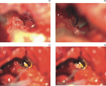
In the first patient (a 10-year-old boy), a small, free, dysmorphic fragment of the incus body was found in the attical space. Neither the stapes superstructure nor the oval or round windows were accessible in the middle ear. After careful inspection of the stapes facial nerve complex, a bony fragment of a different composition was found which was fused with the bony canal of the overlying facial nerve. This structure was carefully detached and mobilised. Although the oval window was not seen under the facial nerve, the detached structure was treated as a rudimentary stapes. Therefore, it was decided not to proceed with subfacial exploration of the round window membrane. Instead, a Vibrant Soundbridge floating mass transducer was attached to the remaining superstructure of the stapes. The clip of the floating mass transducer was reshaped and crimped around the detached, protruding shaft of the stapes superstructure. This contact was not firm enough, so hydroxylapatite bone cement was used to create a solid structure (Figure 2).
Operating microscope views for patient one. (a) The rudimentary stapes, fused with the bony canal of the facial nerve. (b) The same stapes, now detached and mobile. (c) The floating mass transducer clip, reshaped and crimped around the shaft of the stapes superstructure. (d) The floating mass transducer, firmly cemented to the rudimentary stapes with bone cement to ensure adequate contact.

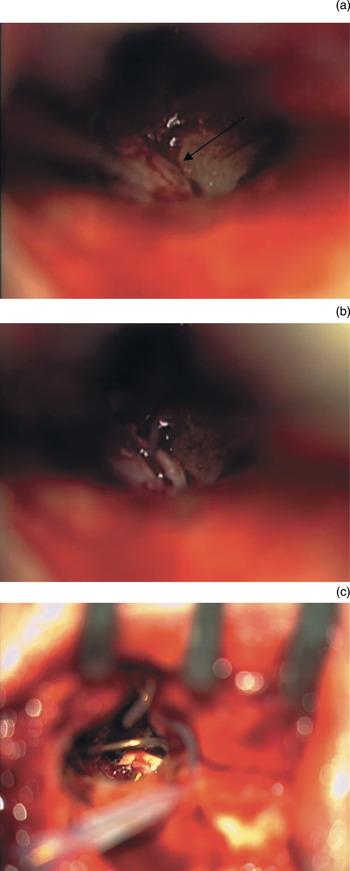
In the middle ear of the second patient (a 19-year-old young man), an almost identical anatomical situation was found. The external ear canal of this patient was well developed, but in the middle-ear space behind the osseous atretic plate no ossicular remnants were found. As in the first patient, we found a thin osseous plate fused to the bony facial canal in the oval window area. This weak structure was detached and treated as a rudimentary stapes, as in our first patient. As this mobile bony fragment was weak, fixation of a floating mass transducer was not possible. Thus, a Vibrant Soundbridge floating mass transducer was positioned adjacent to the released structure and covered in cartilage (Figure 3). A piece of perichondrium was wrapped around the whole arrangement to avoid contact with the surrounding bone.
Operating microscope view for patient two. (a) The rudimentary stapes, fused with the bony canal of the facial nerve. (b) The same stapes, now detached and mobile. (c) The floating mass transducer, positioned adjacent to the stapes superstructure and covered with cartilage.

The CT scans for the third patient (a six-year-old girl) indicated that the external and middle ear structures were even less developed than in the other two patients, with an absent external ear canal and a very hypoplastic middle-ear cavity identified. However, in contrast to the previous patients, the footplate of the stapes was identified in the region of the oval window (Figure 1c). After entering the middle-ear space, the ossicles were found to be absent, but a mobile footplate and remnants of the anterior branch of the stapedial arch were identified. A Vibrant Soundbridge floating mass transducer was positioned on the mobile stapes footplate in the oval niche, with interposed perichondrium. To ensure better contact and stability, the floating mass transducer was covered by and stabilised with cartilage.
The three patients were observed at intervals of one, two and six months after surgery. The minimum follow-up period was 8 months (range, 8–18 months; mean ± SD 11 ± 4.4 months). The audioprocessor was activated four to six weeks post-operatively for all patients.
Statistical analysis used paired, two-tailed t-tests to test for significant differences between pre- and post-operative audiometric measurements, taking a p value of less than 0.05 as statistically significant.
Results
No intra- or post-operative surgical complications were encountered. Wound healing was complete in all patients. Two patients noted an unpleasant feeling of pressure after activation of their device, which resolved when a lower-power magnet was set in.
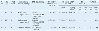
The results of patients’ pre- and post-operative audiological evaluations are presented in Table I. All the patients showed high levels of bone conduction and poor pure tone threshold scores pre-operatively. The patients’ mean bone conduction threshold did not differ significantly due to surgery, being 8.6 ± 12.1 dB HL pre-operatively and −11.7 ± 10.1 dB HL post-operatively (p > 0.05). However, the patients’ mean air conduction threshold was significantly reduced, by 44.5 dB HL, being 67.3 ± 8.9 dB HL pre-operatively and 22.8 ± 5.5 db HL post-operatively (p < 0.001). Moreover, there were statistically significant differences between pre-operative air conduction thresholds and post-operative aided air conduction thresholds for all frequencies. Non-aided air conduction levels improved slightly in the two patients in whom fused, rudimentary stapes were detached from the facial canal and mobilised. In these two patients, the mean post-operative non-aided air conduction threshold was 53.5 dB HL (p < 0.05, compared with pre-operative air conduction thresholds). With the device activated, the three patients’ mean word recognition score increased from 0 to 96.7 per cent, and their mean speech recognition threshold decreased to 46.7 dB SPL. After Vibrant Soundbridge implantation, there was a significant improvement in speech perception in quiet conditions, compared with the best aided situation pre-operatively (p < 0.05).
Pre- and post-operative findings

*Radiological (Rad) score, Jahrsdoerfer classification. †With Vibrant Soundbridge. Pt no = patient number; y = years; FMT = floating mass transducer; pre-op = pre-operative; BC = bone conduction; SD = standard deviation; AC = air conduction; post-op = post-operative; SRT = speech recognition threshold; WRS = word recognition score; – = unable to complete
At the most recent follow-up appointment, each patient's satisfaction with and sense of personal benefit from their device were assessed. All patients were constant users of their device, and all reported a better subjective quality of sound perception, compared with their pre-operative aided hearing.
Discussion
Conventional attitudes to hearing rehabilitation in patients with congenital aural atresia have changed significantly in recent years. Many authors are now of the opinion that conventional reconstructive surgery is less justifiable, and prefer instead to utilise the new implant technologies available. Even with significant efforts at external ear canal reconstruction, most patients still require some form of amplification.Reference Evans and Kazahaya11 In even the most optimistic assessment, conventional reconstructive surgery produces adequate hearing results (i.e. a hearing threshold of less than 30 dB) in only about half of all patients.Reference De La Cruz and Teufert12 However, the use of such new technologies as BAHAs and implantable hearing devices enables functional outcomes in almost all patients. These results have changed the approach to treatment for patients with congenital aural atresia. Now, middle-ear and auditory canal reconstructive surgery is recommended only for patients with mild malformations (i.e. a Jahrsdoerfer score of more than 8). Some surgeons view such surgery as already redundant.
Treacher Collins syndrome, also known as Treacher Collins Franceschetti syndrome and mandibulofacial dysostosis, is an autosomal dominant genetic disorder which affects the head and neck. About 50 per cent of patients with this syndrome have conductive hearing loss caused by characteristic major and/or minor ear anomalies.Reference Marres3 Microtia has been reported in as many as 85 per cent of patients, with one-third of patients presenting with stenosis or complete atresia.Reference Jahrsdoerfer and Jacobson5 In most cases, the middle-ear cavity is hypoplastic and dysmorphic, with dysmorphic or missing ossicles and absent mastoid pneumatisation. The conductive hearing loss of patients with Treacher Collins syndrome is mainly attributable to middle-ear malformations.Reference Pron, Galloway, Armstrong and Posnick4 Severe dysplasia of the stapes facial nerve complex frequently prevents conventional correction of the middle-ear malformations.Reference Jahrsdoerfer, Aguilar, Yeakley and Cole6 The functional outcome of reconstructive surgery of the external ear canal and middle ear is often disappointing.Reference Marres3
Bone-anchored hearing aids provide a good alternative to atresia surgery. However, they may be associated with local inflammation, extrusion, or loosening of the implanted fixture, and patients must constantly maintain good hygiene in order to avoid infection. Generally, BAHA implantation is a routine procedure which enables satisfactory hearing rehabilitation. However, in Treacher Collins syndrome patients, poor or irregular calvarial bone thickness often necessitates additional drill holes, placement of the fixture in contact with the dura, or the use of bone augmentation materials.Reference Marsella, Scorpecci, Pacifico and Tieri2
Within this context, the Vibrant Soundbridge middle-ear implant offers new possibilities for audiological rehabilitation in patients requiring atresia surgery.
Following the introduction of the Vibrant Soundbridge implant, which involves the positioning of a floating mass transducer adjacent to the round window membrane, the use of these implants in patients with aural atresia has expanded substantially.Reference Colletti, Soli, Carner and Colletti9 This surgery is now performed in many clinics, and clinical practice is evolving. However, the amount of published data is still small, as are the number of reported patients. In addition, there is almost no published information on the results of vibroplasty for genetic craniofacial syndromes. Patients with severe birth defect disorders typically have ear malformations related to craniofacial syndromes. However, in many publications the results for craniofacial syndrome surgery are presented and analysed together with those for surgery of aural atresia cases with mild middle-ear malformation. This makes it difficult to separate out possible solutions for each individual condition.
Kiefer and Staudenmaier assessed surgical results for 15 patients with middle-ear malformations with or without external auditory canal atresia, and reported satisfactory hearing outcomes (i.e. hearing threshold <30 dB in 14 of 15 patients, with mean hearing levels of 38 dB for low frequencies and 22 dB for high frequencies).Reference Kiefer and Staudenmaier13 The study group included two patients with genetic syndromes (one with Treacher Collins syndrome and one with the syndrome involving coloboma, heart defects, atresia of the choanae, retardation of growth and development, genital and urinary abnormalities, and ear abnormalities and/or hearing loss (the ‘CHARGE’ syndrome)); unfortunately, these patients’ individual hearing results were not described.
Colletti et al. presented 12 patients with severe external auditory canal and middle-ear malformations, including 7 with bilateral external auditory canal malformations and 4 with associated clinical diagnoses (2 had hemifacial microsomia, 1 had Goldenhaar syndrome and 1 had Churg–Strauss syndrome).Reference Colletti, Carner, Mandala, Veronese and Colletti8 A round window placement was used in all these patients, producing a mean post-operative aided air conduction threshold of 28.67 dB HL. Some patients had severe middle-ear malformations (one patient had a Jahrsdoerfer score of 3, while others had scores of 4 to 7). Four patients had no stapes superstructure, but these patients’ hearing results were not separately reported.
Tringali et al. presented a case study of results using a Carina™ fully implantable device with a MET V transducer for conductive applications, in a 14-year-old boy with Treacher Collins syndrome.Reference Tringali, Pergola, Ferber-Viart, Truy, Berger and Dubreuil14 The authors reported a post-operative mean hearing threshold of 41 dB. They concluded that implantable hearing devices can provide appropriate gain relative to the degree of hearing loss, and that such devices may constitute viable treatment for children with agenesis of the external auditory canal and severe malformations of the middle ear.
Our three cases comprised patients with Treachers Collins syndrome and severe malformations, with a Jahrsdoerfer radiological score of 4. Thus, judging by conventional criteria, all these patients had an unfavourable outlook. None of these patients were candidates for standard surgery, based on both conventional surgical assessment and the Jahrsdoerfer scoring system. In such unfavourable cases, one may question whether a technically complex operation would be worthwhile, or whether immediate referral for BAHA surgery would be preferable. However, if the patient has a good, mobile stapes or footplate, and if contact with a floating mass transducer is possible, then the ultimate hearing result may be very satisfactory and stable. In our series, the most successful auditory results were seen in the patient with maximum contact between the floating mass transducer and the rudimentary stapes, aided by hydroxylapatite bone cement.
The degree of implant fixation has an important effect on hearing results. Supposedly better hearing results are obtained when the floating mass transducer is fixed on the stapes superstructure or positioned on the round window. However, in recent years round window vibroplasty has gained increasing popularity as a safe, effective, stable and universal treatment solution for patients with conductive and mixed hearing loss, who may have few other options, if any.Reference Baumgartner, Böheim, Hagen, Müller, Lenarz and Reiss15 In cases of chronic otitis and cases of ear malformation, the stapes is usually fixed, rudimentary and unidentifiable, so it is difficult to establish stable contact with a floating mass transducer. Nonetheless, it could be an optimal solution if floating mass transducer fixation to the mobile stapes is viable. Experimental and clinical research has indicated that direct contact between the floating mass transducer and the stapes footplate enables better performance, especially for high frequencies, because of the larger vibrating area of the complete footplate.Reference Hüttenbrink, Beutner and Zahnert16, Reference Zahnert, Bornitz and Hüttenbrink17 If the stapes is mobile, electrocochleography recordings indicate that the final performance is better when the floating mass transducer is placed on the stapes rather than the round window membrane.Reference Mandalà, Colletti and Colletti18 In cases of fixed stapes and round window coupling, the stapes fixation may in itself cause some hearing loss; therefore, it is reasonable to perform fenestration of the vestibuli, covering the defect with fascia.Reference Mandalà, Colletti and Colletti18, Reference Lupo, Koka, Holland, Jenkins and Tollin19
• Reconstructive surgery for Treacher Collins syndrome usually has disappointing hearing results
• Use of Vibrant Soundbridge middle-ear implants in Treacher Collins patients with aural atresia and poor radiological scores was assessed
• The implant's floating mass transducer was coupled to the rudimentary stapes or footplate
• Satisfactory hearing results were obtained
Currently, there is insufficient published information on vibroplasty as treatment for aural atresia to draw conclusions regarding the most appropriate fixation mechanism. However, regardless of the fixation method, the available results are similar, with hearing thresholds of 23–35 dB HL.Reference Frenzel, Hanke, Beltrame, Steffen, Schonweiler and Wollenberg7, Reference Colletti, Carner, Mandala, Veronese and Colletti8, Reference Kiefer and Staudenmaier13, Reference Mandalà, Colletti and Colletti18 Roman et al. reported a case in which the round window position was used, with a final mean hearing threshold of 33.75 dB.Reference Roman, Nicollas and Triglia20 Colletti et al. and Mandala et al. recently reported series in which a round window placement was performed for all patients, resulting in mean post-operative air conduction thresholds of 28.67 and 24.1 dB HL, respectively.Reference Colletti, Carner, Mandala, Veronese and Colletti8, Reference Mandalà, Colletti and Colletti18 Frenzel et al. presented a homogeneous group of patients with unilateral bony atresia and satisfactory post-operative hearing results (i.e. a mean hearing level of 23.8 dB HL); these authors used various implant coupling sites, including the stapes, incudomalleolar complex, footplate and round window, but hearing results were unexceptional in every case.Reference Frenzel, Hanke, Beltrame, Steffen, Schonweiler and Wollenberg7 Verhaert et al. reported treatment results for five patients with ear atresia who underwent Carina™ MET or Vibrant Soundbridge coupling to the stapes, and who achieved a mean air conduction threshold of 33.3 dB HL.Reference Verhaert, Fuchsmann, Tringali, Lina-Granade and Truy21 In our series, incudostapedial complex coupling was used for the two atresia patients without craniofacial syndromes, with quite similar results (i.e. a mean post-operative hearing level of 28.2 dB HL). In specific cases of malformation in which it is impossible to achieve satisfactory floating mass transducer contact with a mobile footplate, the recently introduced vibroplasty coupler for the oval window may be used.Reference Hüttenbrink, Zahnert, Bornitz and Beutner22 However, in cases of congenital aural atresia the middle-ear cavity is under-developed and hypoplastic, with little space for a specific coupler, so the application of such vibroplasty couplers may be complicated.
Conclusion
In Treacher Collins patients with severe middle-ear malformations and a poor pre-operative radiological score, middle-ear Vibrant Soundbridge implantation can lead to satisfactory hearing results analogous to those for other ear atresia cases. The Vibrant Soundbridge is a versatile implant, and there are various technical possibilities for the coupling of the floating mass transducer even in patients with severe middle-ear malformations (e.g. those associated with craniofacial disorders). Coupling of the floating mass transducer with a rudimentary stapes or footplate may provide satisfactory audiological results. However, the presented, initial results need to be confirmed by additional studies including a greater number of patients.