Implications
The process of understanding and describing degradation mechanisms in the equine digestive ecosystem in general, and in the hindgut in particular, is essential to provide information for proper feeding practices to be implemented. Regardless of its importance for the nutritional status of the host, the significance of hindgut fermentation has not yet been fully understood, and few reports have focused deeply on the contribution of the hindgut microbial population to the nitrogen and energy requirements of the horse. In this review paper, we intend to gather existing information on the equine ecosystem and to provide future perspectives of research.
Introduction
Horses are free-ranging herbivores of grassland environments adapted to eat large quantities of high fiber feeds (Janis, Reference Janis1976; Bennett, Reference Bennett1980). Nowadays, with the increase of their use in sports and leisure, many horses are housed during long periods of time and fed daily on two or three meals of forages plus concentrate. The forage component of the diet is sometimes characterized by having low-to-medium energy concentration and variable levels of fiber and protein (Micol and Martin-Rosset, Reference Micol and Martin-Rosset1995). This pattern of feed supply is not as expected by the horse’s phylogenetic adaptation to grassland environments (Janis, Reference Janis1976). These management procedures have important implications on the utilization of nutrients from both concentrate and forage components of the total ration, on the digestive tract in general and the hindgut in particular and, of course, on the health and welfare of the horse (Hill, Reference Hill2007).
The process of understanding and describing forage degradation mechanisms in the equine digestive ecosystem in general, and in the hindgut in particular, is essential to provide information for proper feeding practices to be implemented. Despite the anatomical and placement differences between the rumen and the hindgut of horses, comparison between these digestive compartments is routinely discussed. Although with two different fermentative compartments (cecum and colon), comparison has mainly been made between the cecum and the rumen. Despite some similarities, differences between the hindgut of horses and the rumen are known not only concerning different and specific populations of micro-organisms, but also concerning anaerobiosis and fermentative activity conditions. In the different anatomical segments of the hindgut, there seem to be differences concerning microbial population and fermentative activity, namely between the cecum and the colon of horses (Hintz and Schryver, Reference Hintz and Schryver1972; Kern et al., Reference Kern, Slyter, Leffel, Weaver and Oltjen1974; Moore and Dehority, Reference Moore and Dehority1993; Julliand et al., Reference Julliand, de Fombelle, Drogoul and Jacotot2001). More recently, research has been directed toward the different anatomo-physiological segments of the colon, and the implication of the right ventral colon (RVC) ecosystem in forage degradation has been highlighted (de Fombelle et al., Reference de Fombelle, Varloud, Goachet, Jacotot, Philippeau, Drogoul and Julliand2003).
Regardless of these data, the significance of hindgut fermentation has not yet been fully understood, and few reports have focused deeply on the contribution of the microbial population of the hindgut to the nitrogen and energy requirements of the host animal (Vermorel and Martin-Rosset, Reference Vermorel and Martin-Rosset1997; Martin-Rosset and Tisserand, Reference Martin-Rosset and Tisserand2004). Very little is known about the interrelationships between the different microbial populations in the equine hindgut and how these microbial populations interact.
Post-gastric placement of the fermentative activity implies that substrate availability is conditioned by pre-cecal digestibility of the diet (Drogoul et al., Reference Drogoul, Poncet and Tisserand2000). Microbial growth and consequent fiber degradation in the cecum-colon environment rely on the energy and nitrogen availability. Available nitrogen is supplied by alimentary protein, which reaches the hindgut, urea recycling and endogenous protein from cell desquamation or secretion into the lumen. Volatile fatty acids (VFAs) produced in the hindgut by fermentative activity contribute with 60% to 70% of the energy absorbed by the horse (Vermorel and Martin-Rosset, Reference Vermorel and Martin-Rosset1997); yet microbial protein produced has a marginal, if any, contribution to the protein nutrition of its host (Martin-Rosset and Tisserand, Reference Martin-Rosset and Tisserand2004).
The aim of this review work is to present current knowledge of digestive processes in different compartments of the equine digestive tract, with higher focus on the hindgut. It is expected that this review will provide a better understanding of the hindgut ecosystem and the implications to its host.
Pre-cecal digestion
Although several workers have examined the total intestinal tract digestibility of different fibrous feeds in horses using the mobile bag technique (Martin-Rosset et al., Reference Martin-Rosset, Andrieu, Vermorel and Dulphy1984; Macheboeuf et al., Reference Macheboeuf, Maranji, Poncet and Martin-Rosset1995; Moore-Colyer et al., Reference Moore-Colyer, Hyslop, Longland and Cuddeford2002; Hyslop, Reference Hyslop2006), limited information is still available on the site and to the extent of their digestion within the different regions of the gastrointestinal tract. Knowledge of the amount of feed that is digested within a certain region of the gut is particularly useful when considering energy production and use (Vermorel and Martin-Rosset, Reference Vermorel and Martin-Rosset1997). The efficiency with which metabolic energy is used for maintenance (km) resulting from absorbed glucose metabolism is 85%, whereas the one resulting from VFA metabolism, which derived from fiber degradation in the hindgut, ranges between 63% and 68% (Vermorel and Martin-Rosset, Reference Vermorel and Martin-Rosset1997). In addition, knowledge concerning the site of digestion is particularly important when measuring protein availability, as it is mainly the protein digested in the small intestine that is utilized by the horse to meet its own requirements (Hintz et al., Reference Hintz, Hogue, Walker, Lowe and Schryver1971; Hintz and Cymbaluk, Reference Hintz and Cymbaluk1994; Martin-Rosset et al., Reference Martin-Rosset, Vermorel, Doreau, Tisserand and Andrieu1994; Moore-Colyer et al., Reference Moore-Colyer, Hyslop, Longland and Cuddeford2002; Potter, Reference Potter2004), whereas protein that reaches the hindgut will play a role to meet the requirements of the microbial population of this ecosystem (Martin-Rosset and Tisserand, Reference Martin-Rosset and Tisserand2004).
Stomach
In the stomach, most of the digesta is held for a limited period of time, but it is rarely completely empty and a significant proportion of digesta may remain in it for 2 to 6 h. As digesta approaches the pylorus, the pH falls, due to the HCl secretion, potentiating the proteolytic activity of pepsin and stopping that of fermentation. Owing to the small size of the stomach, and the relatively short dwell time, the degree of protein digestion is slight. It seems that the amount of microbial fermentation that occurs in the equine stomach, although limited in extent, can not only play an important role in the energetic promotion of the diet (de Fombelle et al., Reference de Fombelle, Varloud, Goachet, Jacotot, Philippeau, Drogoul and Julliand2003) but also can be responsible, in some cases, for gastric malfunction. Coenen et al. (Reference Coenen, Mosseler and Vervuert2006) alerted to the importance of elucidating microbial changes in the foregut of horses with laminitis. Varloud et al. (Reference Varloud, Fonty, Roussel, Guyonvarch and Julliand2007) confirmed an abundant microbial colonization of the stomach of horses reported by other authors. According to de Fombelle et al. (Reference de Fombelle, Varloud, Goachet, Jacotot, Philippeau, Drogoul and Julliand2003), the stomach had the highest anaerobic count of the total gastrointestinal tract (1.45 × 10⁹ CFU/ml against 7.95 × 10⁷ CFU/ml in the cecum) and high concentration, along with the small intestine, of lactobacillus, streptococci and lactate using bacteria, which suggests an important participation of these microorganisms in the degradation of highly fermentable carbohydrates. Nevertheless, a recent review on microbial ecosystems states that only 11% of bacterial taxa present within the rumen have been recovered in culture (Edwards et al., Reference Edwards, Huws, Kim, Lee, Kingston-Smith and Scollan2008), referring to the rest as uncultivable. The same may happen with hindgut bacteria being cultivated and can explain the lower counts found in the cecum when compared with the stomach.
Small intestine
The digesta passage rate is very rapid in the small intestine, with most of the digesta rate being near 30 cm/min. Nevertheless, enzymatic digestion of digestible components of the cellular content of feedstuffs is very efficient in the small intestine compared to the low digestion of cell wall components (Van Weyenberg et al., Reference Van Weyenberg, Sales and Janssens2006).
With respect to the most recent synthesis carried out by the National Research Council (NRC) and Institut National de la Recherche Agronomique (INRA) to update the energy and nitrogen systems (NRC, 1989 and 2007; INRA, 1990 and in progress) using data obtained from the digesta and external markers collected in the cecum or by the mobile nylon bag technique in fistulated horses or ponies, the true digestibility coefficients of the main components have been stated to be: 100% for cytoplasmic carbohydrates which are digested as glucose and lactate; 85% for starch which supplies glucose in usual conditions; 5% to 15% for incompletely digestible cell wall carbohydrates with glucose production (5% to 10% for forages and 5% for concentrates); 90% to 95% for lipids (ether extract) are digested and the amount of long-chain fatty acids absorbed from the small intestine is 90% to 95% of digested lipids; 30% to 90% for protein: 70% to 80% for cereals and their by-products, 75% to 90% for oil meals, 70% for fresh grass, 60% for dehydrated alfalfa and 30% to 75% for grass hay.
As a result, most of the structural carbohydrates and a significant proportion of starch and crude protein more or less linked to the cell wall can bypass the enzymatic digestion according to the botanical origin, vegetative state, feed processing and conservation, diet composition and feeding level.
Hindgut environment
Digestion in the cecum and colon occurs by the action of the microbial population that colonizes these compartments. This microbial population maintains, under normal feeding conditions, a balance with its host, keeping the integrity of the ecosystem, contributing to the prevention of disorders and forming a barrier against pathogens (Julliand, Reference Julliand1998). Nevertheless, they are also responsible for disorders due to the alteration of this ecosystem under stressful conditions, for instance an abrupt change of the diet.
Anatomical and morphological considerations
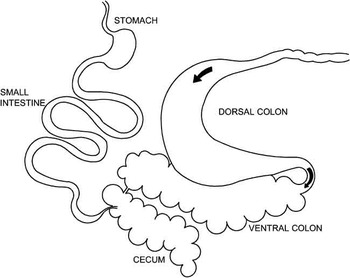
The first appearance of digesta in the cecum is approximately 30 to 45 min after leaving the stomach; within 3 h after feed consumption, most of the digesta reaches the cecum and ventral colon (Van Weyenberg et al., Reference Van Weyenberg, Sales and Janssens2006). The cecum is a blind sac, highly sacculated. At the top of the cecum, the ileo-cecal and the ceco-colic junctions are situated in relatively close proximity to each other, one through which digesta enters from the ileum and the other through which passage from the cecum to the RVC is facilitated (Figure 1). The cecum starts to contract 12 to 15 cm after the ceco-colic junction traps digesta in the cecal base and forces some through the valve to the RVC (Van Weyenberg et al., Reference Van Weyenberg, Sales and Janssens2006). From the RVC, digesta passes to the left ventral colon (LVC); the ventral colon (RVC and LVC) is voluminous, with a diameter up to 25 to 30 cm, while the transition between the LVC and the left dorsal colon (LDC) is narrow, also called the pelvic flexure. This anatomic morphology is responsible for a selective retention of coarse particles (1 cm or more) in the cecum and ventral colon, meaning that in these compartments coarse particles are retained and liquid and fine particles move on to the LDC and right dorsal colon (RDC; Drogoul et al., Reference Drogoul, Poncet and Tisserand2000; Van Weyenberg et al., Reference Van Weyenberg, Sales and Janssens2006; Figure 1). The diameters of these portions of the colon are large, and there are no sacculations in these portions of the dorsal colon. Another selective mechanism at the end of the RDC preferentially retains fuid and smaller particles (below 2 mm) in these compartments (Figure 1), which is known as the colonic separation mechanism (Drogoul et al., Reference Drogoul, Poncet and Tisserand2000).
Localization of main selective retention sites in the hindgut of horses.

Passage rate
The passage rate through the gastrointestinal tract provides indirect information on the extent to which feed is digested and fermented. The type of substrate that arrives in the hindgut is dependent on pre-cecal digestibility, which in turn is related to the passage rate. Higher passage rates will induce a decrease in pre-cecal digestibility. In addition, retention times of digesta in the hindgut will affect digestibility, microbial activity and absorption of water.
Several animal and feed-related factors can affect the passage rate. Miraglia et al. (Reference Miraglia, Poncet and Martin-Rosset1992) reported that mean retention time (MRT) was significantly lower (−6 to 10 h) in light breeds than in draft breeds, regardless of the feeding level. The difference could be ascribed to the lower transit time: TT = −33% and −9% for maintenance and ad libitum feeding levels, respectively, and lower constant rates of the two pool compartments of the digesta tract in the light breed: k 1 = −63% and −30% and K 2 = +41% and +48% for maintenance and ad libitum feeding levels, respectively (Miraglia et al., Reference Miraglia, Poncet and Martin-Rosset2003), but this has no significant effect on digestibility of hay-based diets: 1.6 to 1.9 points for digestibility of organic matter (dOM) (Martin-Rosset et al., Reference Martin-Rosset, Doreau, Boulot and Miraglia1990). In contrast, Udén and Van Soest (Reference Udén and Van Soest1982) could not detect a relationship between MRT and body size. Physiological state will also have an influence on MRT. MRT in late pregnant mares is 10 h lower than that in dry mares fed ad libitum hay-based diets (Miraglia et al., Reference Miraglia, Poncet and Martin-Rosset1992). Lower MRT measured in late pregnant mares explains significant differences in digestibility (5.0 points for dOM) between pregnant and dry mares with the same body weight (BW), both fed ad libitum (Martin-Rosset et al., Reference Martin-Rosset, Doreau, Boulot and Miraglia1990). It could be ascribed again to the constant rates K 1 and K 2 which are 2.3 and 2.1 lower, respectively, in pregnant than in dry mares (Miraglia et al., Reference Miraglia, Poncet and Martin-Rosset2003). Conversely, there is no significant effect on digestibility: 1.5 points for dOM (Martin-Rosset et al., Reference Martin-Rosset, Doreau, Boulot and Miraglia1990) due to lactation; even retention time is lower as K 1 and K 2 are lower: −24% and −38%, respectively, but TT is close (Miraglia et al., Reference Miraglia, Poncet and Martin-Rosset1992 and Reference Miraglia, Poncet and Martin-Rosset2003). The effect of exercise on MRT and digestibility is still very controversial. With respect to the most consistent data, there is only a slight effect of exercise on fiber digestion (see review of Martin-Rosset, Reference Martin-Rosset2008).
Microbiological environment
It is estimated that 30% (Kern et al., Reference Kern, Slyter, Weaver, Leffel and Samuelsons1973) to 80% (Kern et al., Reference Kern, Slyter, Leffel, Weaver and Oltjen1974) of the cecum and colon microbial population is strictly anaerobic. In the cecum, total anaerobes range between 1.85 × 10⁷ and 2.65 × 10⁹c.f.u./ml, according to the literature (Kern et al., Reference Kern, Slyter, Weaver, Leffel and Samuelsons1973 and Reference Kern, Slyter, Leffel, Weaver and Oltjen1974; Mackie and Wilkins, Reference Mackie and Wilkins1988; Julliand et al., Reference Julliand, de Fombelle, Drogoul and Jacotot2001; Medina et al., Reference Medina, Girard, Jacotot and Julliand2002; de Fombelle et al., Reference de Fombelle, Varloud, Goachet, Jacotot, Philippeau, Drogoul and Julliand2003).
Bacteria can be classified into cellulolytic, proteolytic, lactate-using and glycolytic bacteria. From metabolic and genetic studies, it is concluded that the cecal cellulolytic microflora are mainly composed of specific strains of ruminococcus (Julliand et al., Reference Julliand, de Vaux, Millet and Fonty1999). These authors refer to strains of Ruminococcus flavefaciens, Ruminococcus albus and Fibrobacter succinogenes as dominant, showing however genetic differences from congeners found in the rumen; these differences are probably due to an adaptation of these strains to the specificity of the cecal environment. Also, in a study presented by Lin and Stahl (Reference Lin and Stahl1995), two new lineages of F. succinogenes were identified as being equine specific, and these authors refer that, in contrast to ruminants, a sizable proportion of the available protein and carbohydrate is digested and absorbed before reaching the cecum, with plant fiber representing a greater fraction of the incoming substrate. Thus, a greater contribution by fiber-digesting bacteria is anticipated, as was observed in this study.
More recent studies also refer to the specificity of this microbial population (Daly et al., Reference Daly, Stewart, Flint and Shirazi-Beechey2001; Daly and Shirazi-Beechey, Reference Daly and Shirazi-Beechey2003). Using molecular analysis, Daly et al. (Reference Daly, Stewart, Flint and Shirazi-Beechey2001) reported that 89% of the recovered sequences did not correspond to any recorded ones, suggesting that the anaerobic microflora of the equine large intestine are underrepresented and that the equine flora may contain many novel bacterial species. In a latter study, Daly and Shirazi-Beechey (Reference Daly and Shirazi-Beechey2003), using specific oligonucleotide probes, quantitatively analyzed the intestinal microflora and obtained the first data of predominant bacterial populations in the large intestine of horses. The results showed that the strains Spirochaetaceae, Cytophaga–Flexibacter–Bacteroides, the group Eubacterium–Clostridium rectal Coccoidea and an ‘unknown cluster C’ of clostridiaceae were the largest populations in the cecum-colon, each group comprising 10% to 30% of the total microflora in examined horses. Other groups also identified as important were the Bacillus–Lactobacillus–Streptococcus, the Fibrobacter and ‘unknown cluster B’, each representing 1% to 10% of the total flora. Members of the Spirochaetaceae group of bacteria have been shown to be major acetate producers using H2/CO2 as the substrate for acetogenesis. Given the population size detected for the Spirochaetaceae in the equine colon by these authors, it can be suggested that they may compete with other bacteria that use H2 as a terminal electron acceptor, such as methanogens, providing a significant and valuable source of acetate for the horse. In fact, hydrogen recovery in the hindgut is lower than in the rumen in animals fed with hay (0.84 v. 0.61 and 0.59 in cattle rumen and equine cecum and colon, respectively). Also, methane production is lower in the hindgut than in the rumen (250 against 23 mmol/mol in the rumen of cattle and the equine colon, respectively), indicating the existence of other hydrogen sink reactions, making acetogenesis a likely alternative (Demeyer, Reference Demeyer1991). Replacement of methane by acetate as hydrogen sink in a fermentation should increase the VFA yield per unit of the substrate fermented, and thus the energy yield for the animal. Julliand and Tisserand (Reference Julliand and Tisserand1992) found in cecal bacterial flora 2.1 to 15.9 × 10⁶ CFU/ml of anaerobic bacteria. Of these, 1.3 to 13.5 × 10⁶ CFU/ml were cellulolytic bacteria and 0.2 to 6.0 × 10⁶ CFU/ml of bacteria were proteolytic. In a study by Julliand et al. (Reference Julliand, Prevost and Tisserand1993), cellulolytic bacterial population in the cecal microflora was predominant in relation to the proteolytic population with average values of 4.6 to 9.4 × 10⁶ CFU/ml and 0.3 to 1 × 10⁶ CFU/ml, respectively. So far there would be on average 78% and 22% of celluloytic and proteolytic bacteria in the cecum.
The proportion of cellulolytic bacteria among total anaerobes appears to be greater in the cecum than in the lower parts of the hindgut (de Fombelle et al., Reference de Fombelle, Varloud, Goachet, Jacotot, Philippeau, Drogoul and Julliand2003). Kern et al. (Reference Kern, Slyter, Leffel, Weaver and Oltjen1974) reported that the number of viable cellulolytic bacteria per gram of ingesta was six times higher in the cecum that in the terminal colon: 43 × 106 and 7 × 10⁶ CFU/ml, respectively, for the cecum and terminal colon. In addition, the average concentrations of starch-utilizing bacteria (lactobacilli and streptococci) and lactate-utilizing bacteria tend to be lower in the cecum than in other parts of the digestive tract, suggesting that the cecal ecosystem is less exposed to rapidly fermentable carbohydrates than the lower parts of the hindgut (de Fombelle et al., Reference de Fombelle, Varloud, Goachet, Jacotot, Philippeau, Drogoul and Julliand2003). The hypotheses presented by de Fombelle et al. (Reference de Fombelle, Varloud, Goachet, Jacotot, Philippeau, Drogoul and Julliand2003), that is, that particles entering the dorsal colon have lower parietal polysaccharide content because of the longer retention of coarse particles in the cecum and ventral colon due to the pelvic flexure, might explain the decrease in proportion of cellulolytic bacteria, and the higher proportion of starch-utilizing bacteria in the lower parts of the colon. In fact, in a series of studies conducted to investigate the impact of different forage : grain proportions in microbial profile and activity of the cecum and colon, researchers found that the site of major variations on microbial profile, when barley was fed, was the colon (de Fombelle et al., Reference de Fombelle, Julliand, Drogoul and Jacotot2001). The same authors refer that soluble carbohydrates and undigested starch flow quickly through the cecum and have a greater impact on colonic microflora than on the cecal microflora (Drogoul et al., Reference Drogoul, de Fombelle and Julliand2001; Julliand et al., Reference Julliand, de Fombelle, Drogoul and Jacotot2001).
In the hindgut, such as in the rumen, we can clearly divide the microbial population into two distinct ecological sites: (i) liquid phase colonized by bacteria and zoospores; and (ii) bacteria associated with feed particles, mainly responsible for hydrolysis of cellular wall polysaccharides (Merry and McAllan, Reference Merry and McAllan1983). If we consider the information referred to earlier concerning differences in microbial population between digestive compartments, we could expect that in the cecum and ventral colon we should find a higher content of bacteria associated with particles of digesta, solid associated bacteria (SAB), and in the dorsal colon a higher content of bacteria that are in the liquid phase, liquid associated bacteria (LAB).
Recent research in ruminants has pointed out the potential of odd- and branched-chain fatty acids (OBCFAs) as markers to quantify bacterial matter leaving the rumen (Vlaeminck et al., Reference Vlaeminck, Dufour, Van Vuuren, Cabrita, Dewhurst, Demeyer and Fievez2005; Bessa et al., Reference Bessa, Maia, Jerónimo, Belo, Cabrita, Dewhurst and Fonseca2009) to provide a qualitative description of the proportions of different classes of microbes leaving the rumen and to predict rumen ratios of VFAs (Vlaeminck et al., Reference Vlaeminck, Fievez, Cabrita, Fonseca and Dewhurst2006a and Reference Vlaeminck, Fievez, Demeyer and Dewhurst2006b).
The OBCFA of rumen bacteria seem to be largely determined by the fatty acid synthetase of the microorganisms and, to a lesser extent, by physiological and culture conditions. This suggests that variations in the profile of OBCFA from the rumen can be considered to be mainly a reflection of changes in the relative abundance of specific bacterial populations in the rumen (Vlaeminck et al., Reference Vlaeminck, Fievez, Cabrita, Fonseca and Dewhurst2006a). The fatty acid composition of rumen bacteria is characterized by a large proportion of OBCFA in their membrane lipids (C15:0, iso C15:0, ante-iso C15:0, C17:0, iso C17:0, ante-iso C17:0 and C17:1; Kaneda, Reference Kaneda1991).
OBCFAs only occur at trace levels in most plants; they are easy to measure and are stable compounds, fulfilling the requirements for an internal marker (Diedrich and Henschel, Reference Diedrich and Henschel1990). Fatty acid compositions of LAB and SAB are different; total fatty acid content of SAB is higher than that of LAB (Vlaeminck et al., Reference Vlaeminck, Fievez, Demeyer and Dewhurst2006b). Vlaeminck et al. (Reference Vlaeminck, Fievez, Cabrita, Fonseca and Dewhurst2006a) reported that fatty acid content in rumen SAB was 1.6 to 2.8 times higher than that in LAB. Differences between LAB and SAB in chemical composition and in enzyme activity show that the distribution of bacterial species is different in the liquid and solid phases of the rumen (Michalet-Doreau et al., Reference Michalet-Doreau, Fernandeza, Peyrona, Millet and Fonty2001). This is in agreement with the current analysis of OBCFA, which suggests that the species composition of the adherent population differs from that of the liquid phase.
Relationships between LAB and SAB bacterial populations and forage : concentrate (F : C) ratio are also reported in the literature. Increasing the F : C ratio is usually beneficial for the pool of SAB because more fibrolytic bacteria attach to forage particles (Weimer et al., Reference Weimer, Waghorn, Odt and Mertens1999). However, results reported by several authors suggest that LAB are enriched in amylolytic bacteria. Increasing the concentrate proportion increases the LAB phase (Vlaeminck et al., Reference Vlaeminck, Fievez, Cabrita, Fonseca and Dewhurst2006a).
Cellulolytic bacteria contain high amounts of iso-fatty acids with Ruminococcus flavefaciens enriched in odd-chain iso-fatty acids and Ruminococcus albus in even-chain iso-fatty acids. In general, the amylolytic bacteria show low levels of branched-chain fatty acids and are relatively enriched in linear odd-chain fatty acids. These differences in the OBCFA profile observed among rumen bacteria allow its use in the assessment of the composition of, or shifts, in the rumen microbial population (Vlaeminck et al., Reference Vlaeminck, Fievez, Cabrita, Fonseca and Dewhurst2006a and Reference Vlaeminck, Fievez, Demeyer and Dewhurst2006b).
By applying these techniques to the cecum and colon contents, Santos et al. (Reference Santos, Jerónimo, Ferreira, Rodrigues and Bessa2007 and Reference Santos, Jerónimo, Ferreira, Rodrigues and Bessa2008) concluded that the technique was easily executed with these contents. These authors found differences in the fatty acid profile of cecum and colon bacteria. Cecum bacteria presented higher trans C18:1 isomers eventually, indicating a higher SAB activity/population that can be related to a preponderant cellulolytic activity, which is in accordance with what was referred to by de Fombelle et al. (Reference de Fombelle, Varloud, Goachet, Jacotot, Philippeau, Drogoul and Julliand2003). Concerning colon bacteria, results suggest that they have a chemical and fatty acid profile close to that of rumen LAB, mainly starch-utilizing bacteria, which is also in accordance with what was stated earlier by de Fombelle et al. (Reference de Fombelle, Varloud, Goachet, Jacotot, Philippeau, Drogoul and Julliand2003). These preliminary data indicate that these techniques can be useful as a microbial marker in the equine cecum-colon ecosystem (Santos et al., Reference Santos, Jerónimo, Ferreira, Rodrigues and Bessa2007 and Reference Santos, Jerónimo, Ferreira, Rodrigues and Bessa2008).
Fermentation parameters
The feed components undigested in the small intestine (15% for starch, 5% to 10% of ether extract, 10% to 70% of protein, 85% to 95% for cell wall carbohydrates) are fermented by a microbial population in the large intestine with production of VFAs, microbial mass, methane and fermentation heat. The amount of VFA produced can be estimated from the apparently digested organic matter (DOM): VFA (g/kg OM of feed) = (DOM–OM digested in the SI) × 0.92 (Vermorel and Martin-Rosset, Reference Vermorel and Martin-Rosset1997).
Fermentation patterns can serve as an indicator for microbial activity and digestibility of substrates in these compartments. The concentration of VFAs, lactate and pH reflects, to some extent, what is happening in the hindgut environment.
Wolter et al. (Reference Wolter, Gouy, Durix, Letourneau, Carcelen and Landreau1978) described the biochemical activity in the cecum of ponies fed a pelleted complete diet, measuring parameters such as pH, molar concentration in VFAs and lactic acid. According to these authors, the average pH is around 7 at the meal and descends to 6.8, 5 to 8 h after the meal, coinciding with the increase in dry weight of cecal contents. VFA average concentrations vary in the opposite direction to that for pH, and reached a maximum of 74.5 mmol/l 5 to 9 h after the meal, with molar percentages of 72.7 for acetic acid (C2), 21.5 for propionic acid (C3) and 5.6 for butyric acid (C4). Lactic acid met its higher value 3 to 5 h after the meal and average value was 3 mmol/l, just before the maximum peak for the VFAs. Higher levels of lactic acid can be observed in the case of over-consumption of food rich in easily fermentable carbohydrates, including starch, where the massive influx of carbohydrates to the cecum accelerates the acidification of the cecal content and facilitates the action of lactic bacteria (Wolter et al., Reference Wolter, Gouy, Durix, Letourneau, Carcelen and Landreau1978).
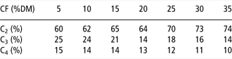
An abrupt dietary change from forage to concentrate modifications in fermentation patterns associated with a decrease in cecal pH and an increase in cecal lactate has been reported (Goodson et al., Reference Goodson, Tyznik, Cline and Dehority1988). In addition, after an abrupt incorporation of barley (50%) in a hay diet, de Fombelle et al. (Reference de Fombelle, Julliand, Drogoul and Jacotot2001) reported that total VFA significantly increased from 94.7 mmol/l to 98.3 mmol/l, but with a significant decrease in acetate (73.2 to 64.0 molar %) and an increase in propionate (18.9 to 28.0 molar %), leading to a decrease in the [(C2 + C4)/C3] ratio from 4.3 to 3.8, meaning a higher starch degradation and a decline in fiber degradation activity. Studying the influence of different hay : barley ratios (100/0; 70/30 and 50/50) on microbial profiles and activity, Julliand et al. (Reference Julliand, de Fombelle, Drogoul and Jacotot2001) also reported a significant decrease both in the pH (from 6.7 to 6.3) and [(C2 + C4)/C3] ratios (4.2 to 3.5) and an increase in lactate concentration (53.2 to 435.9 mmol/l) as well as in total VFA (85.2 to 93 mmol/l). The composition of the VFA mixture is related to the crude fiber (CF) content of the diet (Table 1) and the molar proportion of acetate (C2%) varies with the CF content of the feed or diet: C2 (%) = 0.54 CF (% dry matter (DM)) + 57 (r 2 = 0.80, n = 18; Vermorel and Martin-Rosset, Reference Vermorel and Martin-Rosset1997).
Variation of the molar composition in the large intestine with the CF content of feeds (Vermorel and Martin-Rosset, Reference Vermorel and Martin-Rosset1997)
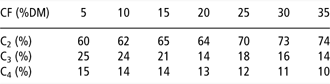
CF = crude fiber; DM = dry matter.
Estimation of the results from Hintz et al. (Reference Hintz, Hogue, Walker, Lowe and Schryver1971), Kern et al. (Reference Kern, Slyter, Weaver, Leffel and Samuelsons1973), Tisserand (Reference Tisserand1975), Tisserand and Masson (Reference Tisserand and Masson1976), Wolter et al. (Reference Wolter, Gouy, Durix, Letourneau, Carcelen and Landreau1978) and Martin-Rosset et al. (Reference Martin-Rosset, Doreau and Thivend1987).
Nitrogen metabolism in the hindgut
In ruminant nutrition, one should maximize the proliferation of rumen microorganisms to maximize the energetic use of cell wall constituents. Microbial protein from the rumen accounts for more than half of the total protein entering the intestine, which has an important role in nutrition. As in the rumen, microbial growth and consequent fiber degradation in the cecum-colon environment rely on the energy and nitrogen availability. VFAs produced in the hindgut by fermentative activity contribute with 60% to 70% of the absorbed energy by the horse (Vermorel and Martin-Rosset, Reference Vermorel and Martin-Rosset1997), whereas microbial protein produced has an apparent marginal contribution to the protein nutrition of the animal host (Martin-Rosset and Tisserand, Reference Martin-Rosset and Tisserand2004).
Feed and endogenous proteins that reach the hindgut are degraded as amino acids (AA), peptides and ammonia, and resynthesized to microbial protein according to the available energy (Robinson and Slade, Reference Robinson and Slade1974; Meyer, Reference Meyer1983; Martin-Rosset et al., Reference Martin-Rosset, Vermorel, Doreau, Tisserand and Andrieu1994). Bacteria are known to be able to use AA (and non protein nitrogen) for synthesizing their own protein (Baruc et al., Reference Baruc, Dawson and Baker1983) at a rate estimated to reach 2.5 mg N/kg DM per h in the cecum contents (Slade et al., Reference Slade, Robinson and Al-Rabbat1973). The growth of the proteolytic microbial population can be stimulated by soya supplementation (Julliand and Tisserand, Reference Julliand and Tisserand1992). Nitrogen balance studies have shown that urea is recycled in the equine gastrointestinal tract (Slade et al., Reference Slade, Robinson and Casey1970; Houpt and Houpt, Reference Houpt and Houpt1971; Prior et al., Reference Prior, Hintz, Lowe and Visek1974). When urea is recycled to the large intestine, it is degraded to ammonium ions by urease-catalyzed hydrolysis (Wootton and Argenzio, Reference Wootton and Argenzio1975). Hintz et al. (Reference Hintz, Hogue, Walker, Lowe and Schryver1971) measured urease activity in cecal fluid of 17% to 25% of that reported in bovine rumen fluid. Prior et al. (Reference Prior, Hintz, Lowe and Visek1974) refer that about 60% of the produced urea is recycled in the intestine of ponies that are fed a low protein diet, and that 50% of this nitrogen is recovered. This entero-hepatic cycle can supply nitrogen to meet the microflora requirement of the horse, but with a lower efficiency when compared to ruminants; only 10% to 30% of this nitrogen will be recovered by the microflora of the horse (Slade et al., Reference Slade, Robinson and Casey1970; Houpt and Houpt, Reference Houpt and Houpt1971; Hintz and Schryver Reference Hintz and Schryver1972; Glade, Reference Glade1984).
Cecal bacteria show a proteolytic activity in vitro and cecal isolates were shown to use ammonia or urea as nitrogen sources for growth, (Maczulak et al., Reference Maczulak, Dawson and Baker1985). Meyer (Reference Meyer1983) refers evidence that peptides, AAs and ammonia are absorbed from the large intestine. Assuming that one to two-thirds of the N will be absorbed as peptides or AAs, the amount of valuable protein produced by microorganisms in the hindgut would be equivalent to 1 to 1.5 g of protein/kg BW0.75 (Meyer, Reference Meyer1983). Slade et al. (Reference Slade, Bishop, Morris and Robinson1971) and McMeniman et al. (Reference McMeniman, Elliot, Groenendyk and Dowsett1987) reported that labeled AAs (cysteine) were slightly absorbed from the cecum and the colon, but this occurred in low amounts; only 1% to 6% of the plasma cysteíne was labeled. Using infusion of homoarginine into the cecum, Schmitz et al. (Reference Schmitz, Abrens and Hagemeister1990) detected only a trace of homoarginine in the blood. As a result, no significant AA absorption was detected in the large intestine. Passive diffusion of AA across the wall of the large intestine might occur to explain the slight appearance of isotopes N15 or C14 in the blood, but Bochröder et al. (Reference Bochröder, Schubert and Bodecker1994) pointed out the impermeability of colon mucosa to AA using in vivo experiments. However, in a recent study, Woodward et al. (Reference Woodward, Holcombe, Staniar, Colvin, Liesman and Trottier2009 and Reference Woodward, Holcombe, Steibel, Staniar, Colvin and Trottier2010) refer to the presence of AA transporters in the cecum and colon of horses, indicating that the large intestine might contribute to both cationic and neutral AA uptake and absorption. The topic of peptide and AA absorption in the equine hindgut remains controversial; either it is microbial protein AA or dietary originated AA. In the ruminant, the importance of microbial protein to the nutritional status of the host is mainly due to microbial hydrolysis and subsequent digestion and AA absorption from the SI of the ruminant, rather than its absorption in the rumen. It seems to us that even if there is, to some extent, AA absorption from the equine hindgut, its role on the nitrogen status of the horse is not a significant one. It is estimated that microbial protein accounts for 50% to 60% of fecal nitrogen (Meyer, Reference Meyer1983), of which around 20% to 25% is endogenous nitrogen (Martin-Rosset et al., Reference Martin-Rosset, Vermorel, Doreau, Tisserand and Andrieu1994), 5% to 8% is NH3 (Nicoletti et al., Reference Nicoletti, Wohlt and Glade1980) and the rest is indigested nitrogen. Therefore, most of the microbial protein produced in the hindgut is excreted in the feces. It is now generally assumed that although microbial protein can be digested by microbial enzymes (Baruc et al., Reference Baruc, Dawson and Baker1983), this microbial protein does not contribute significantly to the AA supply of the horse (Martin-Rosset and Tisserand, Reference Martin-Rosset and Tisserand2004).
Final considerations
In the rumen, maximizing microbial growth in order to achieve an adequate relationship between energy (VFA) and protein is an objective. The composition of bacterial cells is fairly uniform and the energetic cost of their synthesis from different precursors can be estimated. If, for example, microbial cells are synthesized from glucose, growth is highly efficient (347 × 104 mol ATP are used to form 1 g of microbial cells); if, however, acetate is used, the synthesis of microbial cells is much lower per unit of organic matter fermented (995 × 104 mol ATP/g cells). Microbial growth efficiency can be expressed in terms of YATP: weight (g) of dry cells that is produced per mol of ATP available (Hespell and Bryant, Reference Hespell and Bryant1979; Preston and Leng, Reference Preston and Leng1987; Russell and Cook, Reference Russell and Cook1995).
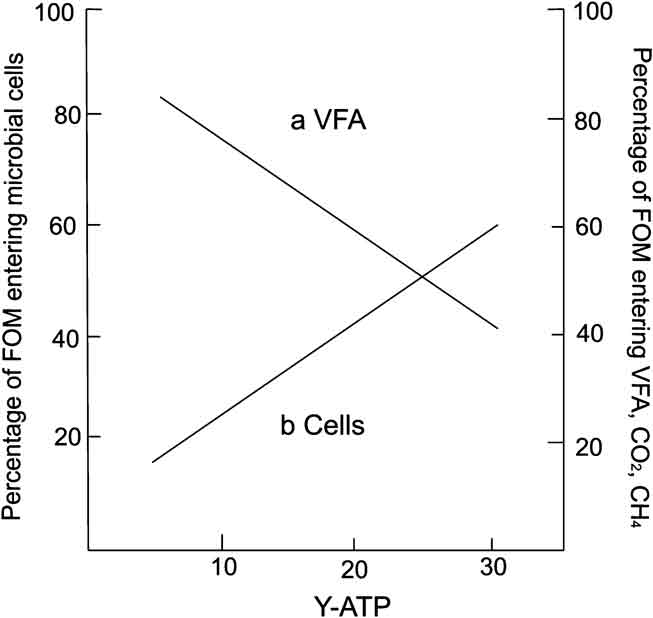
One of the major factors that affect microbial cell synthesis in the rumen is the availability and/or concentration in rumen fluid of precursors (glucose, AA, nucleic acids, peptides, ammonia and minerals). When bacteria are limited for energy sources, the free energy change of catabolic reactions is generally tightly coupled to the anabolic steps of cellular biosynthesis, and total energy flux can be partitioned into growth and maintenance functions of microorganisms (Russell and Cook, Reference Russell and Cook1995; Russell, Reference Russell2007). However, if growth is limited by nutrients other than energy (e.g. nitrogen), a situation of energetic uncoupling occurs, and bacteria can spill ATP in reactions that cannot be readily categorized as maintenance per se, and can be energy spilling reactions (Russell and Cook, Reference Russell and Cook1995; Russell, Reference Russell2007). In the rumen, in a situation of energetic uncoupling, there is a higher heat production, a large increase in VFA production and a decrease in cell yield (i.e. a decrease in YATP; Preston and Leng, Reference Preston and Leng1987; Figure 2).
Relationship between microbial growth efficiency (Y-ATP) and the percentage of fermentable organic matter that is partitioned into VFAs and gases (methane and carbon dioxide) and that entering into microbial cells (adapted from Preston and Leng, Reference Preston and Leng1987).
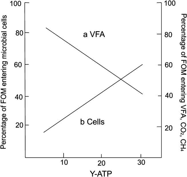
Assuming that the cecum-colon environment functions like the rumen seems to us wrong, as substrate that reaches the hindgut highly depends on pre-cecal digestibility of feeds, which means that cytoplasmic protein (nitrogen) and soluble sugars reach the hindgut in low amounts. However, the majority of cell wall carbohydrates and linked nitrogen will reach the hindgut since very low hydrolysis of these constituents occurs either in the stomach or in the pre-cecal environment. If we consider the type of substrate that gets to the hindgut, we can expect it to be nitrogen limiting, and a situation of energetic uncoupling seems feasible. The hindgut environment would aim to favor VFA’s production by promoting microbial activity but without microbial growth.
In the hindgut of horses, two circumstances should be analyzed:, is the coupling of energy and protein availability requested to optimize the synthesis of bacterial protein in order to support maximum VFA and energy production for the benefit of the host? Or is the hindgut environment adapted ‘to work’ in a permanent uncoupling situation, with fiber degradation being maximized but not microbial growth? These issues need further clearance. Compared to ruminal activity, very little is known about hindgut ecosystem activity in the horse. Our studies and hypotheses point to a very complex environment, very different from the rumen, where the microbial population is adapted to maximize energy production in benefit of the host as stated by others (Martin-Rosset and Tisserand, Reference Martin-Rosset and Tisserand2004), but probably in a permanent uncoupling situation.
Additional research is being conducted by our research group to characterize the activity and nutritional requirements of the bacterial population of the equine hindgut to achieve a more complete understanding of gut microflora populations in the horse and to clarify its contribution to meet the nutritional requirements of the horse.
Acknowledgments
A.S. Santos was funded by the Portuguese Foundation for Science and Technology doctoral Grant SFRH/BD/31294/2006.