Introduction
Lyme borreliosis is the commonest tick-borne disease in Europe and the USA. It is caused by the spirochaete Borrelia burgdorferi sensu lata.Reference Davidson, Evans and Ho-Yen 1 It is a multi-system disease which affects mainly the skin, joints, heart and nervous system, and which may cause both acute and chronic illness.Reference Davidson, Evans and Ho-Yen 1 It often presents solely with primary cutaneous lesions; head and neck lesions are fairly common, occurring in up to 75 per cent of patients with Lyme borreliosis.Reference Shapiro, Gerber and Lyme 2
Humans acquire Lyme borreliosis mainly from infected tick nymphs, usually in late spring or early summer, and occasionally from adult ticks during the cooler months.Reference Shapiro, Gerber and Lyme 2 A recent study from the National Lyme Borreliosis Testing Service suggested that the combination of warm and wet weather conditions would increase tick survival, but that dry conditions would probably favour increased human exposure to ticks due to outdoor activities.Reference Mavin, Joss and Ho-Yen 3 Geographical differences in Lyme borreliosis incidence are dependent on tick density and the proportion of infected ticks.Reference Shapiro, Gerber and Lyme 2 Lyme borreliosis occurs throughout Scotland, especially in the Highlands and Tayside, and those visiting Scotland may be at risk.Reference Davidson, Evans, Ling, Wiseman, Joss and Ho-Yen 4 , Reference Slack, Mavin, Yirrell and Ho-Yen 5
We report a case of B burgdorferi induced lymphocytoma on the earlobe of a child, and we also discuss the management of this condition.
Case report
A healthy, two-year-old boy was referred to the ENT out-patient clinic in Raigmore Hospital with a five- to six-month history of a slightly swollen, erythematous left earlobe. There was a vague history of a minor injury to that ear shortly before the swelling was first noticed. The mother denied any knowledge of a recent insect or tick bite. The lesion had initially been crusted, and had been treated by the child's general practitioner with steroid creams and antibiotic and Epaderm ointments, without improvement. According to the child's mother, the affected, inflamed earlobe had fluctuated slightly in size and hue but the inflammation had persisted. There was no associated systemic illness nor development of other skin lesions.
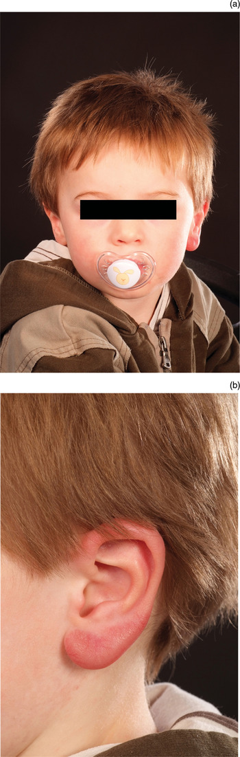
On physical examination, the boy's left earlobe was significantly enlarged (Figure 1). The brawny infiltration and light erythema were confined mainly to the earlobe, with some induration of the skin overlying the antitragus. The lesion was firm, rubbery in consistency and mildly tender on palpation. There was no associated lymphadenopathy. The remainder of the ENT examination was normal, as was otoscopic examination.
Clinical photographs: (a) frontal view demonstrating that the left earlobe is twice the size of the right earlobe; and (b) side view giving a closer view of the left earlobe.

Initial laboratory investigations (including full blood count, urea and electrolytes, liver function and C-reactive protein tests) were normal.
A wedge incision biopsy of the lesion, extending down to the cartilage, was performed.
On histopathological analysis, there was a florid dermal lymphoid infiltrate with many large C-reactive lymphoid follicles with well developed mantles and T zones, showing no reactivity to the overlying epidermis. Immunohistochemical analysis confirmed normal lymphoid architecture and the presence of reactive follicles. There was no aberrant expression of cyclin D1 or cluster of differentiation 5 or 23 proteins. The plasma cells and B lymphocytes were polyclonal. Staining for the MIB1 enzyme was extremely positive in the follicle centre, providing evidence of a reactive process, but outside the germinal centre there was little positivity. These appearances indicated the presence of a benign reactive lymphoid infiltrate, the cause of which was not clear from the sample.
Further blood tests were normal, including assessment of thyroid function, angiotensin-converting enzyme, complement levels and peripheral T-cell subsets. However, borrelia-specific serological tests for B burgdorferi antibody (using an enzyme-linked immunosorbent assay and western blot) were positive, and consistent with current infection. Subsequent polymerase chain reaction testing for B burgdoferi, using stored biopsy material from the ear lesion, was negative.
The patient was diagnosed with primary cutaneous Lyme borreliosis. He was treated with a three-week course of high-dose oral amoxicillin. His left earlobe swelling resolved completely within a few days of commencing this treatment.
He remained well during a further six months of follow up.
Discussion
Borrelia lymphocytoma is the most common cause of cutaneous B-cell pseudolymphoma, but the least common skin manifestation of Lyme borreliosis. It presents as an erythematous, dusky or violaceous nodule, most usually on the earlobe, nipple or scrotum.Reference Sewnaik, Kamarashev, Peters and van Rijswijk 6 , Reference Mullegger 7
The epidemiological characteristics and clinical presentation of B. burgdoferi appear to differ between continents.Reference Davidson, Evans and Ho-Yen 1 Hengge and colleagues observed a difference in the clinical presentations of Lyme disease between North America and Eurasia, due to the different pathogenicity of distinct genospecies of B burgdorferi.Reference Hengge, Tannapfel, Tyring, Erbel, Arendt, Ruzicka and Lyme 8 Late skin manifestations of Lyme borreliosis, such as acrodermatitis chronica atrophicans and borrelia lymphocytoma, occur almost exclusively in Europe following B afzelii infection, transmitted by Ixodes ricinus.Reference Hengge, Tannapfel, Tyring, Erbel, Arendt, Ruzicka and Lyme 8 By contrast, Lyme borreliosis in North America is caused by an infection with B burgdorferi sensu stricto and transmitted by Ixodes scapularis. The true incidence of Lyme borreliosis in the Scottish Highlands is estimated to be 37.24 to 49.69 per 100 000 population, which is proportionally much higher than in other Scottish regions.Reference Slack, Mavin, Yirrell and Ho-Yen 5
Our patient had no definite history of a tick bite. However, he lived in an area highly endemic for ticks and Lyme borreliosis, and he had serological evidence of current Lyme borreliosis. His ear lesion most probably arose following a tick bite sustained during the late summer months.
The clinical differential diagnosis in this case included infiltration with an inflammatory process (e.g. granulomatous contact dermatitis), a foreign body granuloma, sarcoidosis, substance deposition (e.g. amyloid) and primary cutaneous malignancy (e.g. lymphoma, sarcoma or carcinoma). However, all of these differential diagnoses were unlikely due to the clinical history, lack of associated symptoms, clinical signs and investigative findings. The initial swelling had begun at least two weeks after an episode of possible minor trauma, making a post-traumatic reaction most unlikely. Non-specific papular proliferation at the site of a tick bite can also histologically resemble an early dermatofibroma, lymphocytoma or interstitial myositis of the musculus erector pili, but none of these features were present in this case.
In our patient, the diagnosis of borrelia lymphocytoma was supported by the positive B burgdorferi western blot result and the patient's response to amoxicillin therapy.Reference Aberer and Klade 9 In Scotland, our laboratory uses local isolates for western blot analysis, rather than US or European isolates, and this improves the specificity of Lyme borreliosis testing.Reference Evans, Mavin and Ho-Yen 10
In patients presenting with erythema chronicum migrans, culture can detect spirochaetes in tissue specimens with nearly 100 per cent specificity and a sensitivity of 57–86 per cent, compared with a sensitivity of less than 50 per cent for serological testing.Reference Tugwell, Dennis, Weinstein, Wells, Shea and Nichol 11 Although culture has been recommended as the investigation of choice, this can take several months and requires adequate skin biopsy material or large volumes of plasma, which were not available in our case.
Alternatively, polymerase chain reaction testing for B burgdorferi DNA can be used.Reference Davidson, Evans and Ho-Yen 1 In our case, polymerase chain reaction testing of biopsy material was negative. This was probably because lymphocytoma is predominantly an inflammatory process, compared with erythema chronicum migrans which is due to bacterial infiltration. In erythema chronicum migrans, skin biopsies for polymerase chain reaction testing must be taken from the advancing edge of the lesion, where the bacteria are concentrated, to avoid false negative results. Nevertheless, skin biopsy is useful to rule out cutaneous malignancy and lymphoma.
In early Lyme borreliosis, the immune response is critical for the control of local disease and spread of infection. It has been shown that epidermal Langerhan's cells are invaded by the spirochaetes in early Lyme borreliosis, which may cause either a reduction or an increase in the number of Langerhan's cells positive for cluster of differentiation 1a protein, and also a reduction in these cells' surface expression of major histocompatability complex class II antigens.Reference Aberer, Koszik and Silberer 12 While this down-regulation may protect the auto-antigen reaction, it may also lead to an impaired capacity of Langerhan's cells to eliminate the spirochaetes, resulting in chronicity of the infection. Insufficient elimination of borrelia spirochaetes by complement and antibodies has also been reported due to down-regulation of the immune response.Reference Liang, Jacobs, Bowers and Philipp 13 A case report has shown that borrelia spirochaetes may be cultured from skin biopsy material as long as 10 years after the onset of disease.Reference Asbrink and Hovmark 14
In a child younger than eight years of age with early localised or disseminated infection, evidence-based treatment recommendations specify treatment with amoxicillin, ceftriaxone or cefuroxime, for a duration of 14–21 days.Reference Aberer and Klade 9 , Reference Garbe, Stein, Dienemann and Orfanos 15 Erythromycin is used if the patient is allergic to penicillin. Doxycycline is often the first-line treatment for older children or adults.Reference Hengge, Tannapfel, Tyring, Erbel, Arendt, Ruzicka and Lyme 8 , Reference Aberer and Klade 9
-
• Head and neck Lyme borreliosis can present without a classic history or recent tick bite
-
• Specific laboratory testing aids the diagnosis of atypical skin lesions
-
• This condition is a differential diagnosis of a persistent, inflamed earlobe in a well child
If the disease is left untreated, long-term follow up has shown that a small percentage of cases of borrelia lymphocytoma cutis progress to cutaneous B-cell lymphoma or systemic disease.Reference Garbe, Stein, Dienemann and Orfanos 15 In addition, untreated patients present especially with joint manifestations of the disease.Reference Hengge, Tannapfel, Tyring, Erbel, Arendt, Ruzicka and Lyme 8 Interferon-α therapy has also been shown to be effective in the treatment of B-cell lymphoma and idiopathic lymphocytoma cutis; most recently, intralesional interferon-α-2A has been used.Reference Aydogan, Karadogan, Adim and Tunali 16
Lyme borreliosis occurs throughout Scotland, and our case is a timely reminder of an unusual clinical presentation.
Conclusion
Lyme borreliosis can present on the head and neck without a history of erythema chronicum migrans, systemic illness or a history of a recent tick bite, as shown in our case. Better understanding of the disease variation found between different countries enables patients to be better informed. Although Lyme borreliosis is often regarded as a clinical diagnosis, specific laboratory testing is helpful in confirming the diagnosis, especially when the clinician is confronted with an atypical skin lesion. The presented case highlights the fact that borrelia lymphocytoma should be included in the differential diagnosis of a persistent, unilateral, inflamed, swollen earlobe in an otherwise healthy child.
Acknowledgement
We would like to thank the staff of the Medical Illustration Department of Raigmore Hospital for their assistance.