Introduction
Angiostrongylus cantonensis, also known as rat lung worm, is the most common cause of parasitic eosinophilic meningitis worldwide (Tsai et al., Reference Tsai, Liu, Kunin, Lee, Chen, Lin, Tsai, Lin, Huang, Yen and Yen2001, Reference Tsai, Liu, Kunin, Lai, Lee, Chen, Wann, Lin, Huang, Ger, Lin and Yen2003, Reference Tsai, Lee, Huang, Yen, Chen and Liu2004). The typical clinical presentations include headache, hyperesthesia with cerebrospinal fluid (CSF) eosinophilic pleocytosis and acute meningitis (Tsai et al., Reference Tsai, Liu, Kunin, Lee, Chen, Lin, Tsai, Lin, Huang, Yen and Yen2001, Reference Tsai, Liu, Kunin, Lai, Lee, Chen, Wann, Lin, Huang, Ger, Lin and Yen2003, Reference Tsai, Lee, Huang, Yen, Chen and Liu2004). Matrix metallopeptidase-9 (MMP-9) is a proteolytic enzyme that has been shown to play an important role in blood–brain barrier (BBB) damage during human and mice infection with A. cantonensis (Tsai et al., Reference Tsai, Chung, Chen, Liu, Susan Lee, Chen, Sy, Wann and Yen2008; Chiu and Lai, Reference Chiu and Lai2013). In these studies, mice models showed that CSF eosinophilia and inflammation induced by A. cantonensis L3 caused an elevation in MMP-9. MMP-9 was also shown to cause claudin-5 degradation in endothelial tight junctions. Furthermore, BBB permeability was shown to be significantly attenuated in MMP-9 knockout mice compared to wild-type mice with A. cantonensis infection. These results support that MMP-9 can cause BBB disruption and promote eosinophil infiltration into cerebral parenchyma via BBB leakage during A. cantonensis infection (Chiu and Lai, Reference Chiu and Lai2013).
Enhanced oxidative stress and production of reactive oxygen species in the central nervous system (CNS) of mice infected with A. cantonensis have been shown to contribute to meningitis-associated brain damage. 8-hydroxy-2'-deoxyguanosine (8-OHdG) is used as a sensitive marker of oxidative DNA damage (Chung et al., Reference Chung, Chen, Wang, Chang and Yen2010a, Reference Chung, Wang, Chen, Lin and Yenb), and previous studies have observed a high level of 8-OHdG due to oxidative DNA damage in patients with CNS infection and parasitic eosinophilic meningitis caused by A. cantonensis infection (Chung et al., Reference Chung, Chen, Wang, Chang and Yen2010a, Reference Chung, Wang, Chen, Lin and Yenb). In addition, several studies have reported that BBB dysfunction in mice infected with A. cantonensis was caused by oxidative DNA damage in the CNS (Chung et al., Reference Chung, Chen, Wang, Chang and Yen2010a, Reference Chung, Wang, Chen, Lin and Yenb).
MMP-9 has been shown to be activated by an oxidative pathway in pneumococcal meningitis (Meli et al., Reference Meli, Christen and Leib2003). We hypothesized that the increased MMP-9 expression in mice with eosinophilic meningitis caused by A. cantonensis infection may also be mediated by an oxidative pathway.
Corticosteroids have been shown to improve capillary function by reducing the activities of urokinase-type plasminogen activator (PA) and MMP-9 and increasing levels of tissue inhibitors of metalloproteinases (TIMPs). Dexamethasone treatment in mice with eosinophilic meningitis caused by A. cantonensis infection has been shown to result in the partial inhibition of PA and inflammation (Hou et al., Reference Hou, Tu, Lee, Chen, Chou and Lai2004; Tu and Lai, Reference Tu and Lai2006). However, dexamethasone was only given for 7 days starting from day 5 post-inoculation in one of the studies (Tu and Lai, Reference Tu and Lai2006). Corticosteroids can potentially stabilize the BBB and ameliorate tissue oedema in neoplastic and inflammatory CNS disorders by inducing occludin and claudin-5 and inhibiting pathologically activated endopeptidases. Thus, corticosteroids have been used as an adjunctive therapy in eosinophilic meningitis (Chotmongkol et al., Reference Chotmongkol, Sawanyawisuth and Thavornpitak2000; Sawanyawisuth et al., Reference Sawanyawisuth, Limpawattana, Busaracome, Ninpaitoon, Chotmongkol, Intapan and Tanawirattananit2004). In these clinical studies from Thailand, the researchers showed that a one- or two-week course of prednisolone was beneficial in relieving headaches in patients with eosinophilic meningitis. In addition, the effect of steroid treatment was dependent on the expression of glucocorticoid receptors (GRs) in the brain endothelium. GRs are expressed in brain vascular endothelium and post-translationally modified and degraded by the ubiquitin–proteasome system, and they are chaperoned by heat shock proteins (Hsps), Hsp90 and Hsp70 (Gu et al., Reference Gu, Qin, Qin and Xu2009; Hue et al., Reference Hue, Cho, Cao, Dale Bass, Meaney and Morrison Iii2015). Therefore, we hypothesized that the beneficial effects of dexamethasone in eosinophilic meningitis caused by A. cantonensis infection may be through an increase in the expression of GRs, interaction with Hsp70, and subsequently by the downregulation of MMP-9 and oxidative stress and improved BBB damage.
In this study, we conducted mice experiments to analyse dynamic changes in GRs, Hsp70, MMP-2, MMP-9, and oxidative stress pathways in CSF and brain homogenates. The messenger RNA (mRNA) levels of GRs were determined by quantitative reverse transcription–polymerase chain reaction (qRT–PCR). The dynamic changes in GRs, Hsp70, MMP-9, MMP-2, nuclear factor kappa B (NF-κB), c-Jun N-terminal kinase (JNK), extracellular signal-regulated kinase (ERK) and 8-OHdG after infection in mice brain/CSF and after steroid treatment were studied by Western blot, gelatin zymography and enzyme-linked immunosorbent assay (ELISA). Immunohistochemistry (IHC) of brain homogenates was done to characterize the dynamic changes in GRs, Hsp70, 8-OHdG, occludin, and claudin-5 in response to steroid therapy.
Material and methods
Infection of BALB/c mice and intra-peritoneum injection of steroids
The detailed methods and protocols have been described elsewhere (Tsai et al., Reference Tsai, Lee, Yen, Wann, Lee and Chen2015). Briefly, 40 BALB/c mice, aged 6–7 weeks, were purchased from the National Laboratory Animal Breeding Research Centre. Third-stage larvae (L3) of A. cantonensis were harvested from infected Biomphalaria glabrata after treatment with artificial gastric juice. Briefly, the snail shells were digested in 0.6% pepsin–HCl solution (pH 2–3, 500 IU pepsin g tissue−1) according to a previously described method with some modifications (Lee et al., Reference Lee, Tsai, Chen, Wang, Hsiao and Yen2006). The mice were orally infected with 50 A. cantonensis L3 via an orogastric tube after ketamine anaesthesia, and 5–7 were then sacrificed every week for 3 consecutive weeks after infection until the end of the study. Dexamethasone at a dose of 500 μg kg−1 day−1 was injected intraperitoneally from the 7th day of infection until the end of the study (21 days post infection). The total duration of dexamethasone treatment was 2 weeks.
Collection of serum and CSF specimens
The detailed methods and protocols have been described previously (Tsai et al., Reference Tsai, Lee, Yen, Wann, Lee and Chen2015). Briefly, blood samples from the experimental mice were collected by heart puncture under ketamine anesthesia. Serum specimens separated from the blood samples after centrifugation at 3500 g (Hermle, Z326K, Germany) for 5 min at 4°C were stored at −70°C until they were measured. Brains were removed and washed with 150 μL 0.15 m phosphate-buffered saline (PBS), and the cerebral ventricles and cranial cavities were washed with 350 μL PBS. The CSF was, thus, harvested with the PBS, which was centrifuged in an Eppendorf tube at 3000 g (Hermle, Z326K, Germany) for 10 min at 4°C to eliminate cells. The supernatant was stored at −70°C until further use.
Measurement of permeability of the BBB using the Evans blue method
The detailed methods and protocols have been described previously (Tsai et al., Reference Tsai, Lee, Yen, Wann, Lee and Chen2015). A volume of 200 μL of 2% (w/v) Evans blue solution in PBS was intraperitoneally injected into a mouse. Two hours later, the brain of the mouse was removed after anesthesia with ketamine. The brain homogenate extract was then centrifuged at 18 000 g (Hermle, Z326K, Germany) for 10 min at room temperature. The optical density of the supernatant was read at 595 nm wavelength using a colorimeter (Thermo Scientific Multiskan FC, USA).
Quantification of albumin in CSF and serum
The concentrations of albumin in the CSF and serum of mice were automatically measured by a biochemical autoanalyser (Beckman LX-20, USA). Albumin in the specimens was reacted with bromocresol green reagent to form bromocresol–albumin complex that has a maximum absorbance at 628 nm. The ratio of CSF/serum albumin was subsequently calculated using the individual concentrations.
Reverse transcription–polymerase chain reaction (RT–PCR) for GRs
Total RNA was isolated from frozen sections containing lateral ventricles and hippocampal tissue with a reagent (TRIZOL-LS; Gibco BRL). Of the total RNA, 2.5–5 mg was used for RT with Superscript II reverse transcriptase (Gibco BRL), according to the manufacturer's instructions. To amplify specific gene products, the following intron spanning primers were used. Mouse glyceraldehyde-3-phosphate dehydrogenase (GAPDH) was co-amplified as an internal control, using the following primer sequences:
GAPDH sense, 5′-CATCACCATCTTCCAGGAGCG-3′;
GAPDH antisense, 5′-GAGGGGCCATCCACAGTCTTC-3′ (357 bp);
GR sense, 5′-CAAAGCCGTTTCACTGTCC-3′;
GR antisense, 5′-ACAATTTCACACTGCCACC-3′ (258 bp).
Of the cDNA product, 0.7–1.4 mL was used for PCR in a reaction mix (40 mL) consisting of 0.12 U of Taq DNA polymerase (Amplitaq Gold; Perkin Elmer), each primer at 1 mm molarity, 0.2 mm deoxynucleotide triphosphate mix, and 2.5 mm MgCl2. Amplification was performed in an Eppendorf thermocycler using the following profile: 94°C for 1 min (denaturation), 56°C for 1 min (annealing), and 72°C for 1 min (elongation), for a total of 40 cycles. Negative control experiments without RT were carried out in parallel for every reaction to exclude amplification of contaminating DNA. The amplified products were separated on a 1.5% agarose gel and stained with ethidium bromide. The gels were dried, digitized, and optical densities were determined using a computer imaging analysis system (VisitronSystems GmbH). RT–PCR was carried out in triplicate, and the mean density of PCR products was determined.
Western blot
The detailed method has been described previously (Tsai et al., Reference Tsai, Lee, Yen, Wann, Lee and Chen2015). The extracts of mouse brains were centrifuged at 12 000 g for 20 min at 4°C to remove debris. The protein concentration was analysed using a protein assay kit (Bio-Rad, Hercules, CA, USA) with BSA serving as the standard. Briefly, 50 μg protein was separated by 10% SDS–PAGE and transferred to polyvinylidene fluoride membranes. The membranes were incubated in blocking buffer for 60 min (PBS containing 5% bovine serum albumin (BSA)) at room temperature. The membranes were then washed three times with Tris-buffered saline with Tween 20, probed with GR (sc-8992), Hsp70 (ab2787), JNK (66210-1-Ig), p-JNK (#4668), NF-κB (10745-1-AP), ERK (#4695), and phosphorylated ERK (p-ERK; #4376) antibodies with β-actin (Sigma, St. Louis, MO, USA) as the control, then incubated with goat anti-mouse (#92798) or goat anti-rabbit (#92322) secondary antibodies and detected by enhanced chemiluminescence. Quantitative analysis of the bands was performed using a computer-assisted imaging densitometer system (UN-SCAN-IT™).
ELISA for MMP-9
Concentrations of MMP-9 were measured using ELISA kits (R&D System, Inc., USA and eBioscience, Inc.), with the assays being performed according to the manufacturers’ instructions. The wells of ELISA plates were first coated with capture antibodies that captured MMP-9 in brain tissue supernatant specimens or CSF. Biotinylated detection antibodies and streptavidin-labelled horseradish peroxidase (HRP) were then added step by step to allow the marker enzyme HRP to bind to the solid phase. TMB substrate solution was used for visualization, and optical density values were read at 450 nm after the addition of 2 N sulphuric acid to stop the reaction.
Gelatin zymography for MMP-2 and MMP-9 activity in CSF
MMP-2 and MMP-9 activities were analysed using modified sodium dodecyl sulfate polyacrylamide gel electrophoresis (SDS–PAGE). The stacking gels contained 4% polyacrylamide and separating gels contained 12.5% polyacrylamide and 0.1% gelatin. CSF was centrifuged at 3000 rpm for 20 min at 25°C in order to remove debris, serum was centrifuged at 3000 rpm for 20 min at 25°C to obtain the supernatant, and tissue was centrifuged at 12 000 rpm for 20 min at 4°C to remove debris. The protein contents of the supernatant were then mixed with an equal volume of 2 × non-reducing sample buffer and loaded at 25 μl per well. The gel was electrophoresed at 90 V and 4°C in running buffer (25 mm Tris, 250 mm glycine and 0.1% SDS) until the bromophenol blue marker dye had reached the bottom of the gel. After electrophoresis, the gel was washed two times with gentle agitation for 30 min in 2.5% Triton X-100 at room temperature. After decanting the washing solution, the gel was equilibrated with developing buffer (50 mm Tris–HCl, pH 7.5, containing 200 mm NaCl, 5 mm CaCl2, 0.02% Brij-35 and 0.01% NaN3) for 30 min at room temperature with gentle agitation, and then fresh developing buffer was added and allowed to incubate at 37°C for 18 h. The gel was stained with 0.25% Coomassie Brilliant Blue R-250 (Sigma) for 1 h and destained in 15% methanol/7.5% acetic acid. Gelatinase activity was detected as unstained bands on a blue background.
IHC for tight junction protein occludin, claudin-5, Hsp70, 8-OHdG and GRs
The detailed method has been described previously (Tsai et al., Reference Tsai, Lee, Yen, Wann, Lee and Chen2015). Mice were transcardially perfused with 20 mL PBS followed by 20 mL of 4% paraformaldehyde, permeabilized by incubating with 0.3% Triton X-100 (Sigma) in PBS for 1 h at room temperature, and then blocked for 1 h using 5% goat serum (Jackson ImmunoResearch) in PBS. Staining with various primary antibodies [claudin-5 (sc-28670), occludin (ab31721), GRs (sc-8992), Hsp70 (ab2787) and 8-OHdG (ab48508)] was conducted overnight at 4°C, followed by incubation with biotinylated secondary antibodies for 2 h at room temperature, and developed using fluorescently labelled streptavidin conjugate. Immunoreactivities of the markers were assessed using a semi-quantitative score, with 0 indicating that the sample was not immunoreactive and 1, 2 and 3 indicating weak, moderate and intense immunoreactivity, respectively (Sulik and Chyczewski, Reference Sulik and Chyczewski2008).
Measurement of 8-OHdG in serum and CSF by ELISA
The CSF and serum concentrations of 8-OHdG were assayed using an 8-OHdG EIA assay kit (Cayman, MI, USA). Briefly, 8-OHdG in CSF and serum was linked to acetylcholinesterase, and specific antiserum to 8-OHdG was added to the wells of a microplate pre-coated with mouse monoclonal antibodies, followed by incubation at 4°C for 18 h. Ellman's reagent was used to develop the wells after washing to remove all unbound reagents. After 90 min development, the plate was read at 405 nm, and the 8-OHdG concentration in each specimen was calculated from the standard curve.
Statistical analysis
The concentrations of Evans blue, CSF albumin, GRs, Hsp70, JNK, phosphorylated JNK (p-JNK), NF-κB, ERK, p-ERK, MMP-2, MMP-9, and 8-OHdG in the different groups of mice were compared using the non-parametric Kruskal–Wallis test followed by post-testing using Dunn's multiple comparison of means. The Mann–Whitney U test was used to compare changes in the markers every week relative to the controls, first week or dexamethasone treatment. All results were presented as median and range. A P value <0.05 was considered to be statistically significant.
Results
Permeability of the BBB measured by Evans blue and CSF albumin methods
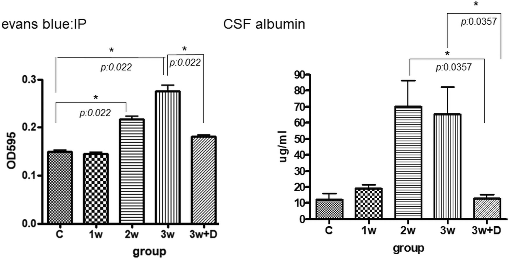
The quantity of Evans blue in brain homogenates gradually increased 2 weeks after infection until the end of this study. In addition, the amount of Evans blue in the mice brains significantly increased 2 and 3 weeks after infection compared to the uninfected mice (P = 0.022). The administration of dexamethasone for 2 weeks significantly decreased the concentration of Evans blue in the mice brains compared to the mice which had been infected for 3 weeks (P = 0.022) (Fig. 1).
Dynamic changes in Evans blue concentrations in brain homogenates and albumin concentrations in CSF in mice with A. cantonensis infection. For each group, n = 5. All data are presented as median and range. Control: no parasitic infection; 1W: 1 week after infection; 2W: 2 weeks after infection; 3W: 3 weeks after infection; 3W + Dex: mice given dexamethasone for 2 weeks (7–21 days post-infection) and sacrificed on day 21.

The concentration of albumin in CSF increased after 2 and 3 weeks of infection. The administration of dexamethasone significantly decreased the albumin concentration compared to 2 and 3 weeks after infection (P = 0.0357) (Fig. 1).
Expression of the mRNA of GRs in the mice with eosinophilic meningitis
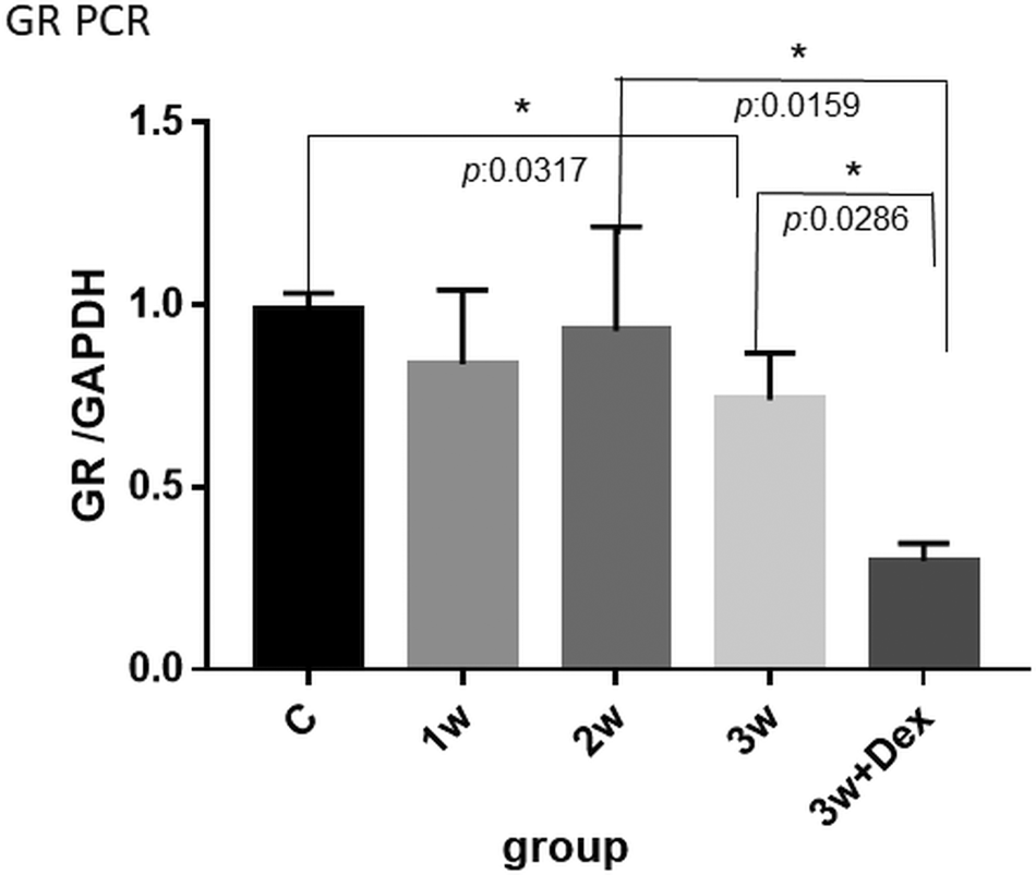
The mRNA expressions of GRs in brain homogenates did not gradually increase after 1–3 weeks of infections; however, dexamethasone administration significantly decreased the mRNA expressions of GRs in mice brains (P = 0.0286) (Fig. 2).
Dynamic changes in GR mRNA expressions in mice with A. cantonensis infection after 3 weeks of infection and after dexamethasone treatment. For each group, n = 5. All data are presented as median and range. The y-axis is expressed as a ratio (GR/GAPDH). Control: no parasitic infection; 1W: 1 week after infection; 2W: 2 weeks after infection; 3W: 3 weeks after infection; 3W + Dex: mice given dexamethasone for 2 weeks (7–21 days post-infection) and sacrificed on day 21.

Western blot for GRs, NF-κβ, Hsp70, JNK, p-JNK, ERK and p-ERK in brain homogenates and CSF in mice with eosinophilic meningitis
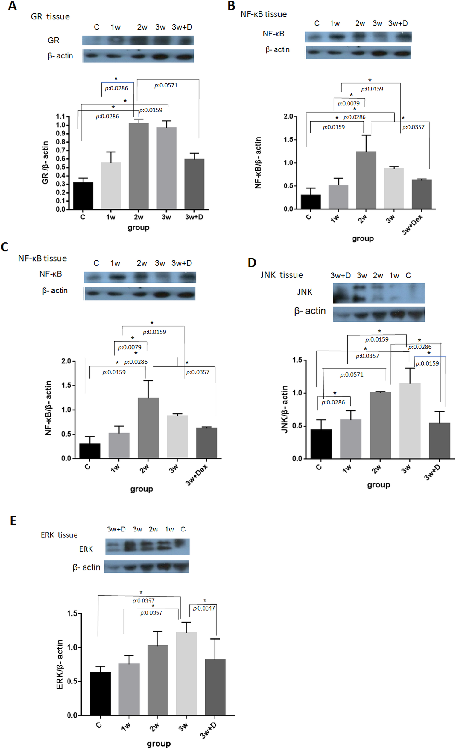
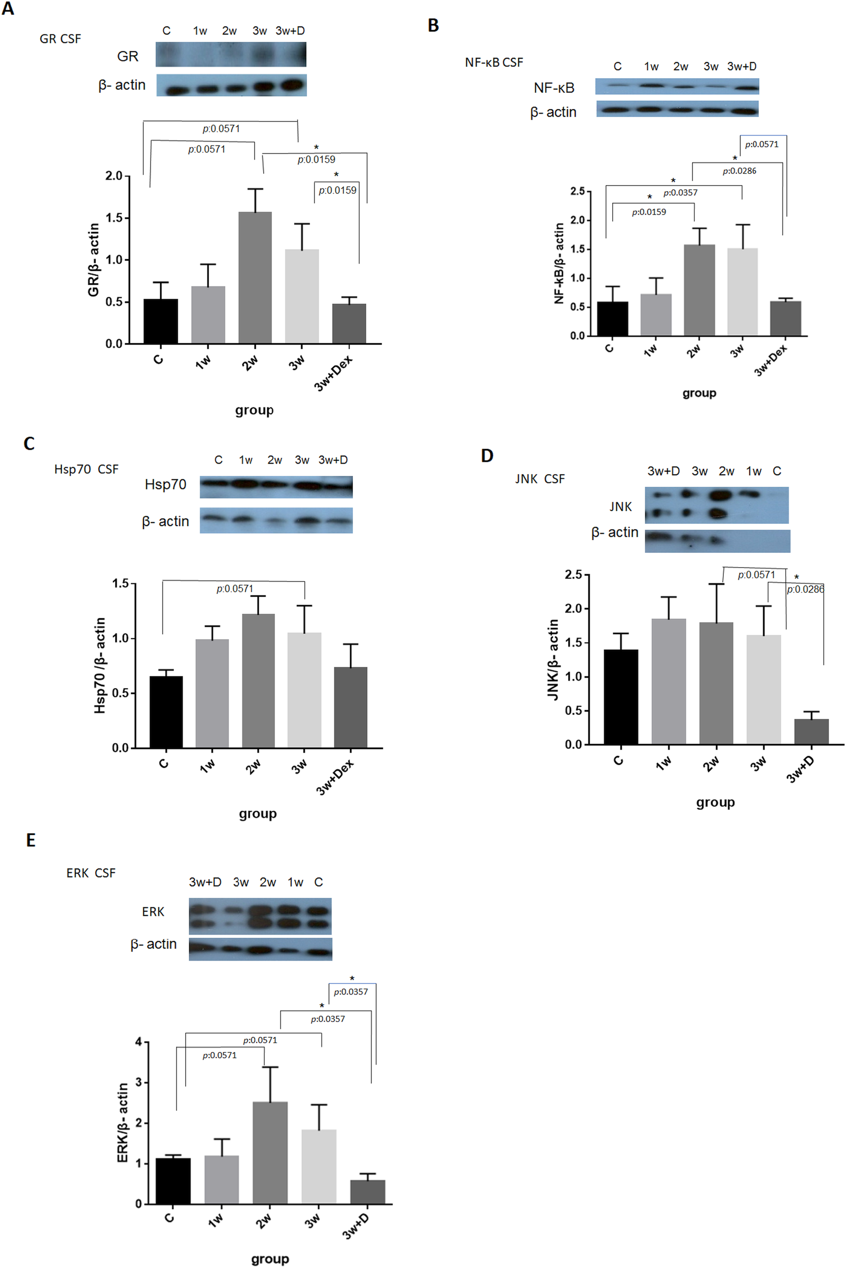
The expressions of GRs, NF-κB, Hsp70, JNK and ERK in brain homogenates gradually increased after 1–3 weeks of infection, and the administration of dexamethasone significantly downregulated the expressions of these proteins (Fig. 3). The expressions of NF-κB, GRs and ERK in CSF gradually increased after 2 or 3 weeks of infection, and dexamethasone administration decreased the expressions of these proteins in the CSF (Fig. 4a, b, e). The CSF expressions of Hsp70 and JNK were not statistically significantly different following 1–3 weeks of infection (Fig. 4c, d).
Dynamic changes in brain homogenate expressions of GRs, NF-κB, Hsp70, JNK and ERK after 1–3 weeks of infection and the administration of dexamethasone in mice with A. cantonensis infection. For each group, n = 5. All data are presented as median and range. The y-axis is expressed as a ratio between Hsp70/GR/NF-κB/ JNK/ERK and β-actin. Control: no parasitic infection; 1W: 1 week after infection; 2W: 2 weeks after infection; 3W: 3 weeks after infection; 3W + Dex: mice given dexamethasone for 2 weeks (7–21 days post-infection) and sacrificed on day 21.

CSF expressions of GR, NF-κB, Hsp70, JNK and ERK after 1–3 weeks of infection and dexamethasone administration in mice with A. cantonensis infection. For each group, n = 5. All data are presented as median and range. The y-axis is expressed as ratio between GR/NF-κB/Hsp70/JNK/ERK and β-actin. Control: no parasitic infection; 1W: 1 week after infection; 2W: 2 weeks after infection; 3W: 3 weeks after infection; 3W + Dex: mice given dexamethasone for 2 weeks (7–21 days post-infection) and sacrificed on day 21.

ELISA for MMP-9 concentration
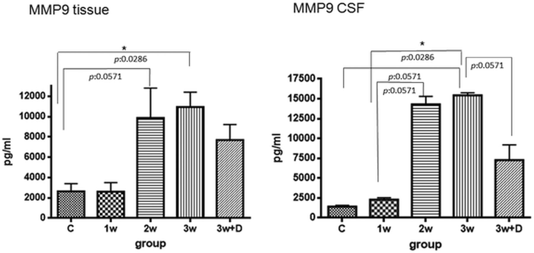
MMP-9 concentrations in brain homogenates were increased following 3 weeks of infection when compared to the control (P = 0.0286). Dexamethasone administration did not decrease MMP-9 concentration in mice brain homogenates. The concentration of MMP-9 in CSF gradually increased after 2–3 weeks of infection (P = 0.0286). Dexamethasone administration slightly decreased the concentration of MMP-9 in the CSF (P = 0.0571) (Fig. 5).
Brain homogenate and CSF MMP-9 concentrations were measured by ELISA following 1–3 weeks of infection and dexamethasone administration. For each group, n = 5. All data are presented as median and range. Control: no parasitic infection; 1W: 1 week after infection; 2W: 2 weeks after infection; 3W: 3 weeks after infection; 3W + Dex: mice given dexamethasone for 2 weeks (7–21 days post-infection) and sacrificed on day 21.

Gelatin zymography for MMP-2 and MMP-9 activity in serum/CSF and brain homogenates
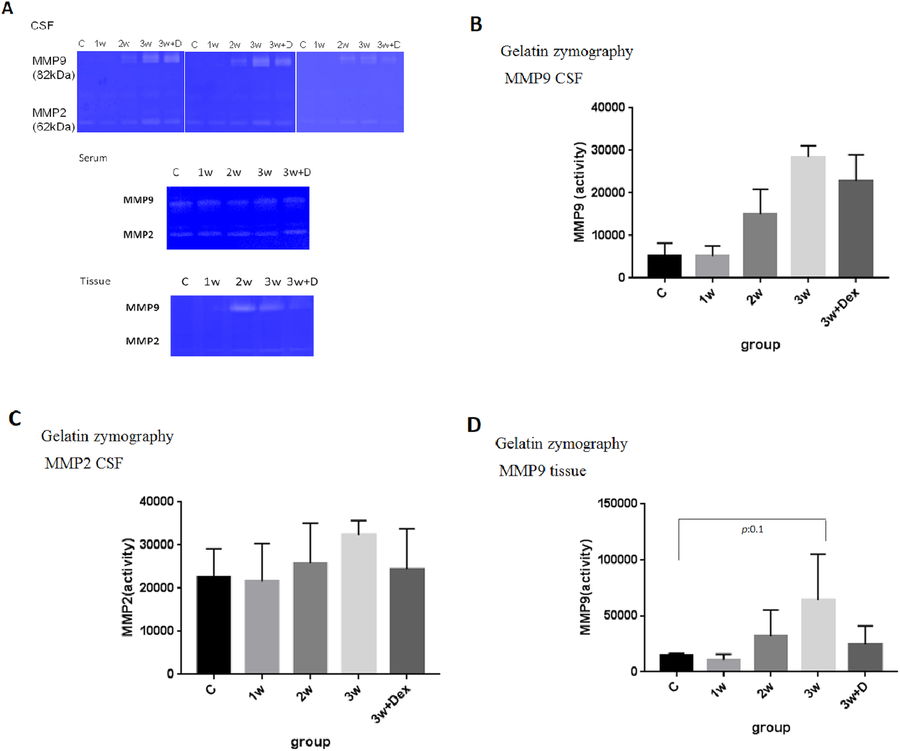
Gelatin zymography showed increases in MMP-9 activities in the CSF and brain homogenates after 2–3 weeks of infection with A. cantonensis L3. No difference was observed in the serum samples. Dexamethasone administration slightly downregulated the activities of MMP-9 in CSF/brain homogenates but not MMP-2 (Fig. 6a–d).
Gelatin zymography changes in MMP-9 and MMP-2 activities in CSF and brain homogenates of mice with A. cantonensis infection. The activities of MMP-2/MMP-9 in CSF/brain homogenates/serum are shown in (a). The serial changes in MMP-9 and MMP-2 activities in CSF are shown in (b) and (c). The serial changes in MMP-9 activities in brain homogenates are shown in (d). For each group, n = 3. All data are presented as median and range. Control: no parasitic infection; 1W: 1 week after infection; 2W: 2 weeks after infection; 3W: 3 weeks after infection; 3W + Dex: mice given dexamethasone for 2 weeks (7–21 days post-infection) and sacrificed on day 21.

Measurement of oxidative stress by 8-OHdG concentration in CSF
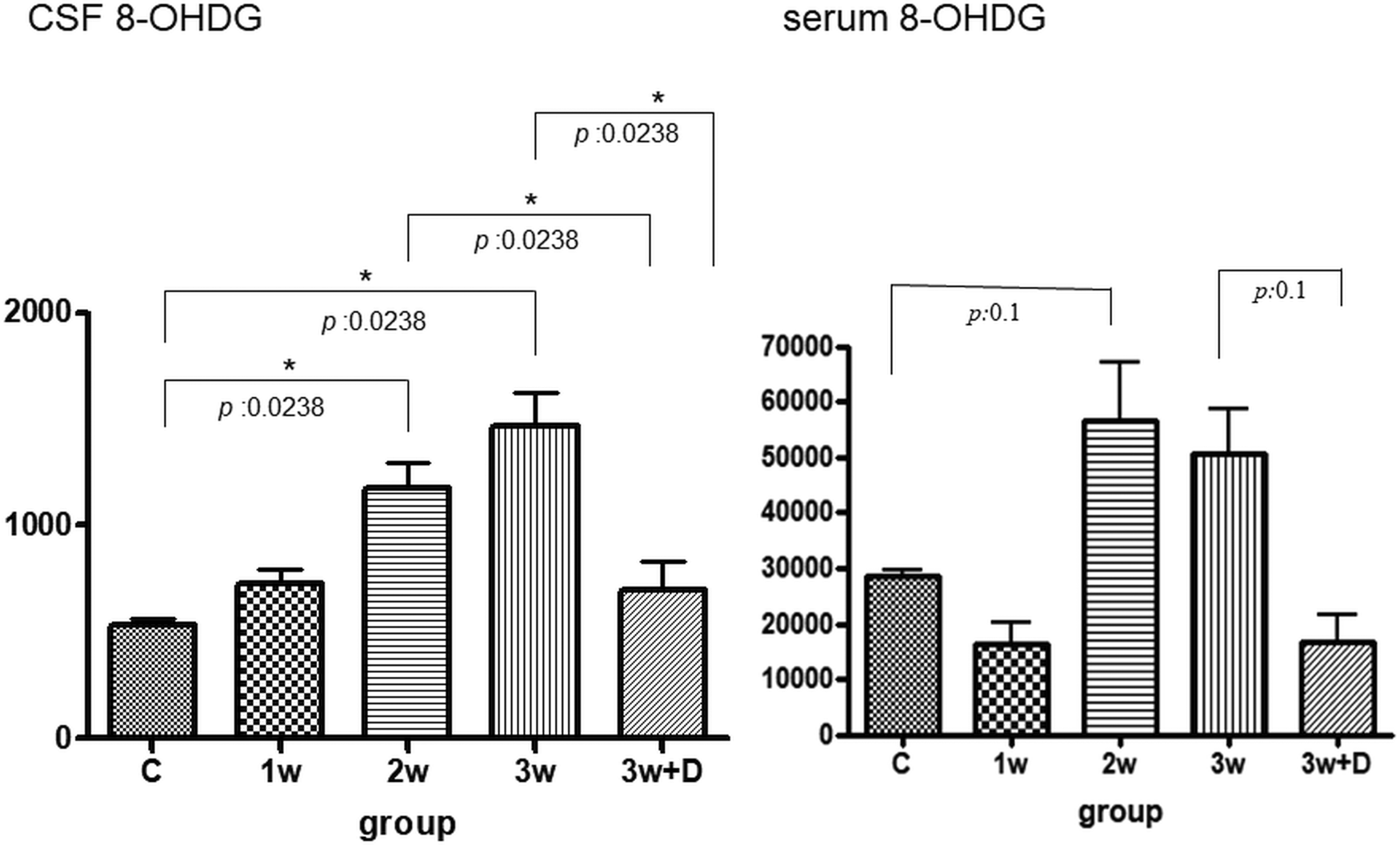
The 8-OHdG concentration in CSF gradually increased after 1–3 weeks of infection (P = 0.0238), which was significantly downregulated by the administration of dexamethasone (P = 0.0238). In addition, there was also an increase in 8-OHdG concentration in serum following 2–3 weeks of infection, although the difference did not reach statistical significance (Fig. 7).
Dynamic changes in CSF and serum 8-OHdG concentrations in mice with A. cantonensis infection. For each group, n = 5. All data are presented as median and range. Control: no parasitic infection; 1W: 1 week after infection; 2W: 2 weeks after infection; 3W: 3 weeks after infection; 3W + Dex: mice given dexamethasone for 2 weeks (7–21 days post-infection) and sacrificed on day 21.

IHC for tight junctional proteins (occludin and claudin-5), GRs, Hsp70 and 8-OHdG
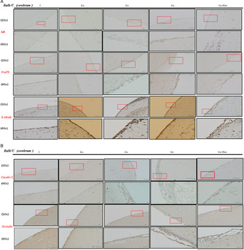
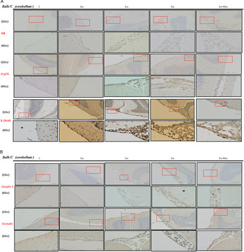
The concentrations of tight junctional proteins (occludin and claudin-5), GRs, Hsp70 and 8-OHdG in mice brain homogenates were detected by IHC staining. The expressions of these protein gradually increased after 2–3 weeks of infection compared to the controls, and dexamethasone administration significantly downregulated the expressions of these proteins both in the cerebrum and cerebellum. Of note, the changes in the expressions were more pronounced in the cerebellum compared to the cerebrum (Figs 8 and 9).
Dynamic IHC changes in cerebral tight junctional proteins (occludin and claudin-5), GRs, Hsp70 and 8-OHdG in mice with A. cantonensis infection. The immunoreactivities of these markers were assessed using a semi-quantitative score, with 0 indicating that the sample was not immunoreactive, and 1, 2 and 3 indicating weak, moderate and intense immunoreactivity, respectively. Scale bar, 10 μm at 10× and 40× magnification. Control: no parasitic infection; 1W: 1 week after infection; 2W: 2 weeks after infection; 3W: 3 weeks after infection; 3W + Dex: mice given dexamethasone for 2 weeks (7–21 days post-infection) and sacrificed on day 21.

Dynamic IHC changes in cerebellum tight junctional proteins (occludin and claudin-5), GRs, Hsp70 and 8-OHdG in mice with A. cantonensis infection. The immunoreactivities of these markers were assessed using a semi-quantitative score, with 0 indicating that the sample was not immunoreactive and 1, 2 and 3 indicating weak, moderate and intense immunoreactivity, respectively. Scale bar, 10 μm at 10× and 40× magnification. Control: no parasitic infection; 1W: 1 week after infection; 2W: 2 weeks after infection; 3W: 3 weeks after infection; 3W + Dex: mice given dexamethasone for 2 weeks (7–21 days post-infection) and sacrificed on day 21.

Discussion
In this study, we found that BBB dysfunction occurred in mice with eosinophilic meningitis 2–3 weeks after infection as evidenced by the increased extravasation of Evans blue and CSF albumin levels. The administration of dexamethasone significantly decreased the amount of Evans blue and CSF albumin. The effect of dexamethasone was possibly mediated by GRs and Hsp70, subsequently resulting in decreased expressions of NF-κB, JNK and ERK in the CSF and brain parenchyma after 2 weeks of steroid administration. Dexamethasone treatment also decreased the expressions of MMP-9 in CSF/brain homogenates but had no effect on MMP-2 levels in CSF, indicating that MMP-9 rather than MMP-2 played a major role in BBB dysfunction in mice with eosinophilic meningitis. In addition, oxidative stress was found in the mice with eosinophilic meningitis. The concentration of 8-OHdG in CSF gradually increased after 1–3 weeks of infection, which was significantly downregulated after the administration of dexamethasone.
Meningitis is associated with dysregulation of the MMP system. Many infectious types of meningitis, and especially bacterial meningitis, have been associated with elevated concentrations of MMP-9 in CSF compared with controls (Tsai et al., Reference Tsai, Shi, Lee, Wann, Tai and Chen2011). In addition, in an experimental pneumococcus meningitis rat brain-slice culture model, Meli et al. demonstrated that MMP-9 was activated by active oxygen species (Meli et al., Reference Meli, Christen and Leib2003). MMP-9 is a proteolytic enzyme which has been shown to play an important role in mouse BBB damage during A. cantonensis infection (Chiu and Lai, Reference Chiu and Lai2013). Chiu and Lai reported that in mice with parasitic eosinophilic meningitis caused by A. cantonensis infection, MMP-9 could cause BBB leakage and promote eosinophil infiltration into cerebral parenchyma. Moreover, the increased BBB permeability was significantly attenuated in MMP-9 knockout mice compared to wild-type mice with eosinophilic meningitis caused by A. cantonensis infection (Chiu and Lai, Reference Chiu and Lai2013). In a previous clinical study of patients with eosinophilic meningitis caused by A. cantonensis infection, we found a significant correlation among BBB damage markers (QAlb values) and CSF MMP-9 concentrations and leucocyte count (Tsai et al., Reference Tsai, Chung, Chen, Liu, Susan Lee, Chen, Sy, Wann and Yen2008).
In the present study, we also found increased expressions of MMP-9 in CSF/brain homogenates after 2–3 weeks of infection. Steroid treatment decreased the expressions of MMP-9 in CSF/brain homogenates, but it did not have any effect on the CSF MMP-2 level, indicating that MMP-9 rather than MMP-2 plays a major role in BBB dysfunction in mice with eosinophilic meningitis.
8-OHdG is used as a sensitive marker of oxidative DNA damage (Chung et al., Reference Chung, Wang, Chen, Lin and Yen2010b). A high level of 8-OHdG resulting from oxidative DNA damage has been observed in C57BL/6 mice with A. cantonensis infection (Chung et al., Reference Chung, Chen, Wang, Chang and Yen2010a, Reference Chung, Wang, Chen, Lin and Yenb), and the amount of 8-OHdG in CSF was in parallel with the severity of BBB damage in mice with eosinophilic meningitis. In the present study, we found that 8-OHdG was also increased after 2–3 weeks of infection in the CSF of BALB/c mice infected with A. cantonensis. Moreover, the administration of dexamethasone significantly decreased the concentration of 8-OHdG. In our previous parasitic eosinophilic meningitis study, we found increased expressions of apoptotic proteins in mice brain homogenates after 2–3 weeks of infection, and treatment with dexamethasone significantly decreased BBB damage and the expressions of these apoptotic proteins (Tsai et al., Reference Tsai, Lee, Yen, Wann, Lee and Chen2015). It is possible that the effect of dexamethasone is mediated by a decrease in oxidative DNA damage in the CSF and decreases in apoptotic protein expressions in brain homogenates in mice with eosinophilic meningitis. Further studies are needed to investigate associations between brain apoptosis and oxidative DNA damage.
In a primary bovine endometrial epithelial cell (BEEC) culture study, Dong et al. investigated the anti-inflammatory effects of cortisol on lipopolysaccharide-induced BEECs. They found that cortisol not only inhibited the activity of NF-κB via blocking the phosphorylation and degradation of IκB, but also suppressed the phosphorylation of the mitogen-activated protein kinase (MAPK) pathway including ERK1/2, p38MAPK and JNK (Dong et al., Reference Dong, Qu, Li, Cui, Wang, Lin and Wang2018). In a Sprague–Dawley rat model of acute lung injury caused by H2S, Wang et al. showed that dexamethasone significantly ameliorated the symptoms of H2S-induced acute lung injury including alveolar oedema, infiltration of inflammatory cells and protein leakage in bronchoalveolar lavage fluid (BAFL) via upregulating GRs to mediate the suppression of MMP-2 and MMP-9 (Wang et al., Reference Wang, Zhang, Su, Chen, Zhu, Zhang, Xiao and Zhang2014). Dexamethasone was also demonstrated to inhibit IκBα phosphorylation and NF-κB DNA-binding activity in a bacterial meningitis cell culture model (Mogensen et al., Reference Mogensen, Berg, Paludan and Østergaard2008). In addition, Chiu and Lai demonstrated that NF-κB and MMP-9 were increased in mice with eosinophilic meningoencephalitis, and that the upregulation of MMP-9 was through the NF-κB signalling pathway (Chiu and Lai, Reference Chiu and Lai2013).
In this study, the expressions of GRs and Hsp70 were increased after 2–3 weeks of infection with A. cantonensis and decreased by the administration of dexamethasone. It is likely that the effect of dexamethasone was mediated by GRs and Hsp70, resulting in decreases in the expressions of NF-κB, JNK, and ERK in the CSF and brain parenchymal after 2 weeks of steroid therapy.
There are several limitations to this study. The best housekeeping gene for PCR experiments in eosinophilic meningitis is currently unknown. GAPDH and β-actin have been widely used as the housekeeping genes in PCR experiments for many years. However, in recent years, some studies have suggested that these housekeeping genes should be used with caution since they may display variable expression levels across various cell types and disease states. For example, the suitability of common housekeeping genes for use in qRT–PCR assays of the cornea in various murine disease models has been evaluated at different time points, and GAPDH and β-actin were shown to be among the least stably expressed markers under most conditions. In the absence of knowledge about situation-specific housekeeping genes, peptidylprolyl isomerase A and ubiquitin C, either alone or in combination with hypoxanthine-guanine phosphoribosyl transferase or ribosomal protein L5 can be used. This highlights the importance of choosing tissue-specific or condition-specific housekeeping genes for each specific experimental system (Ren et al., Reference Ren, Zhang, Li, Jia, Li, Xi, Zhang, Yang and Wang2010).
In conclusion, increased concentrations of 8-OHdG, GRs, NF-κB and MMP-9 were found in mice with eosinophilic meningitis caused by A. cantonensis infection. The effect of dexamethasone was mediated by GRs and not only significantly decreased the levels of 8-OhdG and MMP-9, but also NF-κB, JNK and ERK.
Financial support
This work was supported by a grant from the Kaohsiung Veterans General Hospital (VGHKS 106-099).
Conflicts of interest
None.
Ethics statement
The animal study protocol was approved by the Animal Committee of Kaohsiung Veterans General Hospital. The experiments were carried out following the recommendations of the Animal Protection Act in Taiwan.