Children with cyanotic congenital cardiac disease often develop major aortopulmonary collaterals. This is a cause of significant morbidity in some children. The molecular mechanisms that trigger major aortopulmonary collateral growth are not yet known. In patients with cyanotic congenital cardiac disease, increased serum vascular endothelial growth factor levels have been reported that correlate with the degree of cyanosis.Reference Ootaki, Yamaguchi, Yoshimura, Oka, Yoshida and Hasegawa1 Suda et alReference Suda, Matsumura, Miyanish, Uehara, Sugita and Matsumoto2 noted that elevated serum vascular endothelial growth factor returned to normal after biventricular repair in patients with cyanotic congenital cardiac disease. Vascular endothelial growth factor is a specific mitogen for endothelial cells and a key promoter of angiogenesis. It interacts with two endothelial tyrosine kinase receptors.Reference Klagsbrun and D’Amore3 The soluble vascular endothelial growth factor receptor-1 acts as a high-affinity antagonist, blocking vascular endothelial growth factor-induced cell proliferation and migration.Reference Kendall and Thomas4 The expression of vascular endothelial growth factor is stimulated by hypoxia-inducible factors, transcriptional regulators that mediate cellular response to low oxygen levels. At the same time, hypoxia may induce the abnormal expression of soluble vascular endothelial growth factor receptor-1, which in turn downregulates angiogenesis.Reference Shibuya5 The role of soluble vascular endothelial growth factor receptor-1 in congenital cardiac disease has not been investigated yet.
This study aims to compare the plasma vascular endothelial growth factor and soluble vascular endothelial growth factor receptor-1 levels of infants with cyanotic and acyanotic congenital cardiac disease.
Patients and methods
We studied 30 infants with cyanotic congenital cardiac disease – mean systemic oxygenation under room air is less than 92% – and 27 with acyanotic congenital cardiac disease – mean systemic oxygenation under room air is 95% or higher – who underwent cardiopulmonary bypass from October, 2005 to June, 2007 in a single centre.
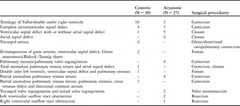
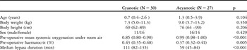
The data collected included demographics, anatomic diagnosis, and details of surgery (Tables 1 and 2). Both groups differed only in median bypass duration, which was significantly longer in the cyanotic group. We used a central venous catheter to obtain haematocrit and blood samples for vascular endothelial growth factor and soluble vascular endothelial growth factor receptor-1. Blood samples for vascular endothelial growth factor and soluble vascular endothelial growth factor receptor-1 were collected pre-operatively, and 24 and 96 hours post-operatively in disodium-ethylenediaminetetra-acetate and immediately centrifuged for 10 minutes at 3000 g. The plasma was removed and stored at −80°C until the assays were performed. Vascular endothelial growth factor and soluble vascular endothelial growth factor receptor-1 were measured using commercially available Enzyme-linked immunosorbent assay kits (human VEGF Quantikine and human sVEGF R1/Flt-1 Quantikine, R&D Systems, Minneapolis, Minnesota, United States of America) according to the manufacturer’s protocol. Statistical analysis was performed using SPSS 12.0 (SPSS, Chicago, Illinois, United States of America). Descriptive statistics are reported as medians with interquartile ranges. We used Mann–Whitney U and Wilcoxon tests to compare age, weight, oxygen saturation, haematocrit, and levels of vascular endothelial growth factor or soluble vascular endothelial growth factor receptor-1. Differences were considered significant at p-value below 0.05.
Diagnoses and surgical procedures.

Description of the study groups (infants with cyanotic and aycyanotic congenital cardiac disease).

Values are shown as median (interquartile range) except for sex
The Institutional Ethics Committee at Charité University Medicine Berlin approved the study, and all participating parents granted informed consent.
Results
Mean systemic oxygenation under room air and haematocrit
Table 2 depicts the pre-operative mean systemic oxygenation under room air and haematocrit in the study groups. Haematocrit was significantly higher in the cyanotic group than in the acyanotic group.
Vascular endothelial growth factor and soluble vascular endothelial growth factor receptor-1 in the cyanotic and acyanotic groups
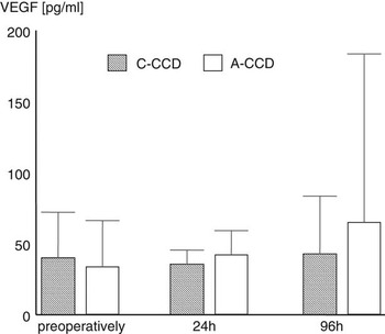
Pre- and post-operatively, central venous vascular endothelial growth factor did not differ between the cyanotic and acyanotic groups (Fig 1). Vascular endothelial growth factor levels, measured 24 hours post-operatively, were comparable to pre-operative levels in both groups – 35.6 (17.5–45.5) picograms per millilitre in the cyanotic group and 42.4 (10.8–59.2) picograms per millilitre in the acyanotic group. There were no significant changes in vascular endothelial growth factor levels measured 96 hours post-operatively – 42.8 (19.7–83.5) picograms per millilitre in the cyanotic group and 65.2 (29.7–183.5) picograms per millilitre in the acyanotic group.
Central venous vascular endothelial growth factor (VEGF) (picogram per millilitre) measured in patients with cyanotic (C-CCD) and acyanotic congenital cardiac disease (A-CCD) pre-operatively, and 24 and 96 hours after surgery. Values are shown as the median and 75th percentile.

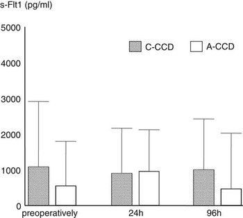
Pre-operatively measured levels of soluble vascular endothelial growth factor receptor-1 tended to be higher in the cyanotic group, 1085.4 (418.6–2920.6) picograms per millilitre, than in the acyanotic group, 552.6 (207.9–1798.5) picograms per millilitre (Fig 2). In the cyanotic group, soluble vascular endothelial growth factor receptor-1 was comparable to pre-operative levels 24 hours after cardiac surgery, 940.2 (652.0–2163.5) picograms per millilitre, and remained unchanged until 96 hours post-operatively, 1002.9 (493.3–2423.3) picograms per millilitre. In the acyanotic group, soluble vascular endothelial growth factor receptor-1 levels were elevated 24 hours post-operatively compared to the pre-operative levels, 1125.2 (357.4–2122.0) picograms per millilitre – and returned to pre-operative levels within 96 hours after surgery, 470.0 (222.4–2025.4) picograms per millilitre.
Central venous soluble vascular endothelial growth factor receptor-1 (s-Flt-1) (picogram per millilitre) measured in patients with cyanotic (C-CCD) and acyanotic congenital cardiac disease (A-CCD) pre-operatively, and 24 and 96 hours after surgery. Values are shown as the median and 75th percentile.

Discussion
In this study, we examined plasma vascular endothelial growth factor and its soluble receptor in infants with cyanotic and acyanotic congenital cardiac disease who underwent open cardiac surgery. In contrast to previous studies,Reference Ootaki, Yamaguchi, Yoshimura, Oka, Yoshida and Hasegawa1, Reference Suda, Matsumura, Miyanish, Uehara, Sugita and Matsumoto2, Reference Starnes, Duncan and Kneebone6, Reference Himeno, Akagi and Furui7 we did not find any differences in vascular endothelial growth factor levels between patients with cyanotic and acyanotic congenital cardiac disease and also observed lower vascular endothelial growth factor levels overall. This may be due in part to our different methodical approach. Plasma is the preferred medium for measuring free-circulating vascular endothelial growth factor levels, since the highly variable platelet-mediated secretion of vascular endothelial growth factor during the clotting process influences the vascular endothelial growth factor levels.Reference Webb, Bottomley, Watson and Brenchley8–Reference Arena and Ferrero10 However, most previous data refer to vascular endothelial growth factor serum values,Reference Ootaki, Yamaguchi, Yoshimura, Oka, Yoshida and Hasegawa1, Reference Suda, Matsumura, Miyanish, Uehara, Sugita and Matsumoto2, Reference Starnes, Duncan and Kneebone6 which might be influenced by platelet degranulation, thus leading to higher vascular endothelial growth factor levels.Reference Arena and Ferrero10 For our study, we followed a recommended protocolReference Hormbrey, Gillespie and Turner9 using plasma samples that were cooled and immediately centrifuged after collection. In order to optimise comparability, we collected blood samples exclusively from central venous lines. Our patients were also younger than those in previous studies.
We found slightly, but not significantly, higher pre-operative plasma levels of soluble vascular endothelial growth factor receptor-1 in infants with cyanotic congenital cardiac disease. This phenomenon is probably explained by the known induction of soluble vascular endothelial growth factor receptor-1 expression under hypoxic conditions, which blocks the physiological vascular endothelial growth factor.Reference Shibuya5 Soluble vascular endothelial growth factor receptor-1 reduces the basal serum angiogenic activity of vascular endothelial growth factor in vitro,Reference Hamada, Ebata and Higashi11 since it acts as a negative regulator by sequestering the vascular endothelial growth factor via its ligand-binding domain. Bound vascular endothelial growth factor is not detected by the Enzyme-linked immunosorbent assay. It might therefore be possible that hypoxia-induced vascular endothelial growth factor expression is counterbalanced by higher soluble vascular endothelial growth factor receptor-1 levels.
Conclusion
In contrast to previous studies, we did not observe elevated vascular endothelial growth factor plasma levels in infants with cyanotic congenital cardiac disease. Levels of soluble vascular endothelial growth factor receptor-1, which are not reported in infants with congenital cardiac disease before, tended to be higher in cyanotic infants.
Acknowledgements
We thank Sarah Smithson-Compton for suggestions regarding English language usage and Evelyn Strauss for technical assistance.