




No CrossRef data available.
Article contents
Behaviour and development of Elasmosoma sp. (Neoneurinae: Braconidae: Hymenoptera), an endoparasite of Formica ants (Formicidae: Hymenoptera)
Published online by Cambridge University Press: 06 May 2004
Abstract
The behaviour and development of Elasmosoma sp., near pergandei Ashm. (Neoneurinae: Braconidae: Hymenoptera), a parasite of the ant, Formica obscuriventris clivia Creighton, was studied in a coastal sand dune habitat in central Oregon over a 2-year period. Female wasps oviposited into the abdomen of major worker ants after alighting briefly on the host. Embryonic development of the wasp occurred inside the serosal membrane or trophamnion of the egg in the abdomen of the ant host. First instar wasp larvae possessed a large head capsule with falcate mandibles, 11 ventral body lobes, an elongate tail and a dorsally located anus. Second instar larvae were unspecialized and lacked the large head capsule, elongate tail and paired ventral body lobes. Third instar larvae were hymenopteriform with a distinct head capsule. Cocoon formation and adult emergence is described. Observations on the percentage parasitism and altered behaviour of parasitized ants are presented. The association between neoneurine wasps and ants is a well-balanced system that has been in existence for at least 40 million years.
Information
- Type
- Research Article
- Information
- Copyright
- 2004 Cambridge University Press
INTRODUCTION
Ants are hosts to a number of parasites, predators and pathogens (Donisthorpe, 1927). In North America, two genera of braconid neoneurine wasps (Neoneurus Haliday and Elasmosoma Ruthe) parasitize adult ants. While a number of neoneurine species have been described (Shaw, 1992), very little is known about their biology and immature stages. The present study discusses the behaviour and development of an undescribed species of Elasmosoma near pergandei Ashm. in a coastal sand dune habitat along the Pacific Northwest coast. Scott Shaw is describing this wasp.
MATERIALS AND METHODS
The observations reported here were made from July 2001 to August 2003 on one colony of Formica obscuriventris clivia Creighton located just north of the city of Waldport (Lincoln County) in central Oregon. Collections of major worker ants, sand from around the nest and nest material were made periodically during the study period. Worker ants and adult wasps were aspirated into glass tubes. The former were transferred to plastic Petri dishes containing a layer of clean (obtained from a dune area away from any Formica nests) sand in the bottom. The ants were provided with water and observed over a period of 2 weeks or until they died. Cocoons were located around the ant nest by sorting through the sand under a dissecting microscope. Dissections of workers were made in 1% Ringer's solution. Developmental stages of the wasps were killed in 70% ethyl alcohol, then carried into glycerin by an evaporation technique and mounted on permanent slides. Observations and photographs were made with a Nikon Optiphot microscope and a Nikon SMZ-10 stereoscopic microscope at magnifications up to ×1000. Micro-dissecting scissors were used to open the cocoons to follow development of the pupae and prepupae.
RESULTS
The climate was maritime since the colony was located in a disturbed dune ecosystem within 300 meters of the Pacific Ocean. The nest, approximately 0·5 meters in diameter and 0·3 meters in height, consisted of plant debris (broken stems and leaves, seeds) and the remains of various arthropods. Dominant introduced plants surrounding the colony were the European dune grass (Ammophila arenaria (L.) Link) and scotch broom (Cytisus scoparius (L.) Link). Native plants included the coast strawberry (Fragaria chiloensis (L.) Duch), shore pine (Pinus contorta Loud.), beach knotweed (Polygonum paronychia C. & S.) and seashore lupine (Lupinus littoralis Dougl.).
Adult wasps appeared at the nest around the first of May, after the winter storms, and populations built up during the dry summer. Peak activity for the wasps was in July and early August, when up to 30 wasps could be seen hovering over the nest in the afternoon sun. Rates of parasitism in worker ants were highest in July and early August, then began to taper off, although adult wasps continued flying into mid-October before cold and wet weather terminated further activity for the season.
Wasp flight activity was dependent in large part on temperature, which was correlated with the rays of the sun reaching the colony. The wasps rarely appeared before the sun reached the ant nest and would retreat when clouds blocked the sun or the nest was shaded. Thus, in the early morning, evening and on cloudy days, few adult wasps appeared. After the sun reached the nest, wasp activity was high and that is when most worker ants retreated into the nest or to the shaded part of the nest. The adult female wasps would harass the ants at the boundary between sun and shade. Most of the male wasps remained around the edge of the nest, attempting to mate with newly emerged females. However, some males followed females over the nest as the latter were chasing ants.
The female wasps pursued the ants continuously during sunny periods, resting only occasionally for several seconds to several minutes on soil particles or grass stems. During pursuit, the wasps continuously advanced and retreated, always attempting to orientate themselves near the rear of the ant and alight on their abdomens. The ants were quite aware of the wasps and would turn about and lash out at them, occasionally catching them in their mandibles (Fig. 4C). Other workers in the vicinity of the ant being chased would often attempt to catch the wasps. The wasps would sporadically make actual physical contact (strikes) with the ants, but actual ovipositon in the abdomen of an ant was only observed twice. In each case, the wasp actually remained on the ant's abdomen for 1–2 sec. The stung specimens exhibited great agitation and, when later dissected, each contained a wasp egg in early stages of embryonic development. During all of the dissections, only in one case, were two wasp larvae in a single ant and one was dead. Otherwise, all infected ants contained only a single wasp egg or larva.
Wasp developmental stages were always found in the abdomen of the host, never in the thorax or head. The newly deposited eggs were very difficult to find since they were placed just under the body wall of the ants' gaster and hidden by a thin layer of host tissue. Each ovary of the female wasps contained from 30–50 broadly elongate eggs measuring 93–120 μm in length, 14–18 μm in greatest width and tapering at the ends (Fig. 1A). When first deposited, they appeared to adhere to the ants' body wall but became free as embryonic development proceeded. The earliest parasite stage found in an ant was an embryo developing within a trophamnion (a layer of cells derived from the serosal membrane of the egg), which was 345 μm long and 170 μm wide (Figs 1B and 7A). The largest trophamnion encountered was 426 μm long and 216 μm wide (Figs 1C and 7B). The trophamnion contained cells with distinct nuclei and nucleoli (Fig. 1B) and a small appendage at the posterior end (Figs 1A,B and 7A,B). Development of the embryonic head capsule in the trophamnion involved the formation of tissue lobes, beginning with a single lobe which then passed through a 3- and 5-lobed stage, until the first instar head capsule was formed (Figs 1B,C and 7A,B).

Fig. 1. Development of Elasmosoma sp. in Formica obscuriventris clivia. (A) An ovary containing a group of elongate ova. (B) Early embryonic development inside the trophamnion. Note nuclei and nucleoli in trophamnion cells. (C) Later embryonic development. Note lobes of tissue (arrow) forming head capsule.
Two distinct larval instars were commonly encountered, the mandibulate or polypodeiform first instar (Figs 2A,B and 7C) and the typical hymenopteriform third instar (Fig. 3B). Another rare instar was also discovered which did not resemble either the first or third instars (Fig. 3A). This instar is tentatively considered the second instar, making 3 instars in the development of Elasmosoma sp.

Fig. 2. Development of first instar larva of Elasmosoma sp. in Formica obscuriventris clivia. (A) Note large head capsule, ventral body lobes, elongate tail and dorsal anus (arrow). (B) Ventral view of head capsule showing paired, simple, falcate mandibles.

Fig. 3. Development of Elasmosoma sp. in Formica obscuriventris clivia. (A) ‘Unspecialized’ second instar larva. (B) Hymenopteroid third instar larva. Note coiled salivary silk glands filling most of body cavity.
When the first larval instar left the trophamnion, it contained 12 body segments, 11 of which contained paired distinct ventral lobes (Figs 2A and 7C). The head capsule was quite pronounced and the mandibles were large and sickle-shaped (Fig. 2B). There was no indication of eyes, antennae or spiracles. The tail was elongate and provided with small retrose spines, especially noticeable on the ventral surface, near the tip. The well-formed gut contained a single layer of columnar cells and the plugged anus opened on the dorsal side of the body. It was possible to determine if an ant was infected with a first instar larva at the time of dissection from the state of its fat body. In non-parasitized hosts, the fat body was compact, whereas in parasitized workers, the fat body was broken up into small particles.
Second instar wasp larvae, which varied from 1·4 to 1·6 mm in length, were much less specialized than the first instar. The head capsule had been shed and replaced with a greatly reduced oral opening (Fig. 3A). There were no eyes, antennae or spiracles and the body segments were, for the most part, indistinct. The anus appeared terminal. The long tail of the first instar was replaced with a small obtuse terminus containing a few spines.
Third instar larvae, which varied from 1·7 to 1·9 mm in length, possessed a small, but distinct head capsule, but lacked a tail (Figs 3B and 5C). The body, composed of 13 segments, was uniformly covered with microscopic setae. Spiracles, absent from the first and second instars, were present on the first thoracic and first 6 abdominal segments. The paired labial silk glands were enlarged and nearly filled the body cavity in mature larvae, imparting them with a light yellowish colour. This instar was a typical hymenopteriform larva.
Exit of mature third instar larvae from ant hosts usually occurred via the anus (Fig. 4A), but occasionally from between the abdominal segments. At the time of emergence, the host ants were usually dead or dying. The exiting parasite larvae were quite active, quickly moving away from the host and attempting to burrow into the substrate by vertical movements of both the anterior and posterior ends. In some cases, the larvae could not completely exit due to the constricted opening and eventually died. Other worker ants ignored the newly emerged wasp larvae.

Fig. 4. (A) Mature larva of Elasmosoma sp. emerging from the anus of Formica obscuriventris clivia. (B) Mature larva (arrow) of a neoneurine wasp emerging from a worker ant in 40 million-year-old Baltic amber. (C) A major worker of F. obscuriventris clivia holding a female wasp in its mandibles. Note extruded ovipositor of wasp (arrow).
After making a shallow depression in the substrate (sand), the newly emerged larvae would proceed to attach sand grains to the future cocoons. The method by which this was accomplished is as follows. The larva first moved around in a circle, touching its head and emitting a strand of silk to some dozen sand grains, thus uniting them all by a long, single silk strand. The larva then rotated and twisted the string of sand grains around its body. It then proceeded to spin a cocoon from its protected site inside the sand grains (Fig. 5A). Newly emerged larvae that were placed on plastic surfaces did not form a cocoon and although they would enter a ‘prepupal’ stage, they eventually died before reaching the pupal stage.

Fig. 5. Development of Elasmosoma sp. (A) Wasp cocoon with sand grains attached to the outer surface. (B) Same cocoon as in (A) with sand grains removed. Same magnification as (A). (C) Mature larva (left) and ‘prepupae’ (right) removed from cocoons.
All of the cocoons were covered with sand in the observation dishes as well as around the periphery of the ant nest. Some cocoons found in the vicinity of the nest had seeds (of scotch broom), bits of rootlets and soil particles attached to them. Since cocoons found inside the ant nest were also surrounded by sand grains, and the nest was composed mostly of plant debris, they were probably formed in the sandy area around the nest and carried in by worker ants. The cocoons were white, smooth, and oval with a small constriction at one end, and measured 2·1–2·3 mm in length (Fig. 5B). Under natural conditions, most cocoons were found in the upper 1 cm of sand within a radius of 500 cm from the nest.
After forming the cocoon, the larva entered what appeared to be a prepupal stage. This stage, identified by the swollen thoracic segments, lasted for at least 48 h before the pupal stage appeared (Figs 5C and 6A). The developing eyes of the adult wasps could often be seen in the older prepupal stage and always in the early pupal stage (Fig. 6A). At the time of emergence from the cocoon (Fig. 6B), the adult wasp used its mandibles to cut a neat circular opening at one end of the cocoon. The cap was completely cut away.

Fig. 6. Development of Elasmosoma sp. (A) Cocoon cut open to reveal developing pupa. Note dark eye spot (arrow) and remains of meconium smeared on abdominal segments. (B) Male adult wasp emerging from its cocoon (cocoon was cut open with a micro-scissors). (C) Adult female wasp. Note extruded ovipositor (arrow).
It was difficult to determine the actual rate of parasitism in the colony, since the percentage of infected ants on or around the colony varied over a 12-h period (Table 1). In July and August, when the wasps were especially abundant, ants harbouring third stage wasp larvae would gather along the edge of the nest in early morning. These parasitized ants received food by returning foragers. This collection of parasitized ants, which facilitated collection, was termed a ‘host assembly’. Apparently a shift in ant behaviour occurred when the parasites reached the third stage. At this time, the ants halted normal activity and became part of the ‘host assembly’. After receiving nourishment, these infected ants either retreated into the nest or wandered off by themselves, the latter possibly indicating that the parasites were ready to emerge. The average developmental time from oviposition to adult emergence was estimated to be 5–6 weeks, which would indicate that some 4–5 generations occurred per summer.
Table 1. Parasitism of Formica workers by Elasmosoma sp. during the late summer of 2003 (All samples taken at exactly the same location adjacent to the ant nest.)

During the course of this study, no other internal parasites were found, nor were any other types of wasps noticed flying around the nest. Occasionally some dauer stages of diplogasterid nematodes were recovered from the head glands of the ants, but they were uncommon and their effect on the ant appeared to be negligible. Some workers had their body cavities filled with iridescent fat globules, a condition suggesting the presence of an iridescent virus.
DISCUSSION
Wasp activity in the spring is apparently initiated from over-wintering cocoons. The apparent dependency of the wasps on sunlight to conduct their ovipositional flights is interesting. It is assumed that the sun provides the warmth required to attain high metabolic levels for maneuvering in flight. This includes not only following an ant, but also avoiding their aggressive behaviour. Such aggressiveness against Neoneurus mantis by Formica podzolica workers was also noted by Shaw (1993).
It is not known if female wasps can oviposit into an ant without alighting on the host since there were many cases of split second contact (strikes) between female wasps and ants. Donisthorpe (1927) implied that Elasmosoma berolinense Ruthe could oviposit in the gaster of Formica rufa during such strikes. However, since the eggs have to be compressed and forced out of the ovipositor, which is extremely fine (the abdomen contained numerous muscle strands associated with the reproductive system), it is difficult to understand how they could be deposited into the abdomen of the ant by an instantaneous strike. Also, the tarsi of female Elasmosoma sp. are extended beyond the vestigial tarsal claws and contain suction-like disks (enlarged pulvilli) at their tips (Fig. 7D). This morphological adaptation, which would allow the wasp to grasp the ant's cuticle, suggests that alighting on the ant is a necessary act to complete oviposition. Such tarsal modifications were also observed in other species of Elasmosoma (Shaw, 1985). In observing the braconid N. mantis ovipositing in F. podzolica, Shaw (1993) noted that the wasp alighted on the ant's abdomen for a short period. In N. mantis, the pulvillus is enlarged and modified for adhering to objects (Shaw, 1992).

Fig. 7. Development of Elasmosoma sp. in Formica obscuriventris clivia. (A) Early embryo developing within the trophamnion. Note terminal appendage (arrow) and three-lobed head region. (B) Later embryo showing further development within the trophamnion. Note terminal appendage and five-lobed head region. (C) First instar larva showing falcate mandibles, 11 paired ventral lobes (only one of each pair of ventral lobes is shown) on thoracic and first 8 abdominal segments, elongate tail, dorsal heart and plugged dorsal anus (arrow). (D) Terminal tarsal segment on foreleg of female wasp. Note preapical claw (arrow) and sucker-like disc (modified pulvillus) on apical extension.
Often a wasp would be following an ant, almost make contact and then turn away. These ants were found to be already parasitized. It is likely that a pheromone produced by the wasp larva inside the ant or a marking pheromone placed on the gaster of the ant by a previous female wasp notifies the ovipositing female that the ant is already parasitized. Mistakes are rare, since only in one case, were two wasp larvae (both first stage) found in a single ant. Under normal circumstances, the smaller larva would be killed, as it was in this instance.
Attempts were made to determine if other behaviour differences, aside from the ‘host assembly’ phenomena, occurred between infected and non-infected ants. Ants parasitized by first and early third instar larvae appeared just as aggressive as non-parasitized ants and would grasp the forceps with their mandibles and eject formic acid, as did non-parasitized ants. However, those ants harbouring mature third stage wasp larvae tended to be more retiring, possibly because their reservoir of formic acid was considerably reduced by the presence of the parasite. No correlation between size of the gaster and the presence of parasites was noted.
The first instar larva could be considered mandibulate, caudate or polypodeiform, as defined by Clausen (1954). Its embryonic development within the trophamnion provided a view to the formation of the various body organs. The tail appendage always appeared on the posterior end of the trophamnion in respect to the head of the developing embryo. This appendage probably represents the still constricted portion of the egg after it passed through the ovipositor.
Body orientation in the first stage larva is interesting. While the paired lobes are on the concave ventral side, as determined by the location of the ventral nerve cord, the anus is located on the convex dorsal side, as determined by the position of the heart. First stage larvae of Aridelus rufotestaceus Tobias, a braconid parasite of the heteropteran, Nezera viridula, have the anus located on the ventral, concave side of the body (Shaw et al. 2001). However, in a study on the caudate first instar larvae of the braconid, Adelura gahani, Baume-Pluvenel (1914) determined the convex side, which contained the anus, to be ventral and the concave side, which contained the body lobes, to be dorsal. In the present case, after the first instar larva loses its tail, the anus shifts to the terminus (in the second instar) and then ventrally subterminal (in the third instar). The large falcate mandibles may be used to macerate the fat body and other non-vital body organs of the host as well as to destroy any other competing larvae. The tail may be used for the same purpose, as well as for locomotion.
What is interpreted here to be the second instar larva is very unusual. When first encountered and because of its rarity, it was suspected of belonging to another wasp species. Its unspecialized character suggests that it is an intermediate form bridging the gap between the morphologically different first and third instar larvae.
Although parasitic wasps are not known to possess a prepupal stage (Quicke, 1997), the mature larva of Elasmosoma sp. was observed to enter a quiescent morphological stage (with a swollen thorax) in the cocoon, which differed from that of both the larva and pupa. The pupa began to form within this stage since the adult eyes could be seen beneath the cuticle of the ‘prepupa’. This ‘prepupal’ stage appeared just before the meconium was eliminated at one end of the cocoon. Since a moult could not be observed between the larval and prepupal stages, the latter could simply represent an early stage of pupal formation.
The purpose of covering the cocoons with sand grains is probably to escape detection and provide protection from enemies. It certainly makes detecting them difficult. While Wassman (cited by Ĉapek, 1970) noted that the cocoons of Neoneurus sp. were attached to the host remains, this was never noted in the present study.
Associations between neoneurine braconids and ants have been in existence for at least 40 million years, as indicated by a mature larva exiting from an ant in Baltic amber (Poinar & Miller, 2002). It is obvious that a balance has been achieved between the ant and wasp, allowing both to survive even with a significant rate of parasitism. Survival of the parasite depends on many attributes, especially skill and precision regarding ovipositional behaviour. However of utmost importance in the survival of the wasp is the total disregard of the emerging third instar larva by other ants. If the workers showed the same aggression against this stage that they demonstrated against the adult wasps, parasite populations would probably be eliminated.
The author is grateful to Scott Shaw for providing encouragement, information and references during the course of this study. Thanks are also extended to Mark DuBois for identifying the ants, Katrine Dailey for help in collecting ants and Roberta Poinar for editorial comments.
References
REFERENCES
BAUME-PLUVINEL, G. (1914). Evolution et formes larvaires d'un braconide, Adelura gahani, parasite interne d'un phytomyzinae. Archives de Zoologie Experimentale et Génerale 55, 47–59.Google Scholar
ĈAPEK, M. (1970). A new classification of the Braconidae (Hymenoptera) based on the cephalic structures of the final instar larva and biological evidence. The Canadian Entomologist 102, 846–875.CrossRefGoogle Scholar
CLAUSEN, C. P. (1962). Entomophagous Insects. Hafner Publishing Company, New York.
DONISTHORPE, H. J. K. (1927). The Guests of British Ants. George Routledge and Sons Ltd, London.
POINAR, Jr., G. O. & MILLER, J.
(2002). First fossil record of endoparasitism of adult ants (Formicidae: Hymenoptera) by Braconidae (Hymenoptera). Annals of the Entomological Society of America 95, 41–43.CrossRefGoogle Scholar
QUICKE, D. L. J. (1997). Parasitic Wasps. Chapman and Hall, London.
SHAW, S. R. (1985). A phylogenetic study of the subfamilies Meteorinae and Euphorinae (Hymenoptera: Braconidae). Entomography 3, 277–370.Google Scholar
SHAW, S. R.
(1992).
Seven new North American species of Neoneurus (Hymenoptera: Braconidae).
Proceedings of the Entomological Society of Washington 94, 26–47.Google Scholar
SHAW, S. R.
(1993).
Observations on the ovipositional behavior of Neoneurus mantis, an ant-associated parasitoid from Wyoming (Hymenoptera: Braconidae). Journal of Insect Behavior 6, 649–658.CrossRefGoogle Scholar
SHAW, S. R.,
SALERNO, G.,
COLASSA, S. &
PERI, E.
(2001).
First record of Aridelus rufotestaceus Tobias (Hymenoptera: Braconidae, Euphorinae) parasitizing Nezera viridula nymphs (Heteroptera: Pentatomidae) with observations on its immature stages and development.
Journal of Hymenoptera Research 10, 131–137.Google Scholar

Fig. 1. Development of Elasmosoma sp. in Formica obscuriventris clivia. (A) An ovary containing a group of elongate ova. (B) Early embryonic development inside the trophamnion. Note nuclei and nucleoli in trophamnion cells. (C) Later embryonic development. Note lobes of tissue (arrow) forming head capsule.
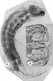
Fig. 2. Development of first instar larva of Elasmosoma sp. in Formica obscuriventris clivia. (A) Note large head capsule, ventral body lobes, elongate tail and dorsal anus (arrow). (B) Ventral view of head capsule showing paired, simple, falcate mandibles.
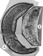
Fig. 3. Development of Elasmosoma sp. in Formica obscuriventris clivia. (A) ‘Unspecialized’ second instar larva. (B) Hymenopteroid third instar larva. Note coiled salivary silk glands filling most of body cavity.
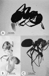
Fig. 4. (A) Mature larva of Elasmosoma sp. emerging from the anus of Formica obscuriventris clivia. (B) Mature larva (arrow) of a neoneurine wasp emerging from a worker ant in 40 million-year-old Baltic amber. (C) A major worker of F. obscuriventris clivia holding a female wasp in its mandibles. Note extruded ovipositor of wasp (arrow).
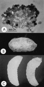
Fig. 5. Development of Elasmosoma sp. (A) Wasp cocoon with sand grains attached to the outer surface. (B) Same cocoon as in (A) with sand grains removed. Same magnification as (A). (C) Mature larva (left) and ‘prepupae’ (right) removed from cocoons.
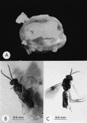
Fig. 6. Development of Elasmosoma sp. (A) Cocoon cut open to reveal developing pupa. Note dark eye spot (arrow) and remains of meconium smeared on abdominal segments. (B) Male adult wasp emerging from its cocoon (cocoon was cut open with a micro-scissors). (C) Adult female wasp. Note extruded ovipositor (arrow).
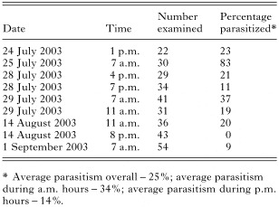
Table 1. Parasitism of Formica workers by Elasmosoma sp. during the late summer of 2003
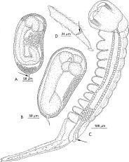
Fig. 7. Development of Elasmosoma sp. in Formica obscuriventris clivia. (A) Early embryo developing within the trophamnion. Note terminal appendage (arrow) and three-lobed head region. (B) Later embryo showing further development within the trophamnion. Note terminal appendage and five-lobed head region. (C) First instar larva showing falcate mandibles, 11 paired ventral lobes (only one of each pair of ventral lobes is shown) on thoracic and first 8 abdominal segments, elongate tail, dorsal heart and plugged dorsal anus (arrow). (D) Terminal tarsal segment on foreleg of female wasp. Note preapical claw (arrow) and sucker-like disc (modified pulvillus) on apical extension.
 You have
Access
- 9
- Cited by