Coronary sinus diverticulum is a rare congenital disorder that may be associated with accessory pathways. Reference Guiraudon, Guiraudon, Klein, Sharma and Yee1 Here, we report a patient with a giant diverticulum of coronary sinus with Wolff-Parkinson-White (WPW) syndrome with multiple accessory pathways and treated by catheter ablation therapy.
Case report
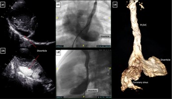
A 5-year-old 14 kg male patient was referred for repeated loss of vision for a very short time. ECG was normal, the echocardiographic evaluation revealed persistent left superior caval vein and a giant diverticulum extending from an enlarged coronary sinus with dimensions of 28x12x28 mm verified by CT 3D reconstruction images. Conventional angiography confirmed the coronary sinus abnormalities and showed no shunting lesions (Fig 1). Low-dose acetylsalicylic acid was started to prevent thrombus formation in diverticulum. Delta waves were detected intermittently on Holter records, and the patient was diagnosed with WPW syndrome. Due to the patient’s complaint about palpitation, antiarrhythmic treatment was started with propranolol (2 mg/kg). Because of tachycardia episodes on Holter records, amiodarone (5 mg/kg) was added to treatment. Catheter ablation was planned due to persistent tachycardia episodes under propranolol and amiodarone combination at 6 months of follow-up.
Images of the coronary sinus diverticulum, dilated coronary sinüs, and persistant left superior caval vein. 2D echocardiography ( a ) and contrast echocardiography ( b ) images from modified parasternal long-axis view demonstrating the diverticulum lying posteriorly by left ventricle free wall. Cineangiograms of persistent left superior caval vein anteroposterior ( c ) and lateral ( d ) views. ( e ) 3D reconstruction images of CT angiography.

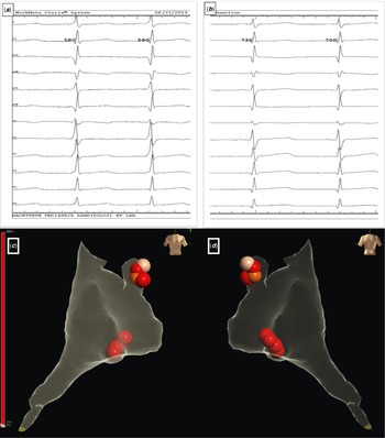
After discontinuing medical treatment for 5 weeks, ablation was performed under general anaesthesia. EnSite PrecisionTM (St Jude Medical, St Paul, MN, United States of America) Cardiac Mapping System was used for electroanatomical mapping procedure. Femoral access was preferred to place decapolar catheter to coronary sinus and quadripolar catheter to right ventricular apex. Irrigated Ablation catheter (FlexAbility™ cardiac ablation catheter, Sensor Enabled™, Abbott Medical, St Paul, MN, United States of America) was used for mapping and ablation procedures. Preexcitation was intermittent and became manifested by atrial pacing from decapolar catheter proximal electrodes. During the electrophysiological study, a narrow QRS complex tachycardia was inducible with 300 ms cycle length and a concentric atrial activation pattern. The earliest atrial activity during the tachycardia was at the proximal electrodes of the decapolar catheter (Fig 2). Right atrium, coronary sinus, and the diverticulum were mapped during atrial pacing and tachycardia for earliest ventricular and atrial activity, which was found to be located at the neck of the diverticulum close to the coronary sinus os. Local bipolar and unipolar recordings showed continuous atrioventricular activity. The earliest ventricular activity was 38 ms earlier than the surface QRS activity. Radiofrequency ablation was performed with 35 W, 45°C on the neck of diverticulum during atrial pacing with continuous preexcitation. The preexcitation disappeared at the 3rd second of energy delivery. Radiofrequency was applied for 100 seconds and then 60 seconds for a bonus lesion. Repeated programmed stimulation revealed a second accessory pathway concealed at the left lateral region far from the posteroseptal area, without decremental conduction. During ventricular pacing, atrial activation pattern was eccentric and the earliest atrial activation was on distal electrodes of decapolar catheter. The presence of negative delta waves in surface ECG lead II was suggestive for an epicardial accessory pathway. Through coronary sinus, left posterolateral ablation was performed for 120 seconds, and ventriculoatrial conduction was dissociated. We demonstrated both atrioventricular and ventriculoatrial conduction blocks after the ablation of both accessory pathways without any preexcitation by adenosine bolus injection. During the ablations, ST interval and T wave were monitored closely on ECG for coronary injury risk. A complete repeated programmed stimulation showed no recurrences in any accessory pathways after the standard 30 minutes waiting time.
( a ) Pre-procedural ECG recording with delta wave. ( b ) Delta wave disappears after ablation of the accessory pathways. Ablation points from anteroposterior ( c ) and posteroanterior ( d ) views.

The patient was discharged the day after the procedure without any complication and without antiarrhythmics, and after 18 months, he is still asymptomatic continuing low-dose acetylsalicylic acid treatment to prevent thrombosis in the diverticulum.
Discussion
Coronary sinus diverticulum is a rare disorder originating from left sinus venous during embryological development, which is primarily asymptomatic and diagnosed incidentally. Thrombosis, compression, or rupture are potential complications, but the most common symptom is palpitation due to arrhythmia disorders. Reference Chen, Nguyen and Dennie2 Coronary sinus malformations can be detected during ablation procedures in patients with posteroseptal accessory pathways. According to the study of Weiss et al, 9% of the patients who underwent electrophysiological study due to various aetiologies, had coronary sinus abnormalities and half of these coronary sinus abnormalities were diverticulums. Reference Weiss, Cappato, Willems, Meinertz and Kuck3
Coronary sinus diverticulums are related to posteroseptal accessory pathways and almost all of these pathways were placed on the neck of diverticulum. Myocardial fibres coating the coronary sinus are accused of producing accessory pathways. Sleeve-like extensions coating the main branches of coronary sinus and sometimes diverticulums originating from here might have been responsible for the connection between the atrium and ventricle. In a study by Sun et al, coronary sinus accessory pathway was identified in 36% of the patients with posteroseptal or left posterior accessory pathway. Retrograde coronary sinus angiography was demonstrated and diverticulums were detected in 21% of patients with coronary sinus accessory pathway. Reference Sun, Arruda and Otomo4 In a study by Schumacher et al, retrograde coronary venography was performed in all patients taken into ablation. Coronary sinus abnormalities were found in 43.6% and coronary sinus diverticulums were found in 17.9% of the patients with posteroseptal accessory pathway. Reference Schumacher, Tebbenjohanns, Pfeiffer, Omran, Jung and Luderitz5 Based upon these findings, coronary sinus imaging should be beneficial for patients with posteroseptal accessory pathways before ablation. Reference Nishimori, Kiuchi and Mori6 Catheter ablation therapy is usually successful when the accessory pathway is on the neck of the diverticulum. Performing the ablation through coronary sinus should be more efficient if the accessory pathway seems to be epicardial. Reference Jang, Rho and Kim7
In epicardial posteroseptal accessory pathways, coronary venous system may be targeted for the ablation site. Because of the close range with coronary arteries, especially branches of the right coronary artery, ablation in coronary sinus may cause severe injuries including heart block. The ablation site’s distance is important, 2 mm or less distant ablations are at high risk (>50%) for coronary artery injuries, 5 mm and more distant ablations are much safer. Reference Stavrakis, Jackman and Nakagawa8 Cryoablation should be considered in posteroseptal accessory pathways due to proximity to coronary arteries and conduction system. Coronary angiography can be performed before ablation to avoid damage. Reference Stavrakis, Jackman and Nakagawa8,Reference Mao, Moriarty, Mandapati, Boyle, Shivkumar and Vaseghi9
Conclusion
Rare malformations like coronary sinus diverticulums are seen more often with tachyarrhythmia disorders. These diverticulums may cause posteroseptal and left posterolateral accessory pathways, mostly on the neck of diverticulum. Catheter ablation therapy is a useful treatment option if the ablation site is not close to coronary arteries.
Acknowledgements
None.
Financial support
This research received no specific grant from any funding agency, commercial, or not-for-profit sectors.
Conflicts of interest
None.
Ethics standards
The procedures performed involving the patient were according to the ethical standards of the 1964 Helsinki declaration and its later amendments.