Introduction
Human cystic echinococcosis (CE), or hydatid disease, caused by the larval stage of Echinococcus granulosus, is increasingly recognized as a significant public health issue (McManus et al., Reference McManus, Zhang, Li and Bartley2003; Craig et al., Reference Craig, McManus, Lightowlers, Chabalgoity, Garcia, Gavidia, Gilman, Gonzalez, Lorca, Naquira, Nieto and Schantz2007). CE remains an important medical problem in western China, especially in Xinjiang, where HIV/AIDS is also highly endemic (MOH & UNAIDS, 2011). Although both diseases are endemic in certain areas, such as in Peru, Argentina, Brazil, India, western China and countries in North Africa, co-infection of CE and HIV/AIDS is rare. In the past 4 years, our two collaborating hospitals (The First Affiliated Hospital of Xinjiang Medical University and Yili Friendship Hospital) received 3750 CE patients for surgery but only four cases were HIV-positive, compared to 650 HIV/AIDS cases received in our hospitals during the same period. Here we report the treatment of the four cases, with three patients receiving surgery for hydatid cyst removal.
Materials and methods
Safety protection of surgical staff
All surgeons wore surgical clothes and double surgical gloves with additional goggles, and extensive care was taken during all surgical procedures and sample collection and testing.
Case reports
Case 1
A 47-year-old male patient presented to our clinic (The First Affiliated Hospital of Xinjiang Medical University) with recurrent abdominal pain, fever and jaundice. He had a long history of drug abuse. The enzyme-linked immunosorbent assay (ELISA)-based HIV antibody specific test (HIV test) showed his serum was positive, which was confirmed by the Xinjiang Center for Disease Control and Prevention (Xinjiang CDC). This patient showed weakness, which was likely due to the HIV infection. Physical examination revealed epigastric tenderness. Laboratory tests showed a low white blood cell count (1.53 × 10⁹/litre), high total bilirubin (88.36 μmol/l), indirect bilirubin (30.77 μmol/l) and direct bilirubin (57.59 μmol/l). However, his CD4+ T-lymphocyte count was 36.6 cells/ml, which is markedly lower than the average level of healthy individuals. Serological tests for echinococcosis using three E. granulosus antigens (crude hydatid cyst fluid, partially purified antigen B and protoscolex proteins) and one E. multilocularis antigen (Em18) (hereafter ‘Echinococcus tests’) (Feng et al., Reference Feng, Wen, Zhang, Chen, Ma, Zhang, Qi, Bradshaw, Vuitton and Craig2010) were negative. Computed tomography (CT) and magnetic resonance imaging (MRI) revealed a 6 × 8 cm hydatid cystic lesion in the right hepatic lobe. The liver hydatid cyst was ruptured, with cyst fluid leaking into the right hepatic bile duct. The physical condition of the patient did not allow surgery for removal of the cyst, so albendazole treatment (15 mg/kg per day) was administered. However, the patient died of septic shock caused by uncontrolled pneumonia after 3 months of treatment.
Case 2
A 44-year-old male was admitted to our clinic with an 8-month history of right upper-quadrant pain. He showed us a CT image taken a year earlier, with a clear hydatid cyst (size 7 × 8 cm) in segment VII of his right hepatic lobe. A new CT scan indicated that the primary cyst was ruptured. There were many cysts in the abdominal and pelvic cavities, with one large cyst 4 × 6 cm in size. These cysts were likely the result of secondary infection. The patient declined surgery but soon returned with severe epigastric pain and jaundice. General laboratory tests showed a white blood cell count of 9.03 × 10⁹/litre; total bilirubin, 54.7 μmol/l; indirect bilirubin, 18.5 μmol/l; and direct bilirubin 36.5 μmol/l. Although he was asymptomatic for AIDS, HIV antibody testing was positive before surgery, which was confirmed by the Xinjiang CDC. His CD4+ T-lymphocyte count was normal (473 cells/ml). Echinococcus tests (Feng et al., Reference Feng, Wen, Zhang, Chen, Ma, Zhang, Qi, Bradshaw, Vuitton and Craig2010) were positive. Both MRI and CT scans showed that hydatid fluid was leaking into the common bile duct. This finding was confirmed by a laparoscopic subtotal pericystectomy, which consisted of common bile duct exploration and T-tube drainage. Albendazole was administered postoperatively for 3 months with a dose of 15 mg/kg per day. The patient refused anti-HIV therapy drugs.
During the 1-year follow-up period, the patient underwent ultrasound every 3 months, which confirmed that there was no residual cavity effusion or recurrence. His CD4+ T-lymphocyte count increased to 520 cells/ml 6 months postoperatively. The patient's general condition also improved significantly.
Case 3
A 41-year-old male was found with a 3-cm hydatid cyst in his liver through a general health check by ultrasonography in a local hospital. His medical record showed that he had undergone surgery locally for removal of a hepatic hydatid cyst when aged 20. The cyst was in the same position as the primary cyst, indicating a secondary infection. The patient was prescribed medication with albendazole (15 mg/kg per day for 3 months), given the small size of the cyst and his good health status. However, he did not return for follow-up evaluation until 3 years later (aged 44), when he was in extreme abdominal pain. He stated that he had a positive diagnosis for HIV infection 2 years previously (when aged 42). The infection was confirmed by the Xinjiang CDC. He had taken anti-HIV drugs since then. Ultrasound imaging revealed that the cyst had grown to 8 × 8 cm in size, with a volume that was 19 times larger than the original cyst diagnosed 3 years previously. He had epigastric tenderness and inguinal lymphadenopathy. Results of general laboratory examinations were within normal levels. His CD4+ count was 290 cells/ml. He was also hepatitis C antibody-positive. Echinococcus tests (Feng et al., Reference Feng, Wen, Zhang, Chen, Ma, Zhang, Qi, Bradshaw, Vuitton and Craig2010) were negative. A contrast-enhanced CT scan of his abdomen showed a typical type III hydatid cyst in segments VII and VIII of the right hepatic lobe. Subtotal pericystectomy was employed to remove the cyst. However, 10 days postoperation, the patient developed right upper-quadrant pain with fever. An abdominal CT scan demonstrated a residual cavity effusion, which was percutaneously drained under ultrasound guidance. One month later, the tube was removed and the CD4+ T-lymphocyte count was shown elevated to 542 cells/ml. Albendazole treatment was given immediately after the pericystectomy, as in the previous case.
The patient was followed-up over 7 months, and his general condition gradually improved. His CD4+ T-lymphocyte count had increased to 640 cells/ml 6 months after the surgery, he had gained 5 kg of body weight and he was less fatigued.
Case 4
A 30-year-old man visited our hospital clinic complaining of intermittent right upper-quadrant abdominal pain that had been present for 6 months. He indicated that he had undergone a hepatic hydatid cyst resection when aged 17 and had a history of 7 years of drug abuse. He was HIV-test positive, as confirmed by the Xinjiang CDC. He had right upper-quadrant tenderness, and CT imaging revealed a cystic lesion measuring 7 × 7 cm with multiple daughter cysts in the left hemi-liver. Echinococcus tests (Feng et al., Reference Feng, Wen, Zhang, Chen, Ma, Zhang, Qi, Bradshaw, Vuitton and Craig2010) were negative. Pericystectomy was performed and pathological examination confirmed that the surgical specimens were hydatid cysts. Following surgery, albendazole was given (the same dose and term as for Case 2 and Case 3). In the following 18 months, Case 4 patient underwent ultrasound every 3 months without recurrence.
Results and discussion
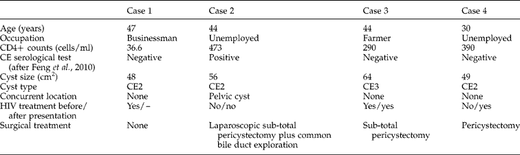
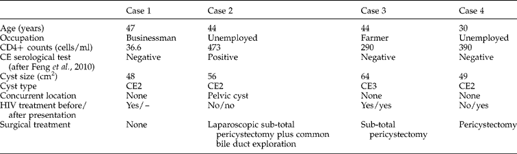
Four CE patients co-infected with HIV/AIDS are reported (table 1); one died of septic shock and uncontrolled infection. We successfully removed the hydatid cysts from three of the patients by surgery. These patients then received albendazole treatment for 3 months. Anti-HIV chemotherapy (200 mg of nevirapine twice a day, 600 mg of lamivudine once a day and 200 mg of zidovudine twice a day) were given to all the patients, except Case 3, who refused to take the medicines. Follow-up checks indicated these patients were remarkably improved in terms of their health status, although they were still HIV-antibody positive. Notably, the combined treatment remarkably increased their CD4+ cell population. Two of the subjects showed a stable and normal level of CD4+ cells during follow-up, indicating that E. granulosus infection may suppress CD4+ cell proliferation, which may be associated with B-cell proliferation and function, given that three of the four patients were serologically negative against four echinococcal antigens (Feng et al., Reference Feng, Wen, Zhang, Chen, Ma, Zhang, Qi, Bradshaw, Vuitton and Craig2010). Antibodies against Echinococcus antigens were still negative in all three followed-up patients in the 3 months after leaving hospital.
Clinical characteristics of four male cases, with symptoms of abdominal pain, co-infected with HIV/AIDS and hepatic cystic echinococcosis.
HIV infection may accelerate hydatid cyst growth. Generally, hydatid cyst growth is slow. Moro et al. (Reference Moro, Gilman, Verastegui, Bern, Silva and Bonilla1999) reported that the rate of growth in natural settings varies from 0.4 to 1.4 cm over a 3-year period. Here, however, CE patients co-infected with HIV had large cysts (mean size 7.5 cm) and, in Case 3, the cyst diameter grew from 3 to 8 cm in 3 years (1.7 cm/year), remarkably faster than previously reported growth rates (Frider et al., Reference Frider, Larrieu and Odriozola1999). In Case 2, cyst rupture occurred and, in only 1 year, a small number of secondary cysts grew to 6 cm in size. The rapid growth of the cysts may be due to the virus suppressing the immune system (Javed et al., Reference Javed, Kalayarasan and Agarwal2012) or, on the other hand, CD4+ T cells may play a role in suppression of cyst growth. Similar reports (including Ramos et al., Reference Ramos, Masia, Padilla, Bernal, Martin-Hidalgo and Gutierrez2007) have shown that giant hydatid cysts and racemose forms of neurocysticercosis are much more frequent in patients infected with HIV than in those who are HIV negative. Sailer et al. (Reference Sailer, Soelder, Allerberger, Zaknun, Feichtinger and Gottstein1997) and Zingg et al. (Reference Zingg, Renner-Schneiter, Pauli-Magnus, Renner, van Overbeck, Schlapfer, Weber, Weber, Opravil, Gottstein and Speck2004) also reported that alveolar echinococcosis patients co-infected with HIV showed rapid parasite lesion development. However, these studies include a very small number of patients, and further studies with a larger number of cases should be conducted to confirm the relationship between immunodeficiency status and rate of parasitic lesion development.
It is noteworthy that two of the four patients had ruptured hydatid cysts with cyst fluid leaking into the bile duct. Cyst rupture is the major complication of hepatic hydatid disease. Various incidence rates of direct rupture have been reported, with a range of 1.75–34.4% (McManus et al., Reference McManus, Zhang, Li and Bartley2003; Craig et al., Reference Craig, McManus, Lightowlers, Chabalgoity, Garcia, Gavidia, Gilman, Gonzalez, Lorca, Naquira, Nieto and Schantz2007). The risk of rupture is likely associated with the increased size of cysts and intracystic pressure (Yuksel et al., Reference Yuksel, Kir, Ercan, Batirel and Baysungur1997). HIV infection increases the growth of cysts, which may increase the risk of cyst rupture. Two cases, Case 2 and Case 4, were HIV-test positive before surgery, and Case 4 was given anti-HIV drug treatment immediately following surgery. This patient did not have ‘immune reconstitution inflammatory syndrome’ (IRIS) at the initiation of anti-HIV therapy, which is observed for other infectious diseases, including viral, mycobacterial, fungal and parasitic diseases, with aggravation of the associated condition (Sharma & Soneja, Reference Sharma and Soneja2011).
In our report, one patient died of septic shock caused by uncontrolled pneumonia. We believe the infection was due to his low level of immunity, given that his CD4+ T-lymphocyte count was only 36.6 cells/ml, indicating severe immunosuppression. We recommend that hydatid patients co-infected with HIV should receive immediate surgery after diagnosis to prevent cyst and intrabiliary rupture.
In addition, one patient underwent laparoscopic surgery, and had an uneventful postoperative course and a normal CD4+ T-lymphocyte count at follow-up. Laparoscopic surgery causes mild surgical trauma compared with conventional surgery, and is a safe approach in patients with HIV infection.
AIDS is one of the most serious human diseases and prevention of iatrogenic infection is crucial. The operating staff should have correct understanding of AIDS, and should take every step of the operation carefully and monitor each other closely during the operation. We believe that close co-ordination, strict implementation of disinfection and isolation systems, with a strict working attitude, can completely avoid iatrogenic infection.
Financial support
This research received no specific grant from any funding agency, commercial or not-for-profit sectors.
Conflict of interest
None.
Ethical standards
All treatment procedures for CE patients in the report were approved by the Ethical Committees of The First Affiliated Hospital of Xinjiang Medical University (Approval No,: 20080731-3) and Yili Friendship Hospital (Approval No.: 2008009). Written informed consent was obtained from the patients for publication of the clinical data.