Introduction
Entomopathogenic nematodes (EPN), which occur naturally in soils around the world, are obligate parasites of the soil stages of insects. Each nematode is in mutual alliance with a specific bacterium and, by working together, they are able to overcome the insect host's immune system and are then able to use the host as a food source for the growth of the bacteria, while the nematodes, in turn, feed on the bacteria and converted host tissue (Kaya & Gaugler, Reference Kaya and Gaugler1993). The life cycle of the nematode occurs mainly inside the insect. The only free-living stage is an arrested third-stage juvenile, which develops as soon as the food in the cadaver is depleted, and which carries the associated symbiont bacteria inside the gut, to be vectored to a new invaded host. The depleted host is abandoned, and the next cohort of infective juveniles (IJs) search for a new host in the soil to invade, and to utilize with the aid of the bacteria they vectored into the host (Nguyen, Reference Nguyen, Nguyen and Hunt2007).
In South Africa, not much EPN diversity has been found during surveys of agricultural soil, for example in deciduous fruit-, grapevine- and citrus-growing areas (Malan et al., Reference Malan, Nguyen and Addison2006, Reference Malan, Knoetze and Moore2011; Hatting et al., Reference Hatting, Stock and Hazir2009). Currently, ten EPN species have been reported to occur in South Africa, of which six are Steinernema – S. citrae Stokwe, Malan, Nguyen, Knoetze & Tiedt, Reference Stokwe, Malan, Nguyen, Knoetze and Tiedt2011; S. khoisanae Nguyen, Malan, Gozel & Tiedt, 2006; S. sacchari Nthenga, Knoetze, Berry, Tiedt & Malan, Reference Nthenga, Knoetze, Berry, Tiedt and Malan2014; S. innovationi Çimen, Hatting & Stock, 2014; S. tophus Çimen, Hatting & Stock, 2014; S. yirgalemense Nguyen, Tesmariam, Gozel, Gaugler & Adams, 2005 – and four species are Heterorhabditis – H. bacteriophora Poinar, 1976; H. noenieputensis Malan, Knoetze & Tiedt, 2013; H. safricana Malan, Nguyen, Knoetze & Tiedt, 2008 (Malan et al., Reference Malan, Nguyen, De Waal and Tiedt2008); and H. zealandica Poinar, 1990 (Stokwe et al., Reference Stokwe, Malan, Nguyen, Knoetze and Tiedt2011; Malan et al., Reference Malan, Knoetze and Tiedt2014; Nthenga et al., Reference Nthenga, Knoetze, Berry, Tiedt and Malan2014). In South Africa, the most common species found in disturbed soil is H. bacteriophora, while Steinernema, in general, has been found to be rare, although it has been found to be more prevalent in undisturbed soil.
During a non-targeted survey, an unknown isolate of Steinernema (J194) was trapped from a soil sample. When tested in laboratory bioassays for pathogenicity against Cydia pomonella (L.) (codling moth) (Lepidoptera: Tortricidae) (De Waal et al., Reference De Waal, Malan and Addison2011), using 50 IJs/insect at 25°C, the isolate was found to be highly infective. The nematode isolate was also tested in a field trial as an aerial application against codling moth last-instar diapausing larvae on apple trees, where it obtained the most effective control of codling moth in comparison with other local species (De Waal et al., Reference De Waal, Addison and Malan2013). These results indicate the importance of isolation and identification of new indigenous EPN species and strains for their potential use as biocontrol agents against key insect pest of apples in South Africa.
The objective of this study was to use the biology, morphology, morphometrics and molecular techniques to describe a new species of Steinernema as Steinernema jeffreyense n. sp., and to add to the body of knowledge concerning the occurrence and diversity of this diverse group of nematodes.
Materials and methods
Nematode collection
Steinernema jeffreyense n. sp., isolate J194 was collected from underneath a guava tree in sandy soil, close to the ocean, in the town of Jeffrey's Bay in the Eastern Cape province (34°02.730′S/024°55.610′E). The nematodes were isolated from soil samples using the Galleria mellonella L. trapping technique (Bedding & Akhurst, Reference Bedding and Akhurst1975).
Morphology
For taxonomic studies, ten G. mellonella were exposed to 100 IJs/insect of the new Steinernema isolate in each of two 9.0-cm Petri dishes, lined with moistened filter paper, and kept in a dark growth chamber at 25°C. The Galleria larvae died within 2 days after inoculation. First-generation males and females were obtained 3 days after inoculation, and second-generation males and females were obtained after 5 days. Cadavers from one Petri dish were dissected in Ringer's solution, whereas those in the other Petri dish were transferred to a modified White trap (Kaya & Stock, Reference Kaya, Stock and Lacey1997), with the IJs being harvested within the first week of emergence. For purposes of direct observation, and to confirm the morphology of certain structures, the different stages were killed with gentle heat. After being fixed in triethanolamine fixative (TAF) (Courtney et al., Reference Courtney, Polley and Miller1955), type specimens were processed to glycerine, using the Seinhorst method (Seinhorst, Reference Seinhorst1959). They were then mounted in glycerine, using paraffin wax rings as support. Measurements were taken by means of a compound microscope (Leica DM2000, Leica Microsystems, Wetzlar, Germany), fitted with a digital camera and Leica Application Suite V3.5.0 software.
Spicules and gubernaculums were prepared by colouring the males for 2 min in a drop of lactophenol, containing 0.02% acid fuchsin, on a glass slide. After transferring the nematodes to a drop of clear lactophenol, the tail was cut from the rest of the body, using the slanted, sharp edge of a syringe needle. Having removed the pieces of nematode severed from the tail, the tails were covered with a cover slip, lightly pressed and moved, so as to free the spicules and the gubernaculum from the tail end. Use of this method enabled the observation of a total of 20 males.
For scanning electron microscopy (SEM), first- and second-generation males and females and IJs were fixed in TAF for a minimum of 3 days, washed three times in 0.05 m cacodilate buffer for 15 min each, and then washed three times in distilled water for 15 min each. After this, they were dehydrated in a graded ethanol series (70%, 80%, 90% and 2 × 100%). The samples were critical-point dried with liquid CO2, mounted on SEM stubs, and sputter coated with 20 nm gold/palladium (66/33%). The samples were viewed with an FEI Quanta 200 ESEM (Duren, Germany), operated at 10 kV under high vacuum.
Molecular characterization
Nguyen's (Reference Nguyen, Hunt, Mráček, Nguyen and Hunt2007) technique was used for DNA extraction. One young female was placed in 30 ml lysis buffer (500 mm MgCl2, 10 mm dithiothreitol (DTT), 4.5% Tween 20, 0.1% gelatine and 3 μl of proteinase K (600 μg/ml)), on the side of a 0.5-μl microcentrifuge tube. After being cut into pieces with the sharp side of a syringe needle, the nematode was frozen for 1 h at − 80°C. The tubes were then incubated in a thermocycler at 65°C for 1 h, followed by incubation at 95°C for 10 min. After centrifugation for 2 min at 12,000 rpm, the supernatant (20 μl) was transferred to a clean microcentrifuge tube, where it was kept at − 20°C.
The 18S (5′-TTGATTACGTCCCTGCCCTTT-3′) and 26S (5′-TTTCACTCGCCGTTACTAAGG-3′) primers, suggested by Vrain et al. (Reference Vrain, Wakarchuck, Lèvesque and Hamilton1992), were used for amplification of the internal transcribed spacer (ITS) regions. For amplification of the D2D3 region of the 28S rDNA gene, primer D2F (5′-CCTTAGTAACGGCGAGTGAAA-3′) and primer 536 (5′-CAGCTATCCTGAGGGAAAC-3′) were used (Nguyen et al., Reference Nguyen, Malan and Gozel2006). The primers were synthesized by Integrated DNA Technologies Inc. (Coraville, Iowa, USA). Nguyen's (Reference Nguyen, Hunt, Mráček, Nguyen and Hunt2007) technique was followed for purposes of polymerase chain reaction (PCR) amplification.
Post-PCR purification was undertaken using the NucleoFast Purification System (Macherey Nagel, Waltham, Massachusetts, USA). In addition to obtaining PCR products by means of the use of internal transcribed spacer (ITS) and D2D3 primers, some were also cloned using M13 and the vector pGem-T-Easy (Promega, Madison, Wisconsin, USA). Sequencing was performed with the BigDye Terminator V1.3 sequencing kit (Applied Biosystems, Foster City, California, USA), followed by the use of electrophoresis on the 3730 × l DNA Analyser (Applied Biosystems) at the DNA Sequencing Unit (Central Analytical Facilities, Stellenbosch University). Sequence assembly and editing was performed using the software CLC Main Workbench 7.3 (http://www.clcbio.com).
Phylogenetic analysis
GenBank numbers of the ITS of the 18S rDNA and D2D3 region of the 28S rDNA gene sequences used in the phylogenetic analyses are indicated in the phylogenetic trees. Sequences were aligned using ClustalW v. 2.75 (Larkin et al., Reference Larkin, Blackshields, Brown, Chenna, McGettigan, McWilliam, Valentin, Wallace, Wilm, Lopez, Thompson, Gibson and Higgins2007), in keeping with default alignment parameters. Caenorhabditis elegans (FJ589008) was used as the outgroup taxon for the ITS, with Panagrellus redivivus (AF331910) being used for the D2D3 sequence analysis. Distance analysis of the closest taxa (number of base differences per sequence) was conducted in MEGA6 (Tamura et al., Reference Tamura, Stecher, Peterson, Filipski and Kamur2013). Ribosomal and mitochondrial DNA sequences for S. jeffreyense n. sp. were deposited in GenBank, under the accession numbers KC897093 and KP164866, respectively.
The evolutionary history of the ITS region of the 18S gene, and of the D2D3 region of the 28S gene, were inferred separately, using the maximum parsimony method (Saitou & Nei, Reference Saitou and Nei1987) to obtain a consensus tree using the subtree-pruning-regrafting (SPR) algorithm (Nei & Kumar, Reference Nei and Kumar2000). The confidence intervals for the various branching patterns in the trees were measured using the bootstrap test, with 1000 replicates (Felsenstein, Reference Felsenstein1985). Evolutionary analyses were conducted in MEGA6 (Tamura et al., Reference Tamura, Stecher, Peterson, Filipski and Kamur2013).
Cross-hybridization
Three morphologically similar South African species, and close relatives of S. jeffreyense n. sp. – S. khoisanae (Nguyen et al., Reference Nguyen, Malan and Gozel2006), S. innovationi (Çimen et al., Reference Çimen, Lee, Hatting, Hazir and Stock2014a) and S. tophus (Çimen et al., Reference Çimen, Lee, Hazir and Stock2014b) – were crossed, by adapting the method of Nguyen (Reference Nguyen, Nguyen and Hunt2007), for purposes of assessing the reproductive compatibility of the new species. A total of ten drops of G. mellonella blood were placed in three 15-cm-diameter Petri dishes, with each drop containing a single IJ of S. jeffreyense n. sp. The same procedure was followed with the other three species. After a period of 2 days, or until the sex could be determined, a male or female was crossed with S. jeffreyense n. sp., and both male and female were added to a fresh drop of G. mellonella blood. Controls consisted of crosses with male and females of the same species. Single females of all species were also tested in single drops of haemolymph for reproduction. The Petri dishes were placed in a 2-litre plastic container, lined on the bottom with moist paper towels, which was then closed with a lid. They were incubated at 25°C for a period of 3 days, before being inspected for breeding. The experiment was repeated with a fresh batch of nematodes at a later test date.
Results and discussion
Description of Steinernema jeffreyense n. sp
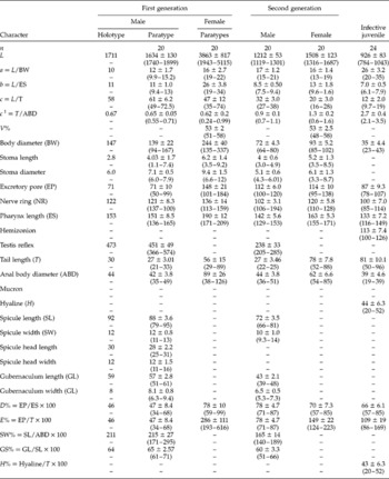
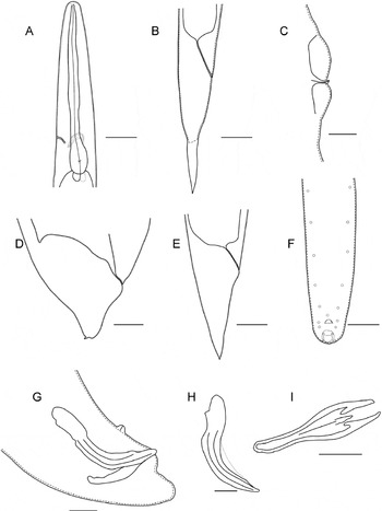
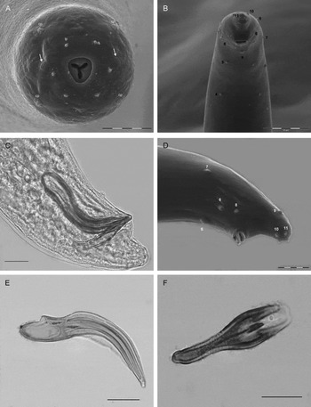
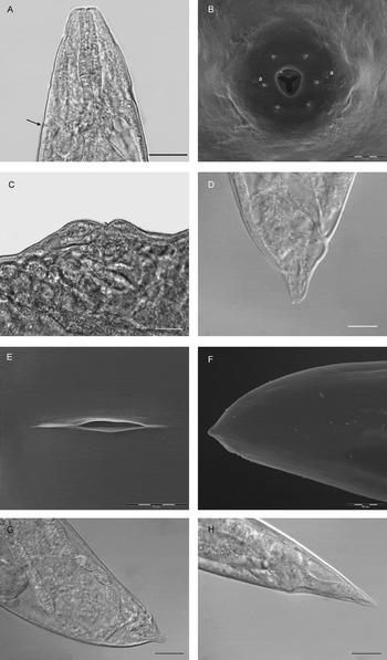
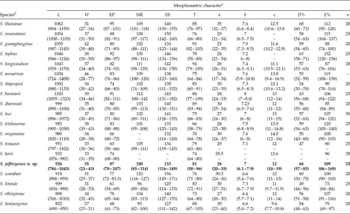
Morphometrics of the holotype (the first-generation male) and paratypes of the first-generation males, IJ and of the first- and second-generation male and female, are listed in table 1. Figures 1–4 illustrate the description and morphometrics of the adult male and female and the third-stage juvenile stages. Steinernema jeffreyense n. sp. is named after the type locality of Jeffrey's Bay, in South Africa.
Morphometrics of different stages of Steinernema jeffreyense n. sp. All measurements in μm and in the form: mean ± SD (range).

V% = distance from anterior end to vulva/body length; EP = distance from anterior end to excretory pore; NR = distance from anterior end to nerve ring; ES = distance from anterior end to end of oesophagus.
Steinernema jeffreyense n. sp. line drawings. (A, B) Infective juvenile: (A) head and pharynx region; (B) tail region. (C, D) First-generation female: (C) short, double-flapped epiptygmata directed to the front; (D) tail shape. (E) Second-generation female tail. (F–I) First-generation male: (F) tail region with papilla arrangement; (G) tail region with spicule and gubernaculum; (H) spicule; (I) gubernaculum. Scale bars: A, B, D, E, G–I = 20 μm; C, F = 50 μm.

Steinernema jeffreyense n. sp. (A) Head region of second-generation male showing mouth opening and six labial papillae, four cephalic papillae (cp) and slit-like amphids (arrows). (B–F) First-generation male: (B) posterior region showing anal aperture and arrangement of papillae; (C, D) ventral view of tails, showing papillae arrangement; (E) spicule; (F) gubernaculum. Scale bars: A = 5 μm; B = 10 μm; C–F = 20 μm.

Steinernema jeffreyense n. sp. (A–G) First-generation female: (A) anterior end, lateral view showing location of excretory canal (arrow) of the first-generation female; (B) en face view, showing six labial papillae, and the openings of the amphids (a); (C) asymmetrical protuberant vulva, with double-flapped epiptygmata directed to the front; (D) tail (lateral view), with dorsal projection on rounded tip; (E) vulva of first-generation female; (F, G) tail regions of the first-generation female (lateral view). (H) Tail (lateral view) of the second-generation female. Scale bars: A, C, F–H = 50 μm; B = 10 μm; D, E = 20 μm.

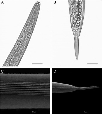
Steinernema jeffreyense n. sp. infective juvenile: (A) anterior of ensheathed infective juvenile, showing the excretory pore (arrow); (B) shape of the tail; (C) eight ridges in midbody; (D) tail region. Scale bars: A, B = 20 μm; C = 10 μm; D = 20 μm.

Morphology
First-generation male
Posterior third of the body slightly curved in a J-shape. Cuticle smooth under a light microscope, but striations visible with SEM. Lateral field absent. Cephalic extremity narrowing gently to the front, with perioral disk. En face view with six labial papillae and four larger cephalic papillae. Amphidial apertures slit-like. Stoma deep, cheilorhabdions prominent, posterior part of the stoma funnel-shaped, moderately cuticularized. Pharynx with a cylindrical corpus, slightly swollen metacorpus, nerve ring surrounds the isthmus or anterior part of the basal bulb, with no visible valve. Excretory pore position anterior of the nerve ring. Cardia prominent. Testis is monorchic, reflexed, spicules paired, gubernaculum 65% of the length of the spicule. Paired spicule with the manubrium of the spicule longer than wide, with a ratio of head length/head width of 4:2, almost no shaft present, blade moderately curved, with two ribs on the arch of the blade, velum prominent, not reaching the tail tip (figs 1H, 2E). Rostrum present on the dorsal side of the manubrium (fig. 2E). Gubernaculum with a short cuneus and capitulum, not reaching the tip of the wings (figs 1I, 2F). Six pairs of genital papillae, with a single precloacal midventral papilla, two pair adcloacal, one pair lateral in line with the cloaca, two pairs subterminal, and one pair subdorsal. Tail tip either rounded or digitate, without a mucron (figs 1G, 2C, D).
Second-generation male
Body length narrower and shorter than the first-generation male. Body more tightly curled posteriorly, sometimes 6-shaped. Tail of the second-generation male same shape as that of the first-generation male, and always without a mucron.
First-generation female
The females are large when compared to males and size varies greatly. Fully reproductive small female pygmies, about the same length as the males, are present on days 3 and 4. Body cuticle smooth, but finely annulated cuticle with SEM. Cephalic extremity rounded, continuous with body; six labial papillae, four cephalic papillae. Lips indistinct. Aphidial apertures present, slit-like (fig. 3B). Stoma shallow, subtriangular anteriorly (fig. 3A). Pharynx with cylindrical muscular procorpus, slightly swollen metacorpus, nerve ring surrounding the slender isthmus, large basal bulb with oesophago-intestinal valve. Excretory pore located just anterior to, or at middle of, basal bulb (fig. 3A). The reproductive system is didelphic, amphidelphic, reflexed, containing many eggs. Vulva at midbody, with an asymmetrical pronounced protuberance in older females, with short double-flapped epiptygmata always forward directed (figs 1C, 3A, C, E). Anus without postanal swelling. Tail shorter than anal body width, conoid-shaped, bluntly pointed with dorsal projection on the rounded tip of the tail (fig. 1D, 3D, F, G).
Second-generation female
Body straight to slightly bent at the vulva, with asymmetrical protuberance, with the highest bump posterior to the vulva, with a double-flapped epiptygmata directed to the front. Tail tapering gently to a sharp point, with no postanal swelling (fig. 3H).
Infective juvenile
Body slender, gradually tapered posteriorly, heat-relaxed specimens straight to slightly ventrally curved. The arrested third-stage IJ has a closed stoma, narrow body, with a second-stage sheath usually not observed after harvesting, cuticle finely annulated. Head region continuous with body, rounded to slightly truncate, not annulated (fig. 1A). Amphidial apertures obscure. Lateral fields of exsheathed larvae with the following formula 2, 4, 6, 8, 7, 6, 2 for smooth ridges from anterior to posterior (fig. 4C). Nerve ring located at isthmus level. Position of excretory pore in the vicinity of the nerve ring just anterior of the basal bulb (fig. 4A). Hemizonion distinct, posterior to excretory pore. Stoma closed. Pharynx long, narrow, with slightly expanded procorpus, narrow isthmus and subpyriform basal bulb (figs 1A, 4A). Cardia prominent. Rectum long, straight; anus distinct. Tail conoid, hyaline portion occupying 54% of tail length (figs 1B, 4B, D).
Cross-hybridization results
Larvae of the different species were grown individually in separate drops of Galleria haemolymph, as S. jeffreyense n. sp. prevented their development when IJs were together in one drop. The different species, therefore, needed to be developed into adults before crossing attempts were made. Adult crosses with the three closely related South African species, S. khoisanae, S. innovationi and S. tophus, were reproductively isolated. Also no progeny were observed in the single-female drops, while crosses with the same species produced progeny.
Biology
Using 100 IJs/G. mellonella, 100% were infected after 48 h, turning the larvae black. Dissected larvae showed well-developed males, and short reproductive pygmy females. After 3 days, developed eggs and larvae were visible inside the females. After 4 days, only a few live males were found, as well as many developing larval stages of the second generation. On day 5, there were many developing larvae and, on day 6, a new cohort of IJs left the cadaver.
Type host and material
The natural host of S. jeffreyense n. sp. is unknown, as the nematode was trapped by baiting a single soil sample with G. mellonella. Holotype male (mounted) and 20 paratypes each of hermaphrodites, females, males and many IJs in TAF, isolated from the haemocoel of G. mellonella, were deposited in the United States Department of Agriculture, Nematode Collection (USDANC), Beltsville, Maryland, USA. In addition, 20 paratypes each of hermaphrodites, males, females and many IJs in TAF were deposited in the National Collection of Nematodes, Biosystematics Division, Plant Protection Research Institute, Agricultural Research Council, Pretoria, South Africa. Slides of one male and of one female of the second generation, as well as of several IJs, were deposited in the California Collection of Nematodes, University of California-Davis Nematode Collection, Davis, California, USA. Several slides of hermaphrodites, males and females, with all stages in TAF, are maintained in the Department of Entomology at Stellenbosch University.
Molecular characterization
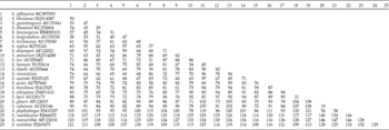
The ITS rDNA regions, flanked by primers 18S and 26S, of S. jeffreyense n. sp. (KC897093) are characterized by the sequence length of 1050 base pairs (ITS1 = 278, ITS2 = 323). Pairwise distances between closely related species (table 2) show that S. jeffreyense n. sp. differs the least from S. khoisanae (50 bp), Steinernema guangdongense (53 bp) and Steinernema dharanaii Kulkarni, Rozvi, Kumar, Paunikar & Mishra, Reference Kulkarni, Rizvi, Kumar, Paunikar and Mishra2012 (54 bp), respectively, in the ITS regions. The new species is most divergent from Steinernema scarabaei Stock & Koppenhöfer, 2003, from which it differs in 121 aligned positions.
Estimates of evolutionary divergence between sequences of the ITS region of S. jeffreyense n. sp. and representatives of Steinernema in Clade V and the glaseri-group.

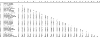
The D2D3 region of the 28S rDNA gene (KP164866) of S. jeffreyense n. sp., flanked by the primers D2F and 536, is characterized by a sequence length of 586 bp. Pairwise distances between closely related species (table 3) show that S. jeffreyense n. sp. differs least from S. innovationi (5 bp), S. tophus (6 bp) and S. khoisanae (6 bp), respectively, in the D2D3 region. The new species is most divergent from S. xueshanense Mráček, Qi-zhi, Nguyen, 2009, from which it differs in 37 aligned positions.
Pairwise distance matrix of the 28 rDNA (inclusive of D2D3 domain) S. jefferyense n. sp. and for representative species in the glaseri-group and Clade V.

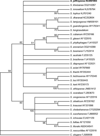
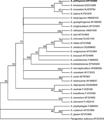
Phylogenetic relationships of S. jeffreyense n. sp. with closely related species inferred from ITS rRNA sequences, by means of the maximum parsimony method, are represented in fig. 5. In the consensus tree, S. jeffreyense n. sp. occupies a basal position in the glaseri group, but it does not cluster together with any of its closest relatives with sufficient bootstrap support to be significant.
Phylogenetic relationship of Steinernema jeffreyense n. sp., with 31 species of Steinernema, based on analysis of the ITS rDNA of the 18S gene, using the maximum parsimony method. The bootstrap consensus tree inferred from 1000 replicates is taken to represent the evolutionary history of the taxa analysed. The percentage of replicate trees in which the associated taxa clustered together in the bootstrap test is shown next to the branches.

Phylogenetic relationships of S. jeffreyense n. sp. with closely related species inferred from the D2D3 region of the 28S rDNA gene sequences, by means of the maximum parsimony method, are represented in fig. 6. In the consensus tree, S. jeffreyense n. sp. forms a monophyletic group with S. innovationi, S. tophus and S. khoisanae, with adequate bootstrap support (89).
Phylogenetic relationship of Steinernema jeffreyense n. sp. with 27 species of Steinernema based on the analysis of the D2D3 region of the 28S rDNA gene, using the maximum parsimony method. The bootstrap consensus tree inferred from 1000 replicates is taken to represent the evolutionary history of the taxa analysed. The percentage of replicate trees in which the associated taxa clustered together in the bootstrap test is shown next to the branches.

From these results, the conclusion can be drawn that the morphological and molecular characteristics that were found were sufficient to allow S. jeffreyense n. sp. to be regarded as a new species.
Diagnosis and relationships
Steinernema jeffreyense n. sp. can be distinguished from other Steinernema species by means of a combination of morphological and morphometric characteristics, which indicate that it belongs to the arenarium–glaseri–karii–longicaudatum group (glaseri-group). Nematodes in this group are characterized by the largest third-stage IJ, having an average total body length that is usually >1000 μm, as well as eight ridges in the midbody region (fig. 4). The IJ of S. jeffreyense n. sp. has a mean body length of 926 μm, which is shorter than that of molecularly closely related species in Clade V (Spiridonov et al., Reference Spiridonov, Reid, Podrucka, Subbotin and Moens2004), except for Steinernema lamjungense Khatri-Chhetri, Waeyenberge, Spiridonov, Manadhar & Moens, Reference Khatri-Chhetri, Waeyenberge, Spiridonov, Manadhar and Moens2011, with an average length of 832 (690–950) μm (table 4). Phylogenetic analysis confirmed the close relationship of S. jeffreyense n. sp. to Clade V, which includes species of the glaseri-group (Nguyen et al., Reference Nguyen, Hunt, Mráček, Nguyen and Hunt2007). Within this clade, the new species is closely related to the three South African Steinernema species: S. innovationi (Çimen et al., Reference Çimen, Lee, Hatting, Hazir and Stock2014a); S. khoisanae (Nguyen et al., Reference Nguyen, Malan and Gozel2006) and S. tophus (Çimen et al., Reference Çimen, Lee, Hazir and Stock2014b). However, S. jeffreyense n. sp. differs mainly from the three previously described South African species in that the vulva of the female has an asymmetrical protuberance, and forward-directed double-flapped epiptygmata (figs 1C, 3C).
Comparative morphometrics of third-stage infective juveniles of Steinernema jeffreyense n. sp. (in bold) and related Steinernema spp. (in descendig order of body length). Measurements are in μm and in the form: mean (range).

a Abreviations as in table 1.
b After the original authors except S. kraussei after Mráček (Reference Mráček1994). – Measurements not available.
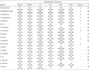
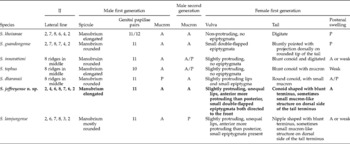
Males of S. jeffreyense n. sp. can be distinguished from the males of S. khoisanae by a D% of 47 (34–68) μm vs. 85 (71–99) μm (table 5). In S. khoisanae, the number of pairs of genital papillae may differ between 11 and 12 pairs. However, in the new species, a constant number of 11 were observed in all specimens examined (table 6). Females of S. jeffreyense n. sp. have a conoid-shaped tail with a blunt terminus (fig. 1D), with a small mucron-like structure on the dorsal side of the tail terminus, while the first-generation female tail of S. khoisanae is digitate. First-generation females of the new species were found to lack a postanal swelling, which is present in the females of S. khoisanae n. sp. (table 6).
Comparative morphometrics of first-generation males of Steinernema jeffreyense n. sp. (in bold) and related Steinernema spp. (in descending order of spicule length). Measurements are in μm and in the form: mean (range).

a Abreviations as in table 1.
b After the original authors except S. kraussei after Mráček (Reference Mráček1994). – Measurements not available. A, absent.
Comparative morphology of Steinenernema jeffreyense n. sp. (bold) and representatives in Glade V (glaseri-group).

A, absent; P, present.
Steinernema jeffreyense n. sp. IJs differ from S. tophus IJs in the length of the oesophagus, being 133 (116–149) μm vs. 146 (134–156) μm, respectively, and in the length of the tail, which is longer in the new species, being 81 (50–96) μm vs. 70 (58–80) μm (table 4). Additionally, the E% of the IJs of S. jeffreyense n. sp. differs from that of S. tophus IJs, being 109 (86–160) vs. 132 (120–156) (table 4). The first-generation male of S. jeffreyense n. sp. differs from that of S. tophus, having D% 47 (34–68) vs. 92 (71–103) and with the SW% being 215 (171–295) vs. 120 (100–160) (table 5). The male S. jeffreyense n. sp. has 11 paired caudal papillae, whereas that of S. tophus has 10. No postanal swelling is present in S. jeffreyense n. sp., whereas, in the case of S. tophus, a weak postanal swelling is noted (table 6).
The IJ of S. jeffreyense n. sp. differs from that of S. innovationi in the length of the oesophagus, being 133 (116–149) μm vs. 150 (142–160) μm, and in the D% being 66 (57–85) vs. 58 (54–63) (table 4). Additionally, the first-generation male of S. jeffreyense n. sp. differs from that of S. innovationi, in that the D% is 47 (34–68) vs. 82 (67–95), and the SW% is 215 (171–295) vs. 140 (110–190) (table 5). Whereas the manubrium of the spicule of S. jeffreyense n. sp. is elongated, the manubrium of S. innovationi is rounded (table 6). The second-generation male of S. jeffreyense n. sp. has no mucron, while in the case of S. innovationi, the mucron can be either absent or present (table 6).
Steinernema jeffreyense n. sp. IJs can be distinguished from those of S. guangdongense (Qiu et al., Reference Qiu, Fang, Zhou, Pang and Nguyen2004) by the presence of a shorter tail, being 81 (50–96) μm vs. 91 (82–103) μm, with the D% being 66 (57–85) vs. 59 (54–65), and the E% being 109 (86–169) vs. 88 (74–100). Males of S. jeffreyense n. sp. can be discriminated from those of S. guangdongense by the values of SW% 215 (171–295) vs. 175 (152–216), respectively (table 5).
The excretory pore of the IJ of S. jeffreyense n. sp. is more posteriorly located than is that of S. lamjungense, being 87 (78–107) μm vs. 68 (61–73) μm, respectively (Khatri-Chhetri et al., Reference Khatri-Chhetri, Waeyenberge, Spiridonov, Manadhar and Moens2011), with differences in the values of D% being 66 (57–85) vs. 54 (48–63), and in those of E%, being 109 (86–169) vs. 79 (69–97) (table 4). Males of the new species differ from those of S. lamjungense, in terms of the values of D% 47 (34–68) vs. 61 (44–91), and with the SW% being 215 (171–295) vs. 173 (151–193) (table 5). Furthermore, the morphology of the spicules in S. jeffreyense n. sp. is different than that in S. lamjungense. In particular, the shape of the manubrium in S. lamjungense is usually round, compared to that of S. jeffreyense n. sp., which is elongated (table 6).
The IJ of S. jeffreyense n. sp. differs from the IJ of S. dharanaii in the position of the excretory pore, which is more posterior – 87 (78–107) μm vs. 80 (70–85) μm, with the D% being 66 (57–85) vs. 56 (53–60), and the E% being 109 (86–169) vs. 85 (84–89) (table 4) (Kulkarni et al., Reference Kulkarni, Rizvi, Kumar, Paunikar and Mishra2012). The length of the spicule of S. jeffreyense n. sp. is longer than that of S. dharanaii, being 87 (79–95) μm vs. 70 (60–81) μm and the length of the gubernaculum is 57 (51–61) μm vs. 40 (38–42) μm, with the D% being 47 (34–69) vs. 74 (64–83), and the GS% being 65 (61–71) vs. 55 (50–60). The females of S. jeffreyense n. sp. do not have a postanal swelling, while in the case of S. dharanaii, the swelling can be either absent or present (table 6).
Financial support
The authors wish to thank the South African Apple and Pear Association (SAPPA), and the Technology and Human Resources for Industry Programme (THRIP–TP2011060100026), for funding.
Conflict of interest
None.