Introduction
The wasp family Braconidae (Hymenoptera) is mainly represented by parasitoid species of larvae of various insect orders (Yu et al. Reference Yu, van Achterberg and Horstman2012; Quicke Reference Quicke2015). Phytophagy, however, has been confirmed to have secondarily evolved in species of three braconid subfamilies: Mesostoinae, Braconinae, and Doryctinae (Wharton and Hanson Reference Wharton and Hanson2005). In the Doryctinae, molecular phylogenetic studies have revealed that gall association, including gall formation but probably also parasitoidism and inquilinism, is restricted to a single clade composed of at least nine morphologically disparate Neotropical genera (Zaldívar-Riverón et al. Reference Zaldívar-Riverón, Belokobylskij, Leon-Regagnon, Martínez, Briceño and Quicke2007, Reference Zaldívar-Riverón, Belokobylskij, Leon-Regagnon, Briceño and Quicke2008, Reference Zaldívar-Riverón, Martínez, Belokobylskij, Pedraza-Lara, Shaw, Hanson and Varela-Hernández2014). Phytophagy in this subfamily has so far only been confirmed in one species of Monitoriella Edqvist and two of Allorhogas Gahan (de Macêdo and Monteiro Reference de Macêdo and Monteiro1989; Infante et al. Reference Infante, Hanson and Wharton1995; Badenes-Pérez and Johnson Reference Badenes-Perez and Johnson2007). Nevertheless, indirect observations suggest that this condition occurs in all species of the former genus, in a number of the second, as well as in members of Labania, Ficobolus Martínez, Belokobylskij, and Zaldívar-Riverón, Psenobolus Reinhard, Sabinita Belokobylskij, Zaldívar-Riverón, and Martínez, and the monotypic Mononeuron Fischer (Zaldívar-Riverón et al. Reference Zaldívar-Riverón, Martínez, Belokobylskij, Pedraza-Lara, Shaw, Hanson and Varela-Hernández2014).
With 45 currently described and an undetermined number of undescribed species, Allorhogas is by far the most speciose gall-associated doryctine genus and perhaps one of the most speciose genera of the subfamily in the Neotropics. Members of Allorhogas have been associated with eight different host-plant families, though whether they are gall inducers, parasitoids, or inquilines still needs to be assessed in most cases (Zaldívar-Riverón et al. Reference Zaldívar-Riverón, Martínez, Belokobylskij, Pedraza-Lara, Shaw, Hanson and Varela-Hernández2014). To date, all known and presumed records of phytophagy in the genus are by gall induction of seeds (Wharton and Hanson Reference Wharton and Hanson2005; Zaldívar-Riverón et al. Reference Zaldívar-Riverón, Martínez, Belokobylskij, Pedraza-Lara, Shaw, Hanson and Varela-Hernández2014). Moreover, other species of Allorhogas have been reared from Cecidomyiidae (Diptera) and Cynipidae (Hymenoptera) galls (Gahan Reference Gahan1912; Penteado-Dias and Carvalho Reference Penteado-Dias and Carvalho de2008; Martínez and Zaldívar-Riverón Reference Martínez and Zaldívar-Riverón2012), and thus they are suspected to be parasitoids of gallers or inquilines.
A recent molecular phylogenetic study of the members of the gall-associated doryctine genera recovered Allorhogas as polyphyletic, with some of its examined species appearing more related to members of other genera, though the relationships involved had low support (Zaldívar-Riverón et al. Reference Zaldívar-Riverón, Martínez, Belokobylskij, Pedraza-Lara, Shaw, Hanson and Varela-Hernández2014). This evidence suggests the necessity of performing further molecular phylogenetic and taxonomic studies that clarify the relationships and classification of this morphologically conserved group.
Based on the relationships obtained in the above phylogenetic study, it was suggested that the adaptations for attacking different gall-forming taxa on various unrelated plant families probably promoted the current species diversification in Allorhogas, presumably resulting in an independent origin of phytophagy on at least three plant groups. However, in order to confirm this evolutionary hypothesis, it is first necessary to investigate the host plant association and natural history (phytophagy, parasitoidism, inquilinism) of additional species.
In this work, we describe nine species of Allorhogas from Brazil and Costa Rica, provide their host plant records and give detailed information for some of them showing that their larvae possibly are phytophagous feeding in seeds. The described species were reared from five different host plant families, two of which are reported for the first time (Asteraceae, Myrtaceae). Moreover, one of the newly described species had been previously confirmed to feed on seeds of the invasive Miconia calvescens de Candolle (Melastomataceae), representing the first case of a braconid that could be employed as a biological control agent of an exotic weed (Badenes-Perez and Johnson Reference Badenes-Perez and Johnson2007). The information provided here considerably expands the biological knowledge of Allorhogas, and will serve as a basis for further studies that seek to investigate the evolution of phytophagy in the group.
Material and methods
Examined material
The type material and specimens of the additional species examined in this study are deposited in the Colección Nacional de Insectos, Instituto de Biología, Universidad Nacional Autónoma de México (IB-UNAM), Mexico City, Mexico; Coleção de Invertebrados do Instituto Nacional de Pesquisas da Amazônia (INPA), Manaus, Brazil; Museo Argentino de Ciencias Naturales “Bernardino Rivadavia”, Buenos Aires, Argentina (MACN); and Museo de Zoología, Escuela de Biología, Universidad de Costa Rica, San Pedro Montes de Oca, Costa Rica (MZUCR). All the examined specimens were identified to genus level using the Marsh (Reference Marsh2002) key.
The terminology employed for the morphological characters follows Sharkey and Wharton (Reference Sharkey and Wharton1997), and terms for surface sculpture follow Harris (Reference Harris1979). Also, the term precoxal sulcus replaces the term sternaulus according to Wharton (Reference Wharton2006). Digital colour images were taken at the Universidad Nacional Autónoma de México with a Leica Z16 APO-A stereoscopic microscope, a Leica DFC295/DFC290 HD camera, and the Leica Application Suite programme (Leica, Wetzlar, Germany). Digital scanning electron microscope (SEM) images were taken with a Philip xl30 electron scanning electron microscope (Philips, Eindhoven, The Netherlands) at the Museo Argentino de Ciencias Naturales “Bernardino Rivadavia”, with the specimens previously being coated with gold–palladium using a Thermo VG Scientific SC7620 coater (Thermo Fisher Scientific, Walthman, Massachusetts, United States of America).
DNA sequences of a fragment (630 base pairs) belonging to the cytochrome oxidase I (COI) mitochondrial DNA gene were generated for five and two specimens belonging to Allorhogas granivorus new species and A. minimus Centrella and Shaw, respectively (GenBank accession numbers MG199221-25 and MG199226-27, respectively). Genomic DNA was extracted with the EZ-10 Spin Column Genomic DNA Minipreps Kit (animal sample) (Biobasic, Markham, Ontario, Canada) using a non-destructive technique. In this technique, the entire specimen is digested overnight in 100 µL of ACL buffer and 20 µL of proteinase k. The supernatant was then transferred to the kit’s column and the rest of the process was carried out using the manufacturer’s instructions. The amplification protocol employed was the one described in Zaldívar-Riverón et al. (Reference Zaldívar-Riverón, Martínez, Belokobylskij, Pedraza-Lara, Shaw, Hanson and Varela-Hernández2014). Unpurified polymerase chain reaction products were sent for sequencing to the DNA sequencing unit at the Universidad Nacional Autónoma de México. Uncorrected pairwise distances were obtained among the sequenced specimens using the programme PAUP version 4.0a151 (Swofford Reference Swofford2003).
Allorhogas clidemiae Martínez and Zaldívar-Riverón, new species
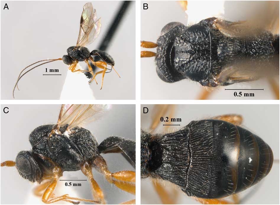
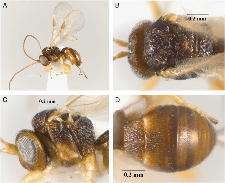
(Fig. 1A–D).
Allorhogas clidemiae new species (female, holotype): A, habitus, lateral, view; B, head and mesosoma, dorsal view; C, head and mesosoma, lateral view; D, metasoma, dorsal view.
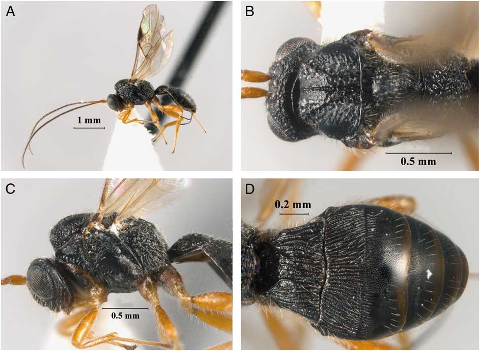
Diagnosis. This species is morphologically similar to A. breviarius Marsh, sharing a similar body colour and sculpture. Allorhogas clidemiae mainly differs from the latter species by having the third tergite costate-coriaceous on basal half and coriaceous on apical half (Fig. 1D) (coriaceous basally and smooth apically in A. breviarius), and entirely coriaceous metasoma beyond the third tergite (Fig. 1D) (smooth in A. breviarius).
Description. Holotype female. Body size 2.6 mm (Fig. 1A), forewing 2.6 mm. Colour: body colour black; scape, pedicel, and legs honey yellow; flagellum dark brown, turning darker apically. Metacoxa dark brown. Wings hyaline, stigma brown, veins light brown. Ovipositor sheaths dark brown to black.
Head: distinctly transverse in dorsal view, 2.0 times wider than its median length (Fig. 1B), and 1.3 times wider than high. Occipital carina complete and reaching hypostomal carina; shortest distance between inner margins of posterior ocelli (POL) 1.2 times longer than ocellar diameter, the maximum diameter of lateral ocellus (OD), 0.6 times shortest distance between outer margin of posterior ocellus and eye (OOL). Face coarsely rugulose-coriaceous; frons, vertex, and gena acinose-coriaceous; frons excavation distinct, without sharp lateral margins. Eye 1.4 times longer than wide; eye width 2.1 times longer than temple in dorsal view. Malar space 0.3 times eye height and as long as width of hypoclypeal depression; mandibles bidentate. Antenna with 25 flagellomeres, first flagellomere about 5.0 times longer than wide, 1.4 times longer than second flagellomere.
Mesosoma: 1.5 times longer than high (Fig. 1C) and 1.6 times longer than wide. Pronotal collar short, not visible in dorsal view; pronotal furrow scrobiculate. Mesoscutum distinctly transverse in dorsal view, its median length 0.4 times its width; median lobe of mesoscutum with a median, rugulose furrow; mesoscutal lobes acinose-coriaceous with extensive rugose-areas surrounding notauli, Notauli present, complete, and rugose-scrobiculate, finishing in a posterior rugose median area; scutellum slightly convex, coriaceous; prescutellar furrow with five transverse carinae. Propodeum entirely rugose-areolate, including basal areas delimited by diverging carinae (Fig. 1B). Mesopleuron mainly coriaceous; subalar sulcus rugose-scrobiculate; precoxal sulcus coriaceous and scrobiculate, 0.6 times as long as mesopleuron.
Wings: forewing 2.7 times longer than wide. Pterostigma 2.8 times as long as wide and 0.8 times as long as R. Vein r 0.5 times as long as 3RSa, 0.2 times as long as 3RSb, and 0.7 times as long as r-m. Vein m-cu almost interstitial with 2RS, vein RS+Mb not distinguishable. Hindwing vein M+CU 0.7 times as long as 1 M, m-cu slightly curved towards wing apex.
Legs: protibia with row of spines along anterior margin. Metacoxa with a small but distinct basoventral tooth. Metafemur 3.3 times longer than wide.
Metasoma: first tergite wider than long, 0.7 times as long as apical width, costate-coriaceous, anterior area smooth and delimited by transverse carina (Fig. 1D). Second tergite costate-coriaceous; suture between second and third tergites distinct; third tergite costate-coriaceous on basal fourth, remaining area entirely coriaceous. Remaining tergites entirely coriaceous. Ovipositor about 0.5 times as long as metasoma.
Variation. Body size 2.6–3.0 mm. Prescutellar furrow with four to five transverse carinae.
Male. Similar to female. Body size 2.2–2.6 mm. Antenna with 22–24 flagellomeres. Metafemur swollen, about 2.6 times longer than wide.
Etymology. The name of this species refers to the host plant genus from which it was reared.
Material examined. Holotype (INPA): female, Brazil, Minas Gerais, Guaraciaba, galled fruits of Clidemia hirta, 21.viii.2015; initial source of colony Allorhogas Vulcão Zuacantin. Paratypes: three females (2 IB-UNAM, 1 MACN), five males (2 IB-UNAM, 1 INPA, 2 MACN), same data as holotype.
Natural history. The type specimens were reared from galled fruits of Clidemia hirta (Linnaeus) Don (Melastomataceae). Because other Allorhogas species are known to be phytophagous in fruits of other Melastomataceae, in particular Conostegia Don (Chavarría et al. Reference Chavarría, Hanson, Marsh and Shaw2009) and Miconia Ruiz and Pavón (Badenes-Pérez and Johnson, Reference Badenes-Perez and Johnson2007), it is likely that A. clidemiae is also phytophagous. However, this remains to be determined.
Allorhogas granivorus Zaldívar-Riverón and Martínez, new species
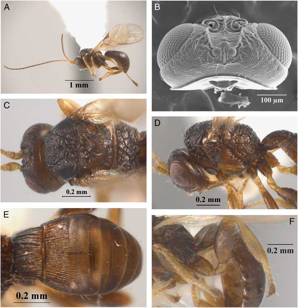
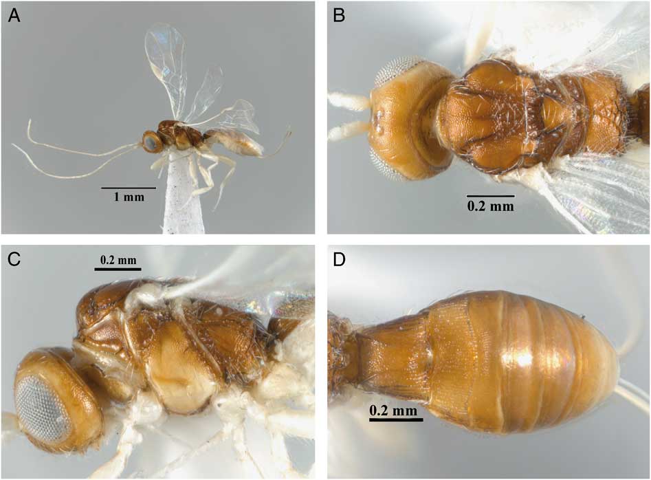
(Fig. 2A–F).
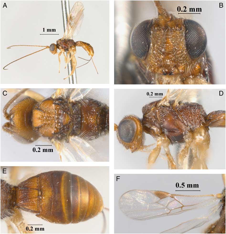
Allorhogas granivorus new species (female, holotype): A, habitus, lateral, view; B, head, dorsal view; C, head and mesosoma, dorsal view; D, head and mesosoma, lateral view; E, metasoma, dorsal view. Male, paratype: F, metafemur, lateral view.
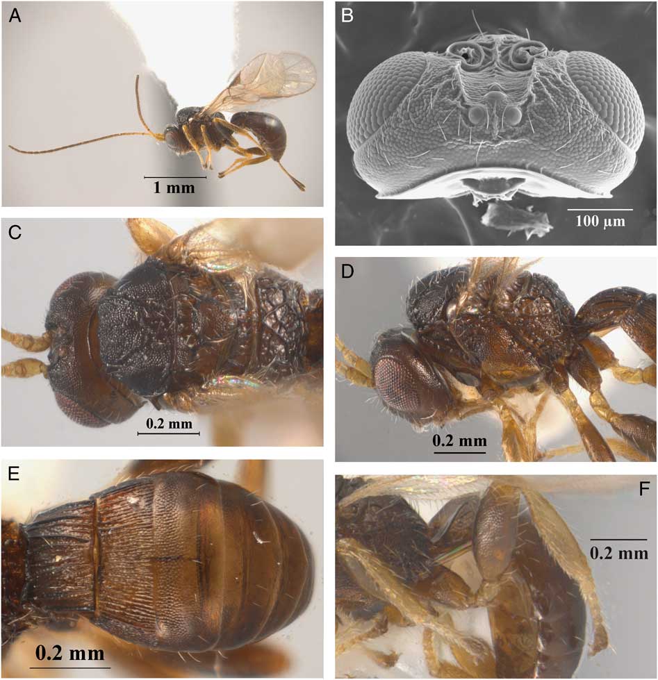
Diagnosis. This species is morphologically similar to A. minimus Centrella and Shaw, which was described from Costa Rica. These two species share a distinctly compact mesosoma (about 1.3 times longer than high) and an almost entirely vertical or oblique propodeum. However, the new species can be morphologically distinguished from A. minimus by its larger body size (1.6–2.0 mm; 0.8–1.4 mm in A. minimus); third tergite slightly striate on basal third, remaining area and remaining tergites mostly coriaceous (mostly smooth in A. minimus); and frons excavation deep, delimited by sharp lateral margins (Fig. 2B) (distinct but not defined by sharp lateral margins in A. minimus). Allorhogas granivorus also differs from A. minimus by its association with Miconia calvescens [M. longifolia (Aublet) de Candolle in A. minimus]. In addition, these two species have a considerable genetic divergence for the barcoding locus (658 base pairs of the cytochrome oxidase I mtDNA gene; interspecific distance=8.0–10.2%; intraspecific distance for within species=0.0–1.6%).
Description. Holotype female. Body size 1.9 mm (Fig. 2A), forewing 1.9 mm. Colour: almost uniformly brown to dark brown; lower part of gena, mandibles, clypeus, area surrounding precoxal sulcus, and legs honey yellow; metafemur and metatibia brown; antenna honey yellow basally, turning brown apically; wings hyaline, stigma and veins brown; ovipositor sheaths brown.
Head: distinctly transverse in dorsal view, 2.0 times wider than its median length (Fig. 2B–C), and 1.7 times wider than high. Occipital carina complete and reaching hypostomal carina; POL 1.3 times longer than OD, 0.4 times OOL (Fig. 2B). Frons, vertex, gena, and face coriaceous; frons excavation deep, delimited by sharp lateral margins. Eye 1.4 times longer than wide; eye width 2.0 times longer than temple in dorsal view. Malar space 0.4 times eye height and as long as width of hypoclypeal depression; mandibles bidentate. Antenna with 19 flagellomeres, first flagellomere about 5.0 times longer than wide, 1.1 times longer than second.
Mesosoma: distinctly short and compact, 1.3 times longer than high (Fig. 2D) and 1.3 times longer than wide. Pronotal collar short, not visible in dorsal view; pronotal furrow scrobiculate. Mesoscutum distinctly transverse in dorsal view, median length 0.6 times its width; mesoscutal lobes coriaceous, median mesoscutal lobe with median longitudinal rugosity. Notauli present, complete, and scrobiculate, finishing in a posterior rugose median area; scutellum coriaceous, prescutellar furrow with three transverse carinae. Propodeum rugose-areolate, with two distinct diverging carinae, almost entirely vertical or oblique (lateral view), dorsal surface short (Fig. 2C). Mesopleuron entirely coriaceous; subalar sulcus scrobiculate; precoxal sulcus coriaceous, 0.6 times as long as mesopleuron.
Wings: forewing 2.9 times longer than wide. Pterostigma 2.5 times as long as wide and 0.8 times as long as R. Vein r 0.4 times as long as 3RSa, 0.2 times as long as 3RSb, and 0.6 times as long as r-m. Vein m-cu interstitial with 2RS, vein RS+Mb not distinguishable. Hindwing vein M+CU 0.7 times as long as 1 M, m-cu slightly curved towards wing apex.
Legs: protibia with row of spines along anterior margin. Metacoxa with distinct basoventral tooth. Metafemur 3.3 times longer than wide.
Metasoma: first tergite wider than long, 0.8 times as long as apical width, costate-coriaceous, anterior area smooth and delimited by transverse carina (Fig. 2E). Second tergite costate-coriaceous. Third tergite weakly costulate-coriaceous on basal third, remaining area coriaceous. Remaining tergites entirely slightly coriaceous. Ovipositor about 0.5 times as long as metasoma (Fig. 2A).
Variation. Body size 1.8–2.0 mm. In some females honey yellow areas brighter. Antenna with 19–21 flagellomeres. Prescutellar furrow with three to five transverse carinae.
Male. Similar to female. Body size 1.6–1.8 mm. Antenna with 18–19 flagellomeres. Metafemur not considerably swollen, 3.1 times longer than wide (Fig. 2F).
Etymology. The name of this species comes from the Latin word granivorus, referring to the spermophagous natural history of this species.
Material examined. Holotype (INPA): female, Brazil: Minas Gerais, Dionisio, 28.viii–5.ix.2013, ex. galled fruits Miconia calvescens, seed code: 6F-19M. Paratypes: three females (2 IB-UNAM, 1 MACN), six males (3 IB-UNAM, 1 INPA, 2 MACN), same data as holotype, DNA voucher numbers CNIN2180-81, CNIN2180-83, CNIN2180-85; GenBank accession numbers MG199221, MG199222, MG199224, MG199225. One female (IB-UNAM), one male (INPA), Brazil: Rio de Janeiro, Cachoeiras de Macacu, 29.viii–2.ix.2013, ex. galled fruits Miconia calvescens, seed code: 2F, 3M. Three females (IB-UNAM), Brazil: Dionisio, D1.3.1, 24.viii.2013, ex. galled fruits Miconia calvescens, seed code: 3F, DNA voucher numbers CNIN2183, CNIN2185. Three males (2 IB-UNAM, 1 INPA), Brazil: Dionisio, D1.4, 26.viii.2013, Miconia calvescens, seed code: 2M, 2F, 1M, 3F, DNA voucher number CNIN2182, GenBank accession number MG199223.
Natural history. The natural history of A. granivorus was described in detail by Badenes-Pérez and Johnson (Reference Badenes-Perez and Johnson2007). The larvae of this species form galls in the fruits/seeds of M. calvescens, which represents a serious threat to natural ecosystems in various Pacific islands (Meyer and Florence Reference Meyer and Florence1996). Due to its host plant preference and its relatively high infestation of seeds, A. granivorus has been evaluated as a potential biological control agent of M. calvescens (Badenes-Pérez and Johnson Reference Badenes-Perez and Johnson2007)
Allorhogas mineiro Zaldívar-Riverón and Martínez, new species
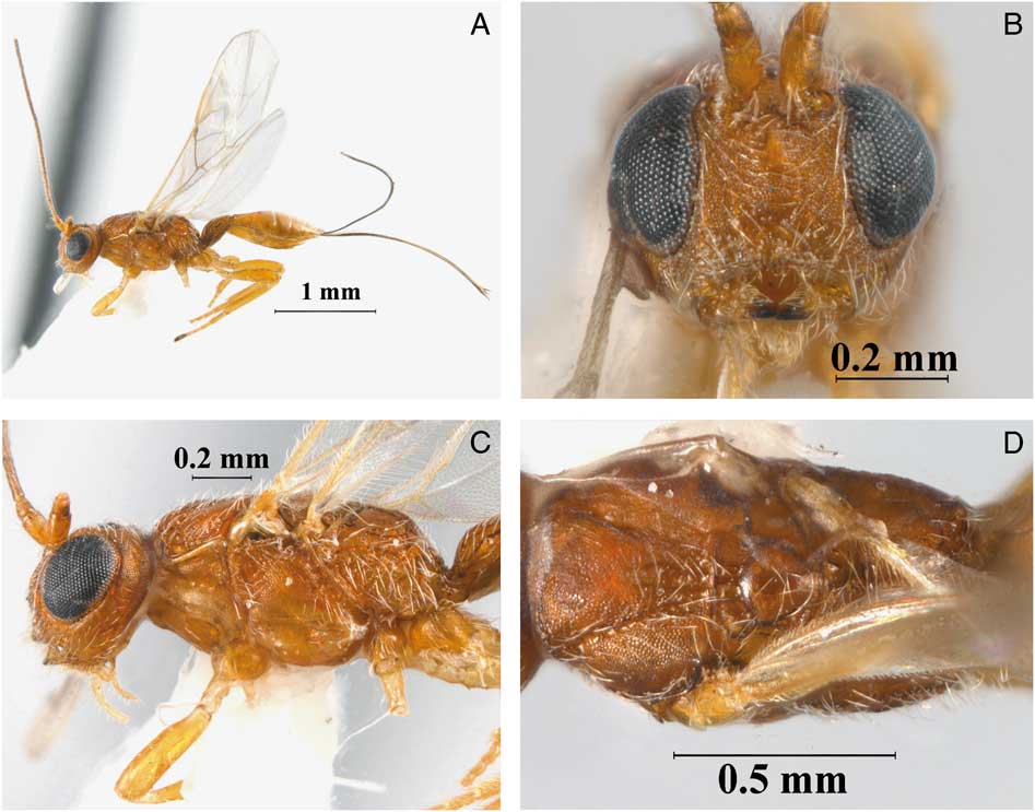
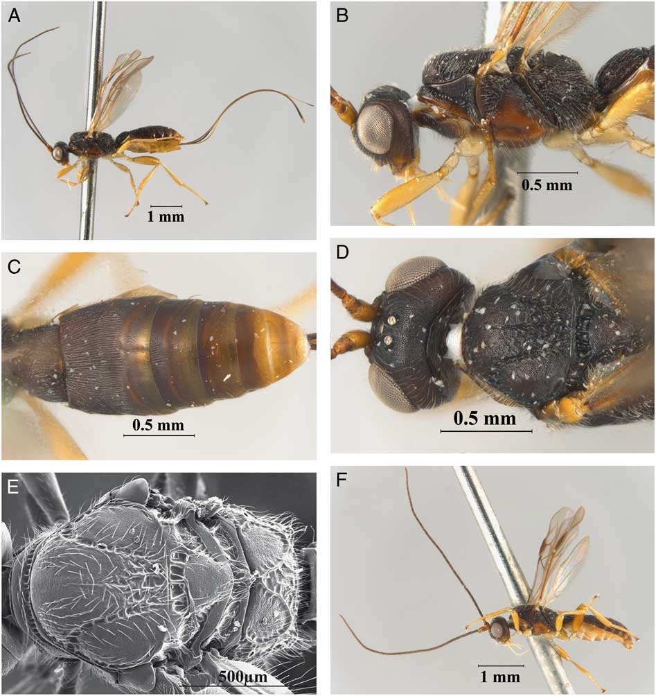
(Fig. 3A–D).
Allorhogas mineiro new species (female, holotype): A, habitus, lateral, view; B, head, frontal view; C, head and mesosoma, lateral view; D, head and mesosoma, dorsal view.
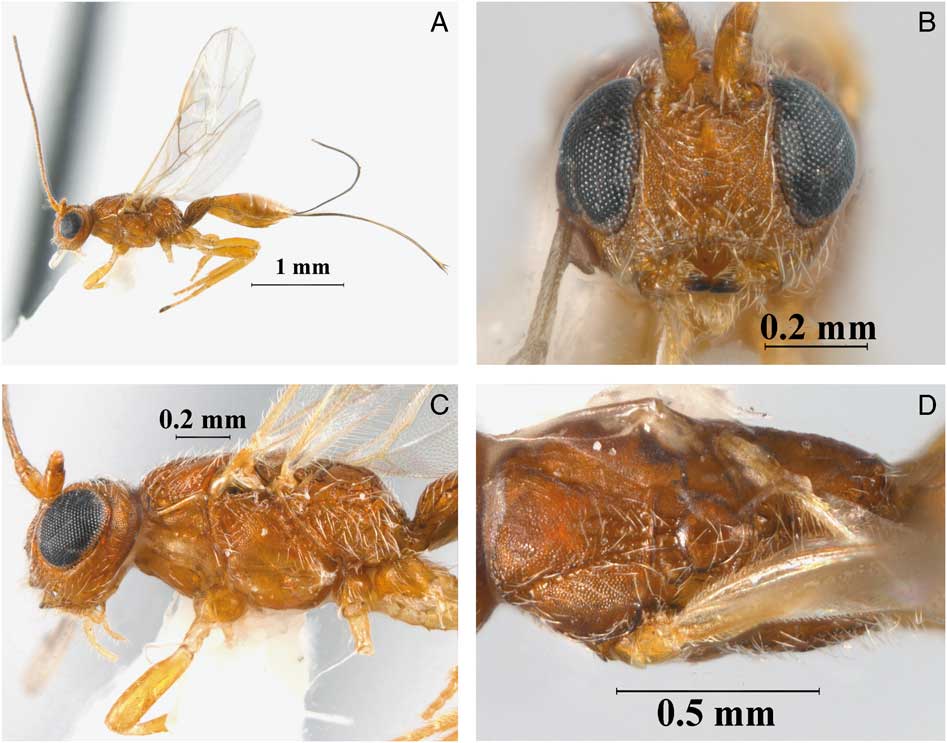
Diagnosis. This species is morphologically and biologically similar to A. spermaphagous Marsh, which was described from Rio de Janeiro, Brazil. The new species and A. spermaphagous were reared from seed pods and seeds of Stryphnodendron adstringens (Martius) Coville and S. polyphyllum Martius, respectively. These two species share an entirely, uniform yellow body colour, though they can be distinguished by the length of the ovipositor (Fig. 3A) (1.1–1.3 and about 0.7 times length of metasoma in A. mineiro and A. spermaphagous, respectively), and the mesoscutum, propodeum, and metsaoma less coarsely sculptured in A. mineiro.
Description. Holotype female. Body size 3.0 mm (Fig. 3A), forewing 2.9 mm. Colour: almost uniformly yellow (Fig. 3A), antenna dark brown; wings hyaline, stigma and veins brown to dark brown; ovipositor sheaths dark brown.
Head: transverse in dorsal view, 1.8 times wider than median length, and 1.2 times wider than high; occipital carina complete and reaching hypostomal carina; POL as long as OD, 0.4 times OOL; frons, vertex, gena, and face coriaceous; frons excavation distinct and coriaceous. Eye 1.6 times longer than wide; eye width 2.0 times longer than temple in dorsal view. Malar space 0.6 times eye height (Fig. 3C) and as long as width of hypoclypeal depression (Fig. 3B); mandibles tridentate, third basal tooth small. Antenna with 25 flagellomeres, first flagellomere about 3.2 times longer than wide, 1.1 times longer than second flagellomere.
Mesosoma: 1.7 times longer than high (Fig. 3C) and 1.8 times longer than wide; pronotal collar short, not visible in dorsal view, pronotal furrow weakly scrobiculate. Mesoscutum transverse in dorsal view, median length 0.8 times width; mesoscutal lobes coriaceous. Notauli present, complete, and weakly scrobiculate, finishing in a posterior slightly rugose median area; scutellum slightly coriaceous, prescutellar furrow with three transverse carinae. Propodeum slightly rugose-areolate, with two distinct divergent carinae, apical area above metasoma insertion slightly rugulose and enclosed by carinae. Mesopleuron entirely coriaceous; subalar sulcus scrobiculate; precoxal sulcus coriaceous, 0.8 times as long as mesopleuron.
Wings: forewing 3.5 times longer than wide. Pterostigma 3.1 times as long as wide and 0.7 times as long as R. Vein r 0.9 times as long as 3RSa, 0.2 times as long as 3RSb, and 0.9 times as long as r-m. Vein 2RS almost interstitial with m-cu, vein RS+Mb almost indistinguishable. Hindwing vein M+CU 0.8 times as long as 1 M, m-cu slightly curved towards wing apex.
Legs: protibia with row of about 16 spines along anterior margin. Metacoxa with distinct basoventral tooth. Metafemur 4.0 times longer than wide.
Metasoma: first tergite wider than long, 0.8 times as long as apical width, costate-coriaceous, anterior area smooth and delimited by transverse carina, with dorsal median area delimited by two lateral carinae. Second tergite costate-coriaceous. Third tergite costate-coriaceous medially and costulate-coriaceous laterally on basal third, remaining area smooth. Remaining tergites coriaceous basally, smooth apically. Ovipositor about as long as metasoma (Fig. 3A).
Variation. Body size 2.7–3.0 mm. Ovipositor 1.0–1.3 times longer than metasoma.
Male. Unknown.
Material examined. Holotype (INPA): female, Brazil: Minas Gerais, Itumirim, Minas Gerais site, A1C01, 21°14'42"S 44°49'43.29"W, ex. Stryphnodendron adstringens fruit 49, 3.v.2015, Juliana Tuller. Paratypes: one female (IB-UNAM), Brazil: Minas Gerais, Itumirim Minas Gerais, site A2C10, 21°13'41.98"S, 44°51'0.43"W, ex. Stryphnodendron adstringens fruit 19, 3.v.2015, Juliana Tuller. One female (IB-UNAM), Brazil: Minas Gerais, Itumirim, Minas Gerais site A2P02, 21°13'43.32"S 44°58'49.99"W, ex. Stryphnodendron adstringens, fruit 60, 3.v.2015, Juliana Tuller. One female (MACN), Brazil: Minas Gerais, Itumirim, Minas Gerais site A2F04, 21°13'41.98"S 44°51'0.43"W, ex. Stryphnodendron adstringens, fruit 5, 3.v.2015, Juliana Tuller.
Natural history. Reared from Stryphnodendron adstringens. This is a broadly distributed plant species in Brazilian savannas, also known as the Cerrado biome. Individuals can reach up to 5 m in height and the bark is used by rural people to treat several ailments. The production of new leaves and inflorescences extends from July to November, and fruit development occurs all year around (Felfili et al. Reference Felfili, Da Silva, Dias and Rezende1999). The pods and seeds harbour a rich insect community composed of 26 species, mostly herbivores and parasitoids. Herbivores attack the seeds at a rate up to 50%, and A. mineiro accounts for almost 10% (n=39) of the herbivorous individuals. No galls were observed in the fruits, the larvae feeds directly on seeds and leave them with little evidence of consumption when compared with Bruchinae (Coleoptera: Chrysomelidae) species.
Allorhogas vulgaris Zaldívar-Riverón and Martínez, new species
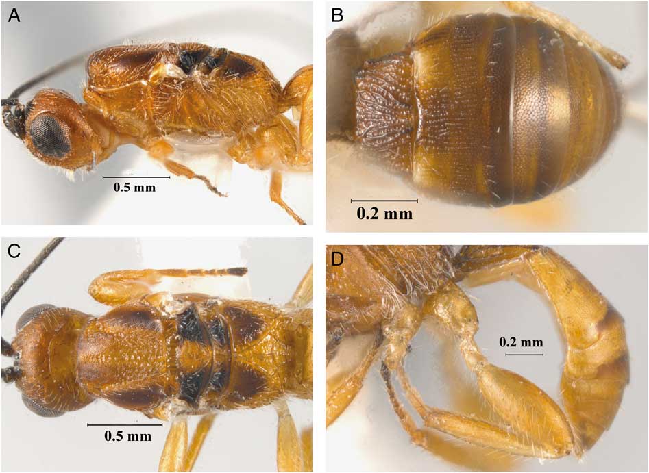
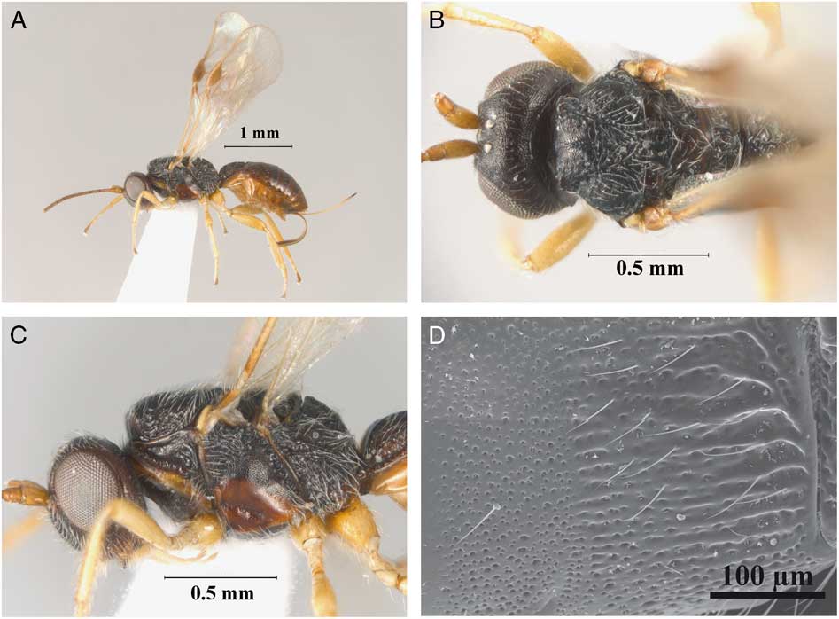
(Fig. 4A–D).
Allorhogas vulgaris new species (female, paratype; wings removed): A, head and mesosoma, lateral view; B, metasoma, dorsal view; C, head and mesosoma, dorsal view. Male, paratype: D, metafemur, lateral view.
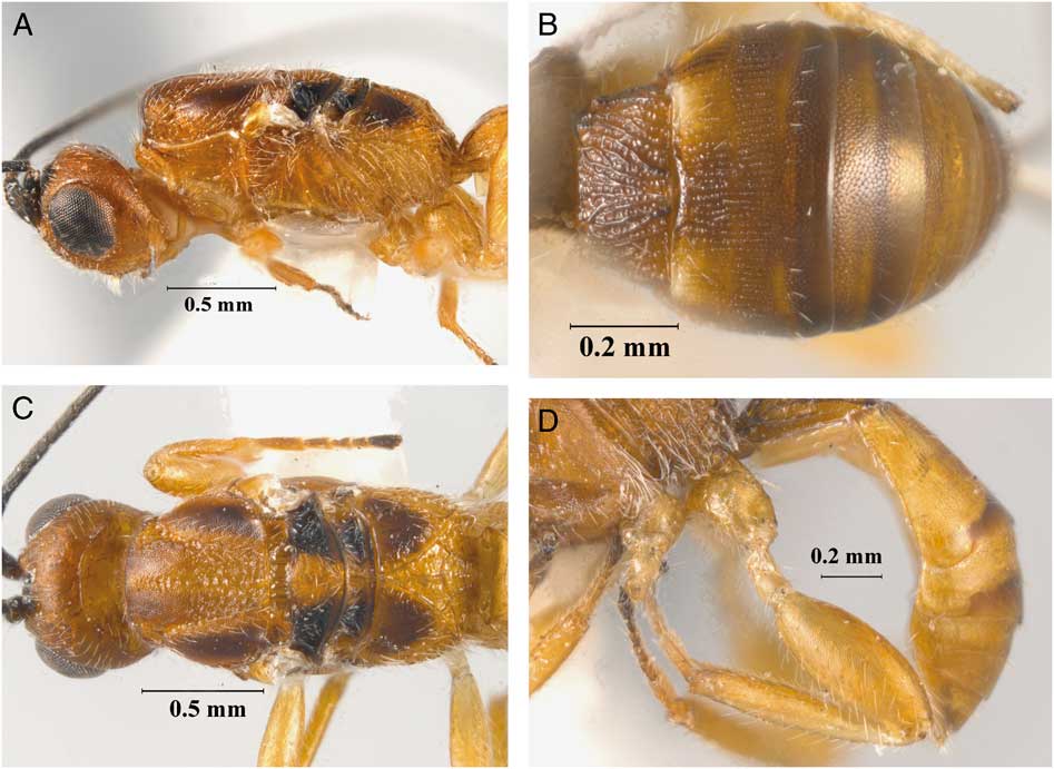
Diagnosis. This species is morphologically similar to A. spermaphagous Marsh, but it can be distinguished from the latter species by having the lateral mesonotal lobes, basal areas of propodeum, and central areas of second to fourth metasomal tergites dark brown (Fig. 4A, C) (entirely honey yellow in A. spermaphagous). Moreover, A. vulgaris has a distinct host legume (Fabaceae) species [Senegalia tenuifolia (Linnaeus) Britton and Rose; Stryphnodendron polyphyllum in A. spermaphagous].
Description. Holotype female. Body size 2.9 mm (Fig. 4A), forewing 2.6 mm. Colour: background colour honey yellow with antennae, lateral mesonotal lobes, basal areas of propodeum, central areas of second to fourth metasomal tergites, and ovipositor sheaths dark brown. Wings hyaline, stigma and veins brown.
Head: transverse in dorsal view, 1.7 times wider than median length, and 1.1 times wider than high (Fig. 4A); occipital carina complete, reaching hypostomal carina; POL as long as as OD, 0.5 times OOL; frons, vertex, gena, and face coriaceous, frons excavation distinct and coriaceous. Eye 1.2 times longer than wide; eye width 2.1 times longer than temple in dorsal view. Malar space 0.5 times eye height and as long as width of hypoclypeal depression; mandibles tridentate. Antenna with 27 flagellomeres, first flagellomere about 4.0 times longer than wide, 1.1 times longer than second flagellomere.
Mesosoma: 1.8 times longer than high (Fig. 4A) and 1.8 times longer than wide. Pronotal collar barely visible in dorsal view, pronotal furrow scrobiculate. Mesoscutum slightly transverse in dorsal view, median length 0.8 times width; median mesoscutal lobe with a median longitudinal, rugose groove; mesoscutal lobes coriaceous. Notauli present, complete, and scrobiculate, finishing in a posterior rugose median area; scutellum coriaceous, prescutellar furrow with three transverse carinae. Propodeum rugose-areolate, with basal areas coriaceous and well delimited. Mesopleuron entirely coriaceous; subalar sulcus scrobiculate; precoxal sulcus coriaceous, long, about 0.8 length of mesopleuron.
Wings: forewing 3.1 times longer than wide. Pterostigma 3.3 times as long as wide and 0.8 times as long as R. Vein r 0.7 times as long as 3RSa, 0.2 times as long as 3RSb, and 0.8 times as long as r-m. Vein m-cu arising slightly basal to 2RS, thus vein RS+Mb present. Hindwing vein M+CU 0.6 times as long as 1 M, m-cu slightly curved towards wing apex.
Legs: protibia with row of 11 spines along anterior margin. Metacoxa with distinct basoventral tooth. Metafemur 3.1 times longer than wide.
Metasoma: first tergite wider than long, 0.8 times as long as apical width, costate-coriaceous, anterior area smooth and delimited by transverse carina, with dorsal median area delimited by two lateral carinae (Fig. 4B). Second tergite costate-coriaceous. Third tergite costate-coriaceous on basal third, remaining area slightly coriaceous. Remaining tergites slightly and irregularly coriaceous. Ovipositor about 0.7 times as long as metasoma.
Variation. Body size 2.9–3.1 mm. One exceptionally small female had a body size of 1.9 mm. Antenna with 25–28 flagellomeres. Prescutellar furrow with three to four transverse carinae. Two females almost uniformly honey yellow. Fourth metasomal tergite ranges from entirely coriaceous to costate basally with remaining area coriaceous.
Male. Higher proportion of males (50%) show uniformly yellow body colour. Body size 2.9 mm. Antenna with 25–27 flagellomeres. Metafemur swollen, 2.5 times longer than wide (Fig. 4D).
Etymology. The term vulgaris refers to the common appearance of this species compared with other species of Allorhogas.
Material examined. Holotype (INPA): female, Brazil, Minas Gerais, Luminarias, Minas Gerais Site Lumi 4, 21°31'5.31"S, 44°52'3.84"W, ex. Senegalia tenuifolia, in fruit number 25, 7.viii.2015, Lucas del Bianco Faria. Paratypes: seven females (3 IB-UNAM, 2 INPA, 2 MACN), same data as holotype except 4, 1, and 1 specimens in fruits number 2, 4, and 16, respectively. Six females (IB-UNAM), two males (1 IB-UNAM, 1 MACN), Brazil, Minas Gerais, Luminarias, Minas Gerais Site Lumi 1, 21°31'1.36"S, 44°53'1.78"W, ex. Senegalia tenuifolia, 7.viii.2015, Lucas Del Bianco Faria, 1, 1, 2, 1, and 1 females in fruits number 7, 12, 14, 20, and 22, respectively; 1 and 1 males in fruits number 8 and 12, respectively. Six females (IB-UNAM), Brazil, Minas Gerais, Lavras, Minas Gerais Site Lav 1, 21°18'3.46"S 44°58'0.53"W, ex. Senegalia tenuifolia, 7.viii.2015, Lucas Del Blanco Faria, 1, 2, 1, and 2 in fruits number 7, 9, 14, and 20, respectively. One female (IB-UNAM), Brazil, Minas Gerais, Luminarias, Minas Gerais Site Aero 3, 21°14'7.87''S 44°58'0.06''W, ex. Senegalia tenuifolia, in fruit number 4, 7.viii.2015, Lucas Del Bianco Faria. One female (IB-UNAM) Brazil, Minas Gerais, Luminarias, Minas Gerais Site Lumi 3, 21°31'5.31"S 44°52'3.84"W, ex. Senegalia tenuifolia, in fruit number 19, 15.vii.2015, Lucas Del Bianco Faria.
Natural history. Reared from seeds of Senegalia tenuifolia, a widely distributed plant species in South America (Queiroz Reference Queiroz2009). It is a pioneer species and can be characterised as a liana or shrub; however, the best characterisation is scandent shrub. The flowers bloom from November to January, the pods develop from January to August then they open and seeds fall to the ground. The pods and seeds support an insect community composed of 15 species, mostly herbivores and parasitoids interacting with each other. The seed feeders affect up to 24% of the seeds, with A. vulgaris accounting for 9% (n=69) of the herbivorous individuals. No galls were observed in the fruits, but instead the larvae appear to feed directly on the seeds and leave them with little evidence of consumption when compared with bruchine seed-feeding behaviour. Besides A. vulgaris, pods and seeds also harboured two bruchine species that possibly compete for resources (Maia et al. Reference Maia, Tuller and Faria2017; Silva et al. Reference Silva, Moneiro, Maia and Faria2017). In addition, an undescribed parasitoid species of Eurytoma Illiger (Hymenoptera: Eurytomidae) was also reared.

Allorhogas brevithorax Zaldívar-Riverón and Martínez, new species
(Fig. 5A–D).
Allorhogas brevithorax new species (female, holotype): A, habitus, lateral, view; B, head and mesosoma, dorsal view; C, head and mesosoma, lateral view; D, metasoma, dorsal view.
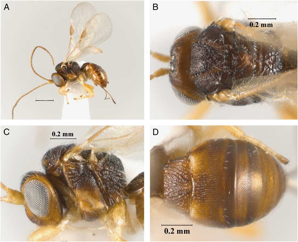
Diagnosis. Allorhogas brevithorax is morphologically similar to the group of species with short and compact mesosoma, A. argentinus sensu stricto, A. granivorus new species, A. minimus Centrella and Shaw, and A. parvus Martínez and Zaldívar-Riverón. The new species can be mainly distinguished from A. parvus by its darker background colour (Fig. 5A) (yellow in A. parvus); from A. minimus by its ovipositor as long as metasoma (Fig. 5A) (<0.5 length of metasoma and slightly shorter than metasoma in the latter two species, respectively); from A. granivorus by having a distinct frons excavation but not defined by sharp lateral margins (deep, delimited by sharp lateral margins in A. granivorus); and from the latter species and A. argentinus by having distinctly pale yellow legs (honey yellow to brown in the latter two species).
Description. Holotype female. Body size 1.6 mm (Fig. 5A), forewing 1.7 mm. Colour: body mostly brown; orbits, area surrounding sternaulus, precoxal sulcus, and antennae pale yellow to light brown; legs pale yellow; wings hyaline, stigma light brown, veins light brown to honey yellow; ovipositor sheaths brown.
Head: distinctly transverse in dorsal view, 2.0 times wider than its median length, and 1.2 times wider than high; occipital carina complete and reaching hypostomal carina; POL as long as OD, 0.3 times OOL. Frons, vertex, gena, and face coriaceous, frons excavation distinct but not defined by sharp lateral margins. Eye 1.4 times longer than wide; eye width 3.0 times longer than temple in dorsal view. Malar space 0.3 times eye height and as long as width of hypoclypeal depression; mandibles bidentate, with an additional small protuberance. Antenna with 19 flagellomeres, first flagellomere about 4.0 times longer than wide, 1.1 times longer than second flagellomere.
Mesosoma: distinctly short and compact, 1.2 times longer than high (Fig. 5C) and 1.1 times longer than wide. Pronotal collar short, not visible in dorsal view; pronotal furrow scrobiculate. Mesoscutum distinctly transverse in dorsal view, median length 0.5 times width; mesoscutal lobes coriaceous, median mesoscutal lobe with median rugosity. Notauli present, complete, and scrobiculate, finishing in a posterior slightly rugose median area; scutellum coriaceous, prescutellar furrow with four transverse carinae. Mesopleuron entirely coriaceous; subalar sulcus scrobiculate; precoxal sulcus coriaceous, about 0.5 times as long as mesopleuron. Propodeum rugose-areolate, with two distinct diverging carinae, almost entirely vertical or oblique (lateral view), dorsal surface short (Fig. 5B).
Wings: forewing 2.5 times longer than wide. Pterostigma 2.8 times as long as wide and 0.7 times as long as R. Vein r 0.7 times as long as 3RSa, 0.2 times as long as 3RSb, and 0.9 times as long as r-m. Vein m-cu interstitial with 2RS, vein RS+Mb not distinguishable. Hindwing vein M+CU 0.7 times as long as 1 M, m-cu slightly curved towards wing apex.
Legs: protibia with a row of 10 spines along anterior margin. Metacoxa with distinct basoventral tooth. Metafemur 3.6 times longer than wide.
Metasoma: first tergite wider than long, 0.7 times as long as apical width, costate-coriaceous, anterior area smooth and delimited by transverse carina (Fig. 5D). Second tergite costate-coriaceous. Third tergite costate-coriaceous on basal half, remaining area coriaceous. Remaining tergites coriaceous. Ovipositor as long as metasoma.
Variation. Body size 1.4–1.6 mm; Antenna with 19–20 flagellomeres. Prescutellar furrow with three to five transverse carinae.
Male. Similar to female. Body size 1.4 mm. Antenna with 18–19 flagellomeres. Metafemur not distinctly swollen, about 3.2 times longer than wide.
Etymology. The name of this species refers to the short and compact mesosoma of this species.
Material examined. Holotype (MZUCR): female. Costa Rica: Punta Arenas, Monte Verde, stem gall on Myrcia, vii–viii.2007, K. Nishida. Paratypes: three females (2 IB-UNAM, 1 MACN), three males (1 IB-UNAM, 1 MACN, 1 MZUCR), same data as holotype.
Comments. Allorhogas brevithorax probably is closely related to the following species, as they share a considerably short and compact mesosoma and a pronotal collar short, not visible in dorsal view: A. argentinus, A. granivorus new species, A. minimus Centrella and Marsh and A. parvus Martínez and Zaldívar-Riverón. Further phylogenetic studies within the genus will test this hypothesis.
Natural history. Reared from stem galls, elongate swellings up to 55 mm long and 5 mm in diameter (K. Nishida, Museo de Zoología, Universidad de Costa Rica, San Pedro de Montes de Oca, Costa Rica and P.E.H., unpublished), on Myrcia de Candolle (Myrtaceae). This is the first record of this plant family for Allorhogas. The trophic position of A. brevithorax in this gall is still unknown. An undescribed species of Cecidomyiidae (Diptera) forms a slightly different gall in the stems, petioles and midveins of Myrcia splendens (Swartz) de Candolle (P.E.H., unpublished), so it is possible that the same is true for the galls from which these Allorhogas emerged, but this remains to be determined.
Allorhogas pallidus Martínez and Zaldívar-Riverón, new species
(Fig. 6A–D).
Allorhogas pallidus new species (female, holotype): A, habitus, lateral, view; B, head and mesosoma, dorsal view; C, head and mesosoma, lateral view; D, metasoma, dorsal view.
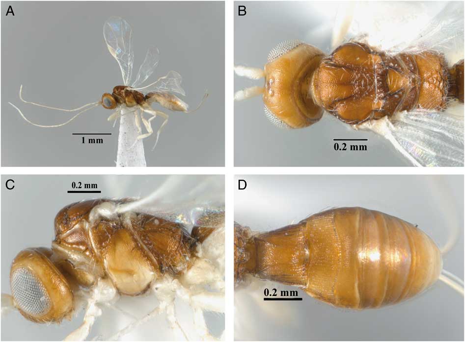
Diagnosis. Allorhogas pallidus can be distinguished from other species of Allorhogas from Costa Rica by having the palps, scape, pedicel, basal flagellomeres, lower part of mesopleuron, and legs pale white (Fig. 6A), and by its association with Palicourea crocea (Swartz) Schultes (Rubiaceae).
Description. Holotype female. Body size 2.2 mm (Fig. 6A), forewing 1.9 mm. Colour: body light brown to honey yellow; palpi, scape, pedicel, basal flagellomeres, lower part of mesopleuron, and legs pale white; wings hyaline, stigma and veins pale yellow; ovipositor light brown, ovipositor sheaths pale yellow, apex sclerotised.
Head: moderately transverse in dorsal view, 1.6 wider than median length, and 1.3 times wider than high (Fig. 6B); occipital carina complete and reaching hypostomal carina; POL as long than OD, 0.4 times OOL. Frons, vertex, gena, and face coriaceous, frons excavation shallow, not defined by sharp lateral margins. Eye 1.3 times longer than wide; eye width 2.0 times longer than temple in dorsal view. Malar space 0.4 times eye height and as long as width of hypoclypeal depression; mandibles bidentate, with additional small protuberance. Antenna with 23 flagellomeres, first flagellomere about 3.7 times longer than wide, and as long as second flagellomere.
Mesosoma: 1.6 times longer than high (Fig. 6C) and 1.7 times longer than wide. Pronotal collar short, slightly visible in dorsal view; pronotal furrow scrobiculate. Mesoscutum transverse in dorsal view, median length 0.8 times its width; mesoscutal lobes coriaceous, median mesoscutal lobe with a median rugosity on posterior half. Notauli present, complete, and scrobiculate, finishing in a posterior slightly rugose median area; scutellum coriaceous, prescutellar furrow with three transverse carinae. Mesopleuron entirely coriaceous; subalar sulcus scrobiculate; precoxal sulcus deep, coriaceous, about 0.5 times as long as mesopleuron. Propodeum rugose-areolate, with two distinct divergent carinae, basal areas coriacoeus (Fig. 6B).
Wings: forewing 2.4 times longer than wide. Pterostigma 2.5 times as long as wide and 0.8 times as long as R. Vein r 0.5 times as long as 3RSa, 0.2 times as long as 3RSb, and 0.7 times as long as r-m. Vein m-cu interstitial with 2RS, vein RS+Mb absent. Hindwing vein M+CU 0.6 times as long as 1 M, m-cu slightly curved towards wing apex.
Legs: protibia with row of seven spines along anterior margin. Metacoxa with distinct basoventral tooth. Metafemur 3.1 times longer than wide.
Metasoma: first tergite slightly wider than long, 0.9 times as long as apical width, median dorsal area largely coriaceous, costate apically; anterior area not delimited by transverse carina (Fig. 6D). Second tergite costate-coriaceous. Third tergite striate-coriaceous on basal half, remaining area slightly coriaceous. Remaining tergites slightly coriaceous to smooth. Ovipositor 0.8 times as long as metasoma (Fig. 6A).
Variation. Body size 2.1–2.2 mm.
Male. Similar to female. Metasoma pale yellow. Body size 1.7 mm. Metafemur not distinctly swollen, about 2.9 times longer than wide.
Etymology. From the Latin, pallidus, which refers to the pale colour of legs, antennae, and wing veins of this new species.
Material examined. Holotype (MZUCR): Costa Rica: Heredia, 3 km S Puerto Viejo, OTS La Selva, 100 m, 27.x.1992, Palicourea, DG018, D. Graham, seeds. Paratypes: 2 females (1 IB-UNAM, 1 MACN), 2 males (1 IB-UNAM, 1 MACN), same data as holotype. 1 female, Costa Rica, Heredia, 3 km S Puerto Viejo, OTS La Selva, 100 m, 2.x.1992, Palicourea crocea fruits (IB-UNAM).
Natural history. Reared from fruits/seeds of Palicourea crocea. It is suspected that this species is phytophagous, but this remains to be confirmed.
Allorhogas psychotria Zaldívar-Riverón and Martínez, new species
(Fig. 7A–F).
Allorhogas psychotria new species (female, holotype): A, habitus, lateral, view; B, head and mesosoma, lateral view; C, metasoma, dorsal view; D, head and mesosoma, dorsal view. Female, paratype: E, mesosoma, dorsal view. Male, paratype: F, habitus, lateral view.
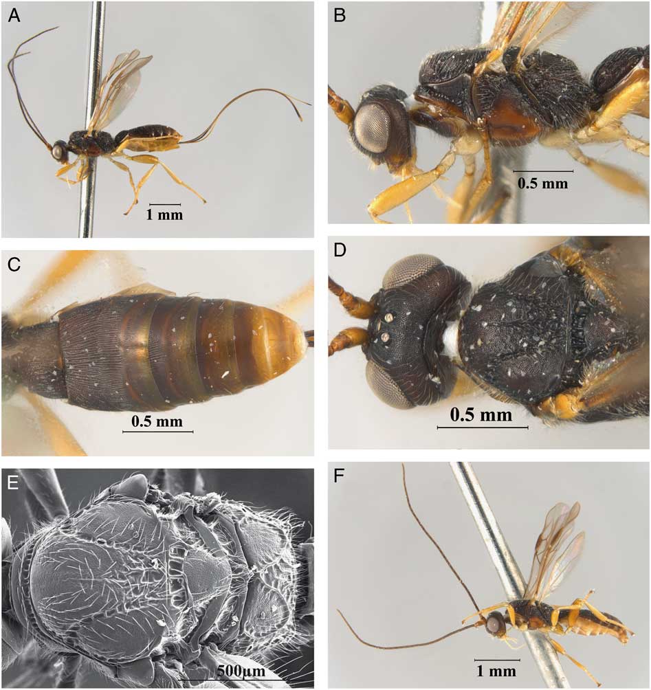
Diagnosis. Allorhogas psychotria can be distinguished from the remaining species of Allorhogas from Costa Rica by its large body size (3.2–4.1 mm) and its considerably long ovipositor (about 2.0 times longer than metasoma). It superficially resembles the species of the Doryctinae genus Barbalhoa Marsh; however, the excavated frons (not excavated in Barbalhoa), vertex coriaceous (striate in Barbalhoa), and the absence of a transverse scrobiculate groove on the third metasomal tergite clearly distinghuish it from the members of the latter genus (present in Barbalhoa).
Description. Holotype female. Body size 4.1 mm (Fig. 7A), forewing 3.3 mm. Colour: body colour dark brown; scape and pedicel, lower parts of pronotum and median and lower areas of mesopleuron light brown; legs pale yellow to light brown; wings hyaline, veins brown, stigma brown with anterior margin ligher; ovipositor sheaths brown.
Head: transverse in dorsal view, 1.8 times wider than its median length (Fig. 7D), and 1.2 times wider than high; occipital carina complete and reaching hypostomal carina; POL as long as OD, 0.4 times OOL. Frons, vertex, gena, and face coriaceous, frons not excavated. Eye 1.3 times longer than wide; eye width 2.5 times longer than temple in dorsal view. Malar space 0.4 times eye height (Fig. 7B) and as long as width of hypoclypeal depression; mandibles tridentate; antenna with 35 flagellomeres, first flagellomere about 5.2 times longer than wide, 1.3 times longer than second flagellomere.
Mesosoma: 1.8 times longer than high (Fig. 7B) and 1.6 times longer than wide. Pronotal collar short, slightly visible in dorsal view; pronotal furrow scrobiculate. Mesoscutum slightly transverse in dorsal view, median length 0.8 times width; mesoscutal lobes coriaceous; median mesoscutal lobe with median rugosity that runs from posterior half and obscures before reaching posterior margin of mesoscutum (Fig. 7E). Notauli present, complete, and weakly scrobiculate, finishing in a posterior rugose median area; scutellum slightly coriaceous, prescutellar furrow with five transverse carinae. Propodeum rugose-areolate, with basal areas coriaceous and well delimited (Fig. 7E). Mesopleuron entirely coriaceous; subalar sulcus scrobiculate; precoxal sulcus coriaceous, 0.5 times as long as mesopleuron.
Wings: forewing 3.6 times longer than wide. Pterostigma 2.7 times as long as wide and 0.8 times as long as R. Vein r 0.4 times as long as 3RSa, 0.2 times as long as 3RSb, and 0.7 times as long as r-m. Vein m-cu arising distal to 2RS, thus vein RS+Mb absent. Hindwing vein M+CU 0.5 times as long as 1 M, m-cu straight, perpendicular to posterior wing margin.
Legs: protibia with row of about 20 spines along anterior margin. Metacoxa with distinct basoventral tooth. Metafemur 3.8 times longer than wide.
Metasoma: first tergite as long as apical width, costate-coriaceous, with two dorsolateral carinae running from the base through anterior half (Fig. 7C). Second tergite costate-coriaceous. Third tergite costate-coriaceous on basal half, remaining area coriaceous. Fourth tergite costate-coriaceous medially on basal third, remaining area coriaceous. Remaining tergites coriaceous. Ovipositor 2.0 times longer than metasoma (Fig. 7A).
Variation. Body size 3.4–4.1 mm. Antennae with 30–35 flagerommeres; vein m-cu basal or interstitial with respect to 2RS.
Male. Similar to females. Body size 3.2–3.9 mm (Fig. 7F); antennae with 30–35 flagerommeres; metafemur 2.6 times longer than wide.
Etymology. The name of this species refers to the host plant genus where the type material was reared, Psychotria (Rubiaceae).
Material examined. Holotype (MZUCR): female. Costa Rica, Puntarenas, Corcovado, Los Patos; Psychotria seeds; 5.ii.2000, J.A. Azofeifa. Paratypes: 13 females (8 IB-UNAM, 3 MACN, 2 MZUCR), seven males (4 IB-UNAM, 2 MACN, 1 MZUCR), same data as holotype.
Natural history. Reared from fruits/seeds of Psychotria. It is suspected that this species is phytophagous, but this requires confirmation.
Allorhogas punctatus Martínez and Zaldívar-Riverón, new species
(Fig. 8A–D).
Allorhogas punctatus new species (female, holotype): A, habitus, lateral, view; B, head and mesosoma, dorsal view; C, metasoma, lateral view; D, detail of second metasomal tergite, dorsal view.
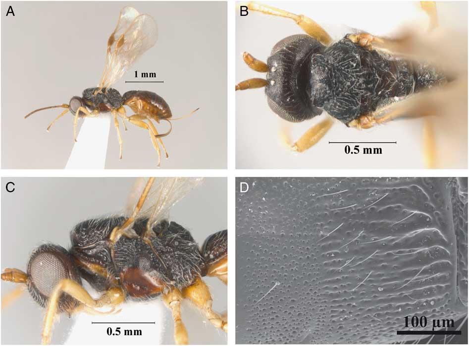
Diagnosis. This species can be distinguished from the remaining species of Allorhogas by having the second and third metasomal tergites largely covered by punctate sculpture (Fig. 8D).
Description. Holotype female. Body size 2.7 mm (Fig. 8A), forewing 2.2 mm. Colour: uniformly dark brown, scape, pedicel, and basal flagellomeres yellow to light brown, turning darker to apex; legs yellow, area surrounding precoxal sulcus light brown; wings hyaline, stigma brown, veins light brown; ovipositor sheaths brown.
Head: transverse in dorsal view, 1.8 times wider than median length (Fig. 8B), and 1.2 times wider than high; occipital carina complete and reaching hypostomal carina; POL as long as OD, 0.5 times OOL. Frons, vertex, gena, and face coriaceous; frons excavation shallow, poorly delimited laterally. Eye 1.2 times longer than wide; eye width 3.2 times longer than temple in dorsal view. Malar space 0.3 times eye height and as long as width of hypoclypeal depression; mandibles bidentate. Antenna with 22 flagellomeres, first flagellomere about 3.5 times longer than wide, 1.3 times longer than second flagellomere.
Mesosoma: 1.5 times longer than high (Fig. 8C) and 1.6 times longer than wide; pronotal collar short, not visible in dorsal view; pronotal furrow scrobiculate. Mesoscutum transverse in dorsal view, median length 0.7 times width; mesoscutal lobes coriaceous, with sharp anterolateral edges. Notauli present, complete, and scrobiculate, obscuring in a posterior rugose median area; scutellum weakly coriaceous, prescutellar furrow with three transverse carinae. Propodeum rugose-areolate, basal areas rugulose and well defined by carinae. Mesopleuron entirely coriaceous; subalar sulcus scrobiculate; precoxal sulcus coriaceous, 0.5 times as long as mesopleuron.
Wings: forewing 2.8 times longer than wide. Pterostigma 2.2 times as long as wide and 0.6 times as long as R. Vein r 0.7 times as long as 3RSa, 0.2 times as long as 3RSb, and as long as r-m. Vein m-cu arising basal to 2RS, then vein RS+Mb present. Hindwing vein M+CU 0.7 times as long as 1 M, m-cu slightly curved towards wing apex.
Legs: protibia with row of 17 spines along anterior margin. Metacoxa with distinct basoventral tooth. Metafemur 3.0 times longer than wide.
Metasoma: first tergite about as long as apical width, carinate-punctate, anterior area smooth and delimited by transverse carina, with a median dorsal area delimited by two dorsal carinae. Second tergites punctate (Fig. 8D), with a few rugae anteromedially. Remaining tergites punctate, with smooth tranverse stripe delineated by setae. Ovipositor about 0.7 times as long as metasoma.
Variation. Body size 2.1–2.8 mm. Some specimens have a slightly lighter metasoma. Antenna with 21–23 flagellomeres. Prescutellar furrow with three to four transverse carinae.
Male. Similar to female. Body size 1.8–2.1 mm. Antenna with 21–23 flagellomeres. Metafemur slightly swollen, 2.7 times longer than wide.
Etymology. The Latin word punctatus refers to the sculpture that is present in most of the metasomal tergites of the species, which is the main diagnostic morphological feature.
Material examined. Holotype (MZUCR): Female. Costa Rica: Cartago, Parque Nacional Barbilla, cecid leaf gall on Piptocarpha, ii.2001, G. Mora. Paratypes: six females (3 IB-UNAM, 2 MACN, 1 MZUCR), four males (2 IB-UNAM, 1 MACN, 1 MZUCR), same data as holotype.
Natural history. This species was reared from leaf galls on Piptocarpha poeppigiana (de Candolle) Baker (Asteraceae), which are induced by an undescribed species of Cecidomyiidae (Diptera). The galls are round swellings of the leaf blade, principally on the underside, about 10 mm in diameter and covered with silky white trichomes. These galls also yielded Euderus Haliday (Hymenoptera: Eulophidae), Eurytoma, Sycophila Walker (Hymenoptera: Eurytomidae), Cecidellis Hanson (Hymenoptera: Pteromalidae), and Lyrcus Walker (Hymenoptera: Pteromalidae). Whether A. punctatus is an inquiline or a parasitoid of one of the other gall inhabitants is unknown.
Allorhogas tico Martínez and Zaldívar-Riverón, new species
(Fig. 9A–F).
Allorhogas tico new species (female, holotype): A, habitus, lateral view; B, head, frontal view; C, head and mesosoma, dorsal view; D, head and mesosoma, lateral view; E, metasoma, dorsolateral view; F, forewing.
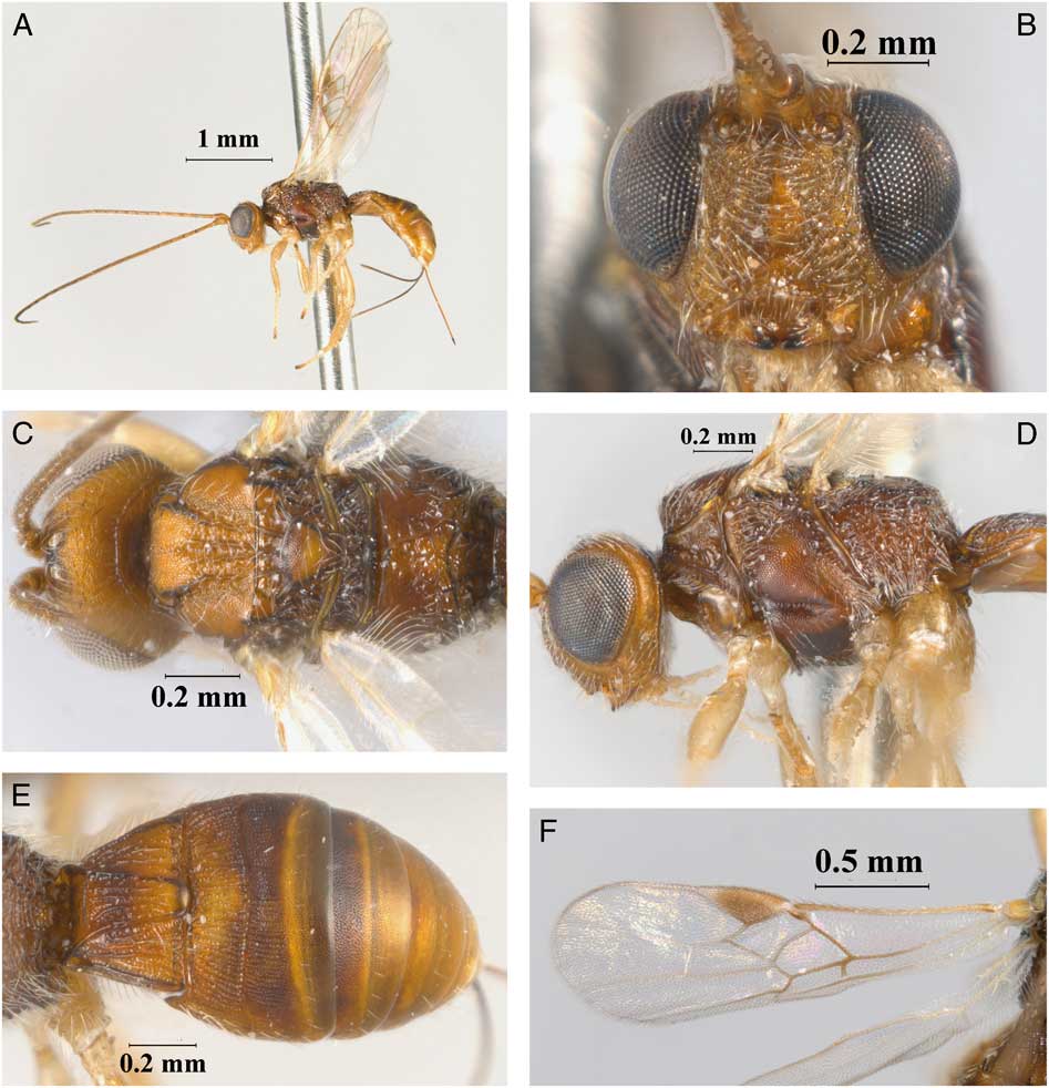
Diagnosis. Allorhogas tico is morphologically similar to A. laselva; however, it can be distinguished from the latter species by having a longer forewing vein r 0.8 length of vein 3RSa (0.5 in A. laselva), m-cu slightly curved towards wing apex (straight in A. laselva), metasoma honey yellow and brown (completelly dark brown in A. laselva), and propodeum rugose-areolate, with basal areas delimited by distinct carinae (strongly rugose with basal median areas indistinctly margined by carinae in A. laselva).
Description. Holotype female. Body size 2.8 mm (Fig. 9A), forewing 2.1 mm. Colour: general body colour brown, head light brown; ocellar triangle, subalar sulcus, precoxal sulcus, and median areas of second and third metasomal tergites darkened; antennae light brown to yellow basally, turning brown apically; legs pale honey yellow. Wings hyaline, stigma brown, veins light brown to brown; ovipositor sheaths dark brown.
Head: transverse in dorsal view, 1.8 times wider than median length, and 1.2 times wider than high; occipital carina complete and reaching hypostomal carina; POL as long as OD, 0.4 times OOL. Frons, vertex, gena, and face coriaceous; frons excavation shallow, poorly delimited laterally. Eye 1.3 times longer than wide; eye width 3.3 times longer than temple in dorsal view. Malar space 0.3 times eye height and as long as width of hypoclypeal depression (Fig. 9B); mandibles tridentate. Antenna with 24 flagellomeres, first flagellomere about 4.0 times longer than wide, 1.2 times longer than second flagellomere.
Mesosoma: 1.5 times longer than high (Fig. 9D) and 1.5 times longer than wide. Pronotal collar short, not visible in dorsal view; pronotal furrow scrobiculate, Mesoscutum transverse in dorsal view, median length 0.7 times width; mesoscutal lobes coriaceous. Notauli present, complete, and scrobiculate, finishing in a posterior rugose median area; scutellum coriaceous, prescutellar furrow with three transverse carinae Propodeum rugose-areolate, with basal areas delimited by distinct carinae (Fig. 9C). Mesopleuron entirely coriaceous; subalar sulcus scrobiculate; precoxal sulcus coriaceous, 0.6 times as long as mesopleuron.
Wings: forewing (Fig. 9F) 2.9 times longer than wide. Pterostigma 3.0 times as long as wide and 0.8 times as long as R. Vein r 0.8 times as long as 3RSa, 0.2 times as long as 3RSb, and as long as r-m. Vein m-cu interstitial or arising basal to 2RS, vein either RS+Mb present or absent. Hindwing vein M+CU 0.7 times as long as 1 M, m-cu slightly curved towards wing apex.
Legs: protibia with a row of about 10 spines along anterior margin. Metacoxa with distinct basoventral tooth. Metafemur 3.3 times longer than wide.
Metasoma: first tergite 0.8 times as long as apical width, costate-coriaceous, anterior area slightly rugulose and delimited by transverse carina (Fig. 9E), with median dorsal area delimited by two dorsal carinae. Second tergite costate-coriaceous; third terigte costate-coriaceous on basal half, coriaceous apically. Remaining tergites coriaceous. Ovipositor about 0.8 times as long as metasoma.
Variation. Body size 2.8–3.0 mm. Some specimens with slightly lighter metasoma. Antenna with 21–24 flagellomeres. Forewing vein m-cu varies from interstitial to basal with respect to 2RS, RS+Mb is absent or present.
Male. Similar to female. Body size 2.4 mm. Antenna with 22 flagellomeres. Metafemur swollen, 2.7 times longer than wide.
Etymology. The name of this species derives from the Spanish demonym for the inhabitants of Costa Rica.
Material examined. Holotype (MZUCR): Costa Rica: Guanacaste, Palo Verde, stem gall on Coursetia caribaea, 11.i.2001, I. Jimenez. Paratypes: 10 females (6 IB-UNAM, 3 MACN, 1 MZUCR), one male (IB-UNAM), same data as holotype.
Natural history. This species was reared from stem galls on Coursetia caribaea (Jacquin) Lavin (Fabaceae). These galls appear to be induced by either Cecidomyiidae or Tanaostigmatidae (Diptera), and thus A. tico is probably a parasitoid or an inquiline.
Discussion
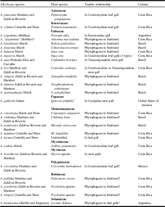
The results of this investigation add to our gradually accumulating knowledge of Allorhogas natural history (Table 1). However, more research is needed to clarify the exact trophic relationships of several described species and to discover additional species. In those species that occur in galls induced by Cecidomyiidae (or other gall formers), it remains unknown whether Allorhogas species are parasitoids, inquilines (feeding on gall tissue), or both. In addition to those included in Table 1, undetermined species of Allorhogas have been reared from cecidomyiid galls on various other plants in Costa Rica. For example on Annonaceae (leaf gall on Anaxagorea crassipetala Hemsley), Asteraceae (stem galls on Mikania Willdenow species), Lauraceae (leaf gall on Nectandra membranacea (Swartz) Grisebach), Rubiaceae (leaf, stem, and flower galls on Sabicea panamensis Wernham), and Tiliaceae (leaf gall on Luehea seemannii Triana and Planchon) (P.E.H., unpublished data).
Summary of host plants and trophic relationships of described species of Allorhogas.

Information from de Macêdo and Monteiro (Reference de Macêdo and Monteiro1989), Marsh (Reference Marsh1991, Reference Marsh2002), de Macêdo et al. (Reference de Macêdo, Pimentel and Vieira1998), Marsh et al. (Reference Marsh, de Macêdo and Pimentel2000), Martínez et al. (Reference Martínez, Zadivar-Riverón and Sáez2008), Penteado-Dias and Carvalho (Reference Penteado-Dias and Carvalho de2008), Chavarría et al. (Reference Chavarría, Hanson, Marsh and Shaw2009), Centrella and Shaw (Reference Centrella and Shaw2010, Reference Centrella and Shaw2013), and this paper. Host plant species previously placed in Pithecelobium are now assigned to Abarema and Chloroleucon.
* Based on the examination of primary types, the figures provided by Marsh (2002) and examined material from Costa Rica identified by P.E.H., the specimens from the latter country assigned therein as A. argentinus represent an undescribed species.
† Not a currently valid name, possibly Quercus palustris Münchhausen.
?=unconfirmed biology.
While some Allorhogas species occur in galls induced by Cecidomyiidae on diverse plant taxa, others are phytophagous and the data thus far suggest that these species are restricted to fruits/seeds of certain plant families, namely Fabaceae, Melastomataceae, and possibly Rubiaceae. In most of these cases there is very little external evidence of gall formation, but upon dissection gall formation can often be noted; e.g. Fabaceae galls are sometimes formed in the parenchymatous cell near the tegument (de Macêdo and Monteiro Reference de Macêdo and Monteiro1989; de Macêdo et al. Reference de Macêdo, Pimentel and Vieira1998), whereas in Melastomaceae it is still unclear exactly which plant tissue is affected (Chavarría et al. Reference Chavarría, Hanson, Marsh and Shaw2009). Many of the potential host plants are very speciose (e.g., Miconia, Palicourea, Psychotria), suggesting that there are numerous undescribed species, especially if gall-forming Allorhogas prove to be as host specific as other gall formers. It is still unclear what proportion of the plant species in these genera harbour Allorhogas, but already additional (probably undescribed) species have been found in Miconia affinis de Candolle, M. appendiculata Triana, M. doniana Naudin, and Clidemia capitellata (Bonpland) Don (Melastomataceae).
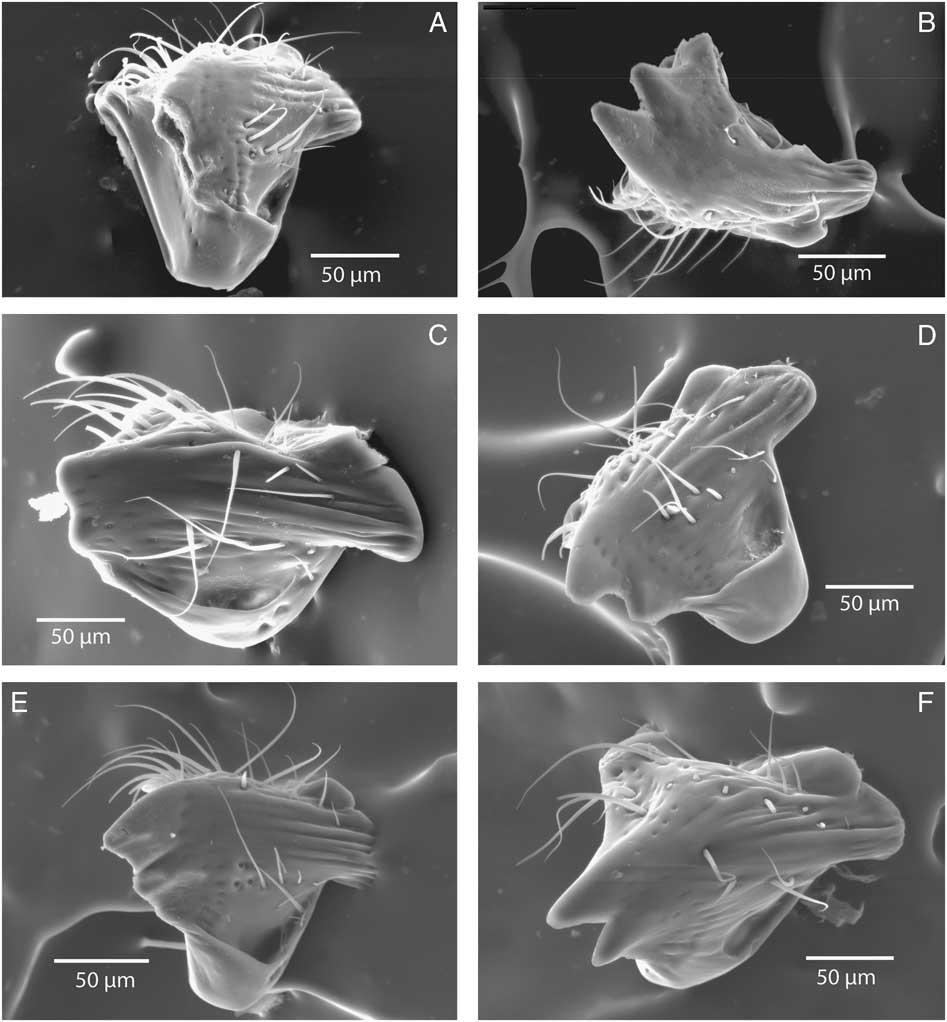
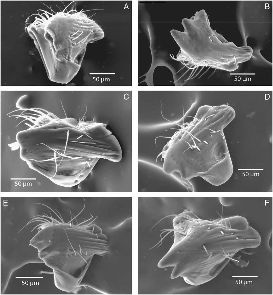
Mandibular morphology in the species of Allorhogas described in this work shows variation that is unusual in the entire family Braconidae (Fig. 10A–F). Bidentate mandibles have been proposed as a synapomorphy of the Ichenumonoidea (Mason Reference Mason1987; Sharkey and Wahl Reference Sharkey and Wahl1992), with an increased number of mandibular teeth in some subfamilies, most notably in members of the subfamily Alysiinae (Quicke Reference Quicke2015). A tridentate mandibular condition had also been previously reported by one of the junior authors for a species of Allorhogas associated with woody stem galls on Lycium Linnaeus (Solanaceae), A. cordobensis (Martínez et al. Reference Martínez, Altamirano and Salvo2011). The gathered evidence thus suggests that this could represent a relatively common feature in members of the genus, which could be related with phytophagy and/or association with lignified plant tissues. However, in this study tridentate species were found to be associated both with galled fruits on Fabaceae and Rubiaceae and also with stem galls on Fabaceae and Myrtaceae. At this stage, we cannot correlate the tridentate condition with the natural history of the examined species of Allorhogas (gall inducers, inquilines, or parasitoids) or with the affected plant organs (fruits/seeds, vegetative stems). Further morphological studies with additional species will help to elucidate whether this feature is associated with a phytophagous condition within the genus.
Scanning electron microscope digital photographs of mandibles for six species of Allorhogas described in this study: A, Allorhogas vulgaris; B, Allorhogas clidemiae; C, Allorhogas brevithorax; D, Allorhogas punctatus; E, Allorhogas tico; F, Allorhogas granivorus.
Acknowledgements
The authors thank Tracy Johnson for donating specimens of A. granivorus; Jose Antonio Azofeifa, Ivan Jiménez, Geovanny Mora and Kenji Nishida for help in collecting specimens in Costa Rica; Scott Shaw for donating comparative material of previously described species of Allorhogas; Laura Márquez for her assistance in the laboratory work; Guillermina Ortega for helping with the curation of the specimens; and Susana Guzmán for her help with the stereomicroscope digital pictures. This study was funded by grants given by the Consejo Nacional de Ciencia y Tecnología (CONACyT, convocatoria Secretaría de Educación Pública (SEP)-Ciencia Básica 2014, number 220454, and Red Temática del Código de Barras de la Vida) and Dirección General de Apoyo del Personal Académico (DGAPA)-UNAM (Programa de Apoyo a Proyectos de Investigación e inovación Tecnológica (PAPIIT) proyecto IN207016) to A.Z.R.; by grants given by the Minas Gerais Research Foundation (FAPEMIG) and the Brazilian National Council for Scientific and Technological Development (CNPq) to L.D.B.F.; and by a grant given by the Consejo Nacional de Ciencia y Tecnología (CONACyT)-Consejo Nacional de Investigaciones Científicas y Tecnológicas (CONICET), proyecto de cooperación bilateral convocatoria CONACyT 2013, number 207072) to J.J.M. and A.Z.R.