Introduction
Streptozotocin-induced diabetes mellitus (STZ-DM) in rats is a well-established model for studying type 1 diabetes. It has been in use since STZ was shown to possess diabetogenic properties in rats and mice almost 40 years ago (Rakieten et al., Reference Rakieten, Rakieten and Nadkarni1963; Junod et al., Reference Junod, Lambert, Stauffacher and Renold1969). STZ-DM is associated with a progressive loss of β-cells, and thus diminished release of insulin after ad libitum feeding, similar to that observed in patients with type 1 diabetes (Burvin et al., Reference Burvin, Armoni and Karnieli1994). By entry and disruptment of the pancreatic β-cells in the islets of Langerhans (Gai et al., Reference Gai, Schott-Ohly, Schulte im Walde and Gleichmann2004), administration of the STZ leads to hyperglycemia, with subsequent polyuria, glucosuria and decreased body weight (BW), all classical signs of type 1 diabetes (Rakieten et al., Reference Rakieten, Rakieten and Nadkarni1963; Junod et al., Reference Junod, Lambert, Stauffacher and Renold1969; Veleminsky et al., Reference Veleminsky, Burr and Stauffacher1970).
However, the STZ-DM model seems to have its limitations. Firstly, STZ-DM is not of genetic origin, but instead an effect of a toxin (Wilson and Leiter, Reference Wilson and Leiter1990; Myers et al., Reference Myers, Laks, Feeney, Mandel, Koulmanda, Bone, Barley, Rowley and Mackay1998). Secondly, rats treated with STZ display greatly increased blood glucose (BG) concentrations, but the biological significance of the BG may, or may not, be the same in rats as it is in humans. Furthermore, STZ is a nephrotoxic substance and has been shown to cause renal tubular injury in patients (Weiss, Reference Weiss1982). The STZ treatment per se may cause urinary protein leakage (Palm et al., Reference Palm, Ortsäter, Hansell, Liss and Carlsson2004), although it does not seem to affect neither renal clearances of inulin nor para-amino-hippuric acid (Churchill et al., Reference Churchill, Churchill, Bidani and Dunbar1993) or cause as marked histological lesions as does diabetes in patients (Gross et al., Reference Gross, Ritz, Schoof, Adamczak, Koch, Tulp, Parkman, El-Shakmak, Szabo and Amann2004).
The aim of the present study was to investigate the effects of suboptimal insulin therapy on insulin response in the STZ-DM rat in vivo, and to develop a suitable protocol for insulin treatment.
Material and methods
Animals and induction of diabetes
The study was performed on 10 age-matched, anaesthetized male Sprague-Dawley rats weighing 264 ± 9 g (M&B, Ry, Denmark). The rats were allowed free access to water and standardized rat chow (R36; Ewos, Södertälje, Sweden, containing 0.3% sodium, 0.8% potassium and 21% proteins) until the time of the experiment. On the first day of the experiment, the rats were made diabetic by means of a single intravenous injection of STZ (55 mg/kg BW; Sigma-Aldrich, St Louis, MO, USA), dissolved in 0.2 ml saline, in order to achieve a BG of about 25 mmol/l (Junod et al., Reference Junod, Lambert, Stauffacher and Renold1969). BW and BG were monitored every 2 days in order to evaluate the degree of diabetes. Blood samples were obtained from the cut tip of the tails and analysed by a glucose oxidase method (MediSense, Bedford, MA, USA). An animal was excluded if it presented with a weight loss more pronounced than 10% of its BW or if the BG exceeded 30 mmol/l for more than two consecutive days. All experiments were approved by the Uppsala Ethical Committee for Animal Experiments and were performed in accordance with national guidelines for the care and use of laboratory animals.
Experimental protocol and animal groups
STZ-DM rats were given saline injections (vehicle) for 14 days. On day 15, insulin response was estimated from the reduction in BG after an i.v. injection of fast-acting insulin (5 IE/kg BW; Eli Lilly and Company, Indianapolis, IN, USA). Thereafter, therapy was initiated with daily suboptimal long-acting insulin (5 IE/kg per day; Insulatard; Novo Nordisk, Hjørring, Denmark) s.c. for 7 days. Again, a single i.v. injection of fast-acting insulin was administrated in order to assess the effect on BG. Thereafter, the rats were treated with vehicle for another 7 days, before the response to fast-acting insulin was once again estimated (day 28). Throughout the study, BW and BG were measured every 2 days.
Statistical analysis
For comparisons of multiple data sets within the same experimental group, repeated measures analyses of variance (ANOVA) were performed and, when appropriate, followed by Dunnett’s post hoc test. All statistical analyses were performed using GraphPad Prism software (GraphPad Software Inc., San Diego, CA, USA). For all comparisons, P < 0.05 was considered statistically significant.
Results
Assessment of insulin response
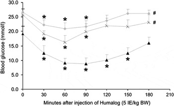
BG decrease after administration of fast-acting insulin was markedly decreased in both groups of vehicle-treated diabetic rats (day 15 and day 28) compared to the group pre-treated with long-acting insulin (day 22) (Figure 1). The vehicle-treated groups had a decreased level of BG with an average of −23% and −34% (from 26.4 ± 1 to 20.3 ± 2 and 25.8 ± 1 to 16.9 ± 2, respectively) after injection of the fast-acting insulin, whereas the insulin-treated group responded to the fast-acting insulin injection with a −61% decrease in BG (from 18.8 ± 3 to 7.3 ± 2). In addition, the group pre-treated with long-acting insulin (day 22) exhibited a longer duration of the effect. The effect of the fast-acting insulin on BG still lasted 150 min after the injection, as opposed to in the vehicle-treated groups, where the last measurable effect was detected after 90 min.
The effect of long-acting insulin pre-treatment on BG response to fast-acting insulin treatment (fast-acting insulin, 5 IE/kg BW). *Denotes a difference compared to the first measurement in the same group. #Denotes a difference in the whole group when comparing to animals treated with long-acting insulin at the corresponding time after injection of fast-acting insulin.

There was no difference between the rats in the first vehicle-treated group (day 15) and the latter (day 28) in their response to the fast-acting insulin injection.
Blood glucose over time
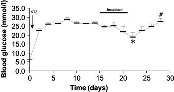
Throughout the study, BG was measured every 2 days (Figure 2). The BG was decreased in the last measurement during the period of treatment with long-acting insulin (day 22, 19.4 ± 3 mmol/l), as compared to the last measurement in the first vehicle-treated period (day 14, 26.1 ± 1 mmol/l). BG was increased in the last measurement in the second vehicle-treated period (day 28, 26.9 ± 2 mmol/l), compared with the last measurement in the period of treatment with long-acting insulin (day 22, 19.4 ± 3 mmol/l).
Changes in blood glucose (BG) over time. The effect of insulin pre-treatment or non-insulin pre-treatment on BG. The line indicates the period of treatment with long-acting insulin (day 15 to day 21). *Denotes a difference compared to the last measurement in the first vehicle-treated period (day 14). #Denotes a difference when compared with the last measurement in the period treated with long-acting insulin (day 22).

Body weight over time
Before the induction of diabetes, the BW of the animals was 240 ± 28 g. Fourteen days after the induction of diabetes, BW was 235 ± 27 g. During insulin treatment all animals gained weight, and after 7 days of insulin treatment they displayed a BW of 294 ± 28 g. When insulin treatment was discontinued, the increase in BW ceased. Seven days after cessation it reached 283 ± 29 g.
Discussion
A profound decrease in response to insulin developed within 2 weeks after the injection of STZ, resulting in a brief and modest response in BG to injections of fast-acting insulin. The decreased response could be reversed by daily pre-treatment with suboptimal doses of long-acting insulin, but rapidly reoccurred when treatment was withdrawn (Figure 1). These findings suggest that pre-treatment with suboptimal doses of long-acting insulin should be a prerequisite in studies of insulin treatment in STZ-DM rats in order to prevent the influence of a decrease in response to insulin.
The STZ-DM rat is a common model for the study of type 1 diabetes, but differs from human diabetic pathophysiology in its insulin resistance (Solomon et al., Reference Solomon, Deaton, Harris and Smoake1989). The average type 1 diabetic patient requires insulin substitution in the range of 0.5 to 1.0 IE/kg BW per day (Weintrob et al., Reference Weintrob, Benzaquen, Galatzer, Shalitin, Lazar, Fayman, Lilos, Dickerman and Phillip2003), in order to achieve normoglycaemia, compared to the insulin doses of 5 IE/kg BW per day, which were administered in the present study without rendering the rats normoglycaemic. The dose used in the present study is about 30 times the dose a diabetic patient would need before a meal (Ahmed et al., Reference Ahmed, Mallias and Home1998). Although rats daily pre-treated with suboptimal doses of insulin tolerated bolus administration of fast-acting insulin without achieving normoglycaemia, the administered insulin caused a more pronounced decrease as compared to the same rats before treatment. Furthermore, the STZ-DM rat differs from other common experimental models of type 1 diabetes, such as pancreatectomized animals, dexamethasone-treated type 1 diabetic rat and alloxan-induced diabetes (Ader et al., Reference Ader, Richey and Bergman1998; Cheta, Reference Cheta1998; Qi et al., Reference Qi, Pulinilkunnil, An, Ghosh, Abrahani, Pospisilik, Brownsey, Wambolt, Allard and Rodrigues2004). An explanation to this could be the different origins of the disease and in the different modes of actions of the diabetes-inducing substances (Cheta, Reference Cheta1998; Peschke et al., Reference Peschke, Ebelt, Bromme and Peschke2000; Gai et al., Reference Gai, Schott-Ohly, Schulte im Walde and Gleichmann2004). To improve the usefulness of the STZ-rat model, it is of vast importance that these differences are taken into account and minimized as much as possible.
The decreased sensitivity to insulin in STZ-DM rats has previously been shown in a number of studies (Karnieli et al., Reference Karnieli, Hissin, Simpson, Salans and Cushman1981; Burvin et al., Reference Burvin, Armoni and Karnieli1994; Cournarie et al., Reference Cournarie, Auchere, Chevenne, Lacour, Seiller and Vauthier2002). In vitro studies suggest that the prolonged insulinopenic state in STZ-DM animals might reduce insulin-stimulated glucose uptake by a depletion of GLUT4 from the intracellular pool, thus diminishing the insulin-responsiveness and causing insulin resistance (Goto et al., Reference Goto, Kida, Kaino, Inoue, Ikeuchi, Miyagawa and Matsuda1988). The STZ-DM has altered levels of insulin-signaling components in liver tissue, due to an altered expression (Bonini et al., Reference Bonini, Colca and Hofmann1995). While insulin-responsiveness is decreased, the rate of insulin clearance remains unchanged (Burvin et al., Reference Burvin, Armoni and Karnieli1994). Recently, it has been demonstrated in perfused hearts from STZ-DM rats that insulin resistance is due to alterations in the kinetics of insulin receptor-binding (Bikhazi et al., Reference Bikhazi, Azar, Birbari, El-Zein, Haddad, Haddad and Bitar2000).
Clinical studies have shown an increased insulin response after intense insulin therapy in diabetic patients (Scarlett et al., Reference Scarlett, Gray, Griffin, Olefsky and Kolterman1982; Lager et al., Reference Lager, Lonnroth, von Schenck and Smith1983; Karnieli et al., Reference Karnieli, Armoni, Cohen, Kanter and Rafaeloff1987). Karnieli and co-workers showed a reversal of insulin resistance in rat adipocytes by insulin therapy, both by restoration of the intracellular pool of GLUT4 and by enhancement of GLUT4 activity (Karnieli et al., Reference Karnieli, Armoni, Cohen, Kanter and Rafaeloff1987). It has also been shown that insulin significantly enhances the activity of GLUT4 in humans, under both normal and diabetic conditions (Karnieli and Armoni, Reference Karnieli and Armoni1990). Furthermore, it seems that insulin treatment of diabetic rats induces partial restoration of the lost insulin effect in hepatocytes (Hussin and Skett, Reference Hussin and Skett1988).
In the present study, the development of insulin response to insulin in vivo was investigated, showing that 1 week without insulin treatment is sufficient for a decreased response to insulin to develop. Since there was no difference either in basal BG or in response to fast-acting insulin between the rats in both vehicle-treated periods, the conclusion was drawn that a decreased response to insulin had redeveloped already within 7 days after withdrawal of insulin treatment (Figure 1). Thus, it seems that a prolonged insulinopenic state is not necessary for the development of a considerably reduced responsiveness to insulin in the STZ-DM rat.
The present study also showed that insulin treatment during 7 days suffices to markedly increase the BG-lowering effect of insulin. Rats pre-treated with suboptimal doses of long-acting insulin displayed a more pronounced BG decrease after administration of the fast-acting insulin, compared to both periods of vehicle-only treatment (Figure 1). The alteration in sensitivity to the injected insulin occurred with regard to both with reference to the duration of response and with reference to the magnitude of effect. Hyperglycemia has been shown to contribute to the development, maintenance and progression of insulin resistance in diabetic subjects, as well as in animal models (Lisato et al., Reference Lisato, Cusin, Tiengo, Del Prato and Jeanrenaud1992; Cameron-Smith et al., Reference Cameron-Smith, Collier and O’Dea1994). Hence, the lowering of the BG by treatment with long-acting insulin (Figure 2) could at least partially explain the findings regarding the lowered insulin response.
Basal BG decreased after the onset and increased after the withdrawal of insulin treatment (Figure 2). Basal BG measurements should not be influenced by the injected long-acting insulin, since the measurements were made 24 h after the administration of long-acting insulin. However, the STZ-DM rats have a remaining basal insulin secretion. Hence, the observed changes in basal BG could be due to an alteration in the sensitivity to the endogenous secretion. However, the decrease in response to insulin did not occur until after approximately 4 days of treatment with long-acting insulin. Furthermore, after 7 days of pre-treatment with long-acting insulin, the rats were still far from normoglycaemic (Figure 2). Thus, it is possible that continued treatment with long-acting insulin could further improve insulin sensitivity.
It has been shown that chronic exposure to elevated glucose in diabetes mellitus results in irreversible alterations in peripheral and central cellular function, as well as leads to further deterioration of the beta-cell function, a phenomenon referred to as glucotoxicity (Rolla, Reference Rolla2004). Also, insulin is known to stimulate peripheral glucose uptake (Rolla, Reference Rolla2004). It is possible that the effects observed in this paper are to some extent mediated by an altered glucose uptake profile and reduced glucotoxicity.
The major type of diabetes mellitus observed in dogs and cats is insulin-dependent at the time of diagnosis (Feldman and Nelson, Reference Feldman and Nelson2004; Rand and Marshall, Reference Rand and Marshall2005), and in cattle, insulinopenic diabetes is known to develop after infection with bovine viral diarrhea virus (Taniyama et al., Reference Taniyama, Hirayama, Kagawa, Kurosawa, Tajima, Yoshino and Furuoka1999). Thus, the fact cannot be excluded that these results could also be beneficial in clinical veterinary settings.
In summary, a decrease in insulin response developed rapidly after the onset of STZ-induced diabetes, resulting in a brief and modest BG-lowering effect of insulin. Partial restoration of insulin sensitivity was observed after 7 days of treatment with suboptimal doses of long-acting insulin, but the insulin response rapidly decreased again after the treatment was withdrawn. The potential consequences of this decreased insulin response and a potential insulin resistance must be taken into consideration when studying diabetes in STZ-DM rats. The findings in the present study suggest that a prerequisite for performing studies with insulin treatment of diabetes in the STZ-DM rat is that the rats are pre-treated with long-acting insulin in order to eliminate this influence.
Acknowledgements
This study was supported by the Wallenberg foundation, The Swedish Pharmaceutical Society and Stiftelsen Petersenska hemmet. The excellent assistance of Britta Isaksson is gratefully acknowledged.