INTRODUCTION
A key feature of saccadic eye movement is that competing visual stimuli result in distortions in trajectory (Ludwig & Gilchrist, 2002; Yarbus, 1967). The extent and direction of deviation has been shown to be modulated as a function of stimulus relevance. In visual search situations, where attention is allocated to multiple stimuli for processing of relevance, saccades may curve towards non-target stimuli (McPeek & Keller, 2001). Conversely, either the allocation of attention away from a saccade target (Sheliga et al., 1997; Sheliga et al., 1995; Tipper et al., 2001; Van der Stigchel & Theeuwes, 2005) or the presentation of an irrelevant visual distractor (Doyle & Walker, 2001; Tipper et al., 2001), results in curvature away from the attended or distractor location, for either horizontal and vertical saccades, with the magnitude of curvature greatest when allocated to or presented in the same hemifield as the target (Doyle & Walker, 2001; Tipper et al., 2001).
Although precise mechanisms are unresolved, modulation of saccadic trajectory is typically attributed to competitive interactions of activity over a saccade motor map (Godijn & Theeuwes, 2002; McSorley et al., 2004), between representations of potential target locations within the neural structures involved in saccade production (Doyle & Walker, 2001; Doyle & Walker, 2002; McPeek et al., 2003; B.M. Sheliga et al., 1995). Curvature away from a distractor location is believed to reflect the active inhibition of a saccade program, which is generated automatically by the distractor (Doyle & Walker, 2001; Sheliga et al., 1995; Tipper et al., 2001). Several models propose that the neurophysiological correlate of the saccade motor map exists in the midbrain structure, the superior colliculus (SC), which provides the final programming of a saccade (Dorris et al., 1997; Sheliga et al., 1995; Tipper et al., 2000). Notably, the SC sends major projections to pre-motor command areas within the brainstem (Andersen, 1972; Moschovakis et al., 1996), with direction and amplitude of a saccade a direct function of the site of stimulation and peak of activity over the SC motor map (Munoz & Wurtz, 1993).
Initiation of any saccade by the SC requires that a population of activity must be selected as the target and that activity associated with a distractor location must be inhibited, whether by lateral interactions within the salience map Sheliga et al., 1995) or by other external sources of modulation including the frontal eye fields (FEF) (Schlag-Rey et al., 1992) and dorsolateral pre-frontal cortex (DLPFC) (Ploner et al., 2005). The consequence of regional inhibition appears to be that the peak of activity over the salience map shifts away from the distractor region, and the saccade deviates away from the location of the distractor.
Although the SC receives inhibitory inputs from frontal and parietal cortices, it also receives powerful inhibitory input from the substantia nigra pars reticulata (SNr) (Hikosaka & Wurtz, 1983). It has been proposed that the existence of long-range inhibitory connections in the SC, revealed by extracellular electrical stimulation (Munoz & Istvan, 1998), actually result from activation of passing inhibitory SNr neurons (Ozen et al., 2004). Whilst the resolution and form of spatial tuning of SNr neurons are unclear (Basso & Wurtz, 2002; Handel & Glimcher, 1999), they may inhibit a broad region of the SC, interacting with activity already induced by visual inputs (Arai & Keller, 2005).
Profound degenerative changes occur within the cortex in people with Huntington's disease (HD) (Aylward et al., 1998), however, the striatum is subject to the most significant atrophy, in particular those neurons projecting to the external segment of the globus pallidus and the SNr. A number of ocular motor abnormalities have already been identified in HD (Lasker & Zee, 1997) that are consistent with basal ganglia dysfunction. Difficulty is experienced in initiating more voluntary types of saccades, with dysmetria (over or under shooting the target), especially to remembered targets, and poor suppression of unwanted reflexive behavior (Avanzini et al., 1979; Lasker et al., 1988; Tian et al., 1991). These deficits have been attributed not only to characteristic FEF pathology but also disruption of BG circuitry involving SNr and FEF, in particular their projection to SC (Avanzini et al., 1979; Lasker & Zee, 1997; Leigh et al., 1983; Tian et al., 1991). It is also conceivable, although as yet undisclosed, that saccadic trajectory may be compromised in HD, given the degeneration of the inhibitory BG pathway, which incorporates the SNr.
Importantly, the involvement of the SC in the subsequent correction of saccade trajectory has not been supported by neurophysiological findings (Soetedjo et al., 2002). A number of models propose a role for the cerebellum as a displacement integrator projecting directly to burst-generating circuits in the brainstem, to adjust the directional drive signal and compensate for initial directional errors (Arai & Keller, 2005; McSorley et al., 2004; Quaia et al., 1999). Thus our interest focused upon the initial deviation of saccadic trajectory, assuming that the SC is pivotal in regulating the earliest component of a saccade motor program. The following two experiments were adopted to investigate the impact of task-irrelevant distractors on both volitional and more reflexive saccadic trajectories in HD.
Experiment 1
The first of two experiments presented to participants, required the generation of a vertical saccade to a visual target suddenly presented in one of two defined spatial locations above or below fixation. A visual distractor was presented simultaneously with the target and appeared in either the upper or lower visual field, in either the left or right visual field. Saccades could be regarded as reflexive, or “exogenous” in nature and the distractors entirely task irrelevant (Doyle & Walker, 2001).
METHOD
Participants
Eleven adults (10 men and 1 woman) with mild to moderate HD participated voluntarily in this study, and were aged between 42 and 64 years (M = 51.00, SD = 7.17). All were clinically diagnosed with HD by a neurologist (A.C.) and underwent genetic testing with confirmation of CAG triplet repeats within the expanded range for HD. Clinical manifestations of the disease had been evident from .5 to 11 years, and the severity of disease was evaluated with the motor sub-scale of the Unified Huntington's Disease Rating Scale (HDSG, 1996). Scores, where available, ranged between 10 and 42 (M = 21.50, SD = 10.95). Clinical data for this group, as well as medication status, are shown in Table 1.
Clinical data Huntington's disease (hd) patients

Eleven neurologically healthy subjects (5 women and 6 men) also participated in this study, aged between 39 and 67 years (M = 51.00 years, SD = 9.91 years). There were no significant differences between neurologically healthy and HD groups in age or years of education (M = 11.4 (2.01) and M = 13.27 (2.70), respectively). Previous investigations have demonstrated no gender differences with respect to automatic orienting behaviors (Bayliss et al., 2005; Lum et al., 2002), therefore these participants were not gender-matched to patient subjects. Our own preliminary analyses confirmed that males and females performed equally on all indices of performance.
No participant demonstrated visual impairment, other than refractive error, or gross clinical ocular motor pathology, and none reported a history of neurological impairment. Participants were screened for cognitive decline using the mini-mental state examination (MMSE) (Folstein et al., 1975), with scores <24 out of 30 considered indicative of abnormal cognitive functioning. Although all scores fell within a normal range, a significant difference was found between HD participants (M = 27.30, SD = 1.77 for 10 participants only) and neurologically healthy participants (M = 29.73, SD = .65), t(19) = −4.101, p < .05. Depressive symptoms were evaluated using the Beck Depression Inventory (BDI) (Beck et al., 1961), revealing no significant difference between HD (M = 7.00, SD = 5.12) and neurologically healthy (M = 5.73, SD = 4.38) groups. The digit span (DS) task, a component of the Wechsler Adult Intelligences Scale (Wechsler, 1955), was administered to ascertain recall of an increasing number of digits both forward and backward; these tasks are considered to reflect attention to a given task and short term working memory. No significant difference was found between HD (M = 10.80, SD = 1.40) and neurologically healthy (M = 10.73, SD = 5.95) groups in scores scaled for age effects.
Apparatus
Both vertical and horizontal displacement of the eye were recorded using the magnetic field scleral search coil technique (Robinson, 1963), with output sampled at 1 kHz. Screen based stimuli were generated using E-Prime software and data were subsequently analyzed offline, using interactive Matlab software. Participants were seated with their heads positioned on a stable chin rest 65.2 cm directly in front of the screen. Stimuli comprised a green central fixation cross (+), with target crosses (x) appearing in one of two white boxes 10° directly above or below fixation. Distractor stimuli (white squares) appeared in one of four grey boxes situated to the left or right of the vertical midline in the same or opposite hemifield as the target (5° above/below and 5° left/right of fixation). Output was displayed alongside a control signal generated by E-Prime, which indicated stimulus change. A photodiode was placed directly over a non-visible portion of the screen to concurrently record stimulus change in real time.
Procedure
Ethics approval was granted by the North Western Behavioural and Psychiatric Research and Ethics Committee and all participants gave their informed consent prior to inclusion in the study, in accordance with the Helsinki declaration. All participants continued with their normal medication regime, and each was tested in a single session.
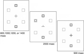
Participants were required to generate a vertical saccade to an upper or lower target cross located in one of the two white boxes located directly above or below fixation. A single distractor was presented in one of the four peripheral boxes, simultaneous with the target, in 80% of trials. The target/distractor remained on screen for 2000 msec, with presentation of a white cross at center for 500 msec summoning a return of gaze to fixation. Intertrial intervals varied randomly from 800 to 1400 msec (see Figure 1). A total of 80 trials was randomly presented to each participant, comprising 16 trials for each distractor location.

Schematic diagram of a target summoning a downwards saccade, in conjunction with a distractor in the lower left hemifield. Following a variable fixation period, a target cross appears in either the upper or lower designated boxes for 2000 msec, in conjunction with a “distractor” stimuli in one of four peripheral boxes. Participants are required to shift gaze to the target as soon as it appears.
Data Analysis
Trials corrupted by blinks or poor fixation (eye movement less than 90 msec following target presentation) were excluded from analysis. Saccadic latency, horizontal deviation, and error rate were determined for remaining trials. Saccadic latency reflected the stimulus-saccade onset differential, with stimulus onset calculated using a real-time trace of on-screen presentation (diode) and saccade onset using a position trace, confirmed by a change in the velocity profile (>30°deg/sec). Horizontal deviation reflected the peak horizontal displacement of the eye immediately following target/distractor presentation, prior to on-line correction. Thus it refers to the initial movement of the eye and not its subsequent return to target. Latency and horizontal deviation data, derived for analysis, were calculated by subtracting saccadic latency and peak initial horizontal deviation in trials without distractors (baseline or inherent performance/deviation) from those with distractors present, for each participant, for both upward and downward saccades, thus deriving a relative latency or horizontal deviation value independent of idiosyncratic variations between participants. These values will be reported as “D” scores.
Horizontal deviation and latency data, baseline and relative, were subjected to 4-way ANOVA, with the between-subjects variable Group (HD, neurologically healthy) and within-subjects variables Saccade direction (up, down), Distractor side (left, right) and distractor and target upper or lower hemifield Compatibility (compatible, incompatible). Error rate for each group was calculated as the proportion of erroneous responses to distractor stimuli, to total number of trials presented, and was compared using independent-measures t-tests. A series of product moment correlations were also performed (Pearson's r) evaluating relationships between disease severity (UHDRS scores), number of CAG repeats, proportion of erroneous responses, proportion of anticipatory or intruding saccades, and deviation/latency as a function of saccade direction (up or down).
RESULTS
Latency
For baseline trials (no distractor present) no significant differences were found in saccadic latency between HD participants (M = 302 msec, SD = 18 msec) and neurologically healthy participants (M = 298 msec, SD = 18 msec).
For neurologically healthy participants, saccade latencies to targets presented with a distractor in the same hemifield (D = 8 msec), were significantly shorter than those presented with distractors in the opposite hemifield (D = 38 msec). However, no such response time advantage was conferred by spatially Compatible target/distractor presentation for HD participants (D = 32 and 34 msec respectively). ANOVA of latency revealed both a main effect of compatibility [F(1,20) = 7.95, p < 0.05] and a significant Compatibility × Group interaction [F(1,20) = 5.82, p < .05]. Main effects of Group, Saccade direction, and Distractor side, and all other interactions were not significant.
No significant correlations were found between these saccadic latencies and any other measure of performance or disease status.
Horizontal Deviation
For baseline trials (no distractor present) no significant difference was found in peak initial horizontal deviation between HD participants (M = .26, SD = .20° right) and neurologically healthy participants (M = .12, SD = .28° right).
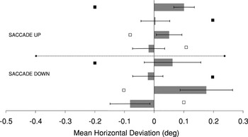
As Figure 2a illustrates, horizontal deviation followed an expected pattern of response for neurologically healthy participants, with curvature consistently in the direction opposite to distractor stimuli. However, this pattern of deviation was not consistently found for saccades by HD participants, clearly evident in Figure 2b. ANOVA revealed a significant Group × Saccade Direction interaction [F(1,20) = 6.17, p < .25, η2 = .24], a significant Group × Distractor Side interaction [F(1,20) = 5.33, p < .05, η2 = .21], and a significant Group × Distractor Side × Saccade Direction interaction [F(1,20) = 6.13, p < .025, η2 = .26].

Mean horizontal deviation by distractor condition for Control subjects—EXOGENOUS saccades. Relative distractor position is indicated by black and white squares, with black representing the upper hemifield and white representing the lower hemifield. Negative values represent deviation to the left and positive values deviation to the right.

Mean horizontal deviation by distractor condition for HD patients—EXOGENOUS saccades. Relative distractor position is indicated by black and white squares, with black representing the upper hemifield and white representing the lower hemifield. Negative values represent deviation to the left and positive values deviation to the right.
Subsequent Group analyses for HD patients revealed a main effect of Distractor Side [F(1,10) = 6.13, p < .05, η2 = .38] and a significant Distractor Side × Saccade Direction interaction [F(1,20) = 8.22, p < .025, η2 = .45]; upward saccades with both left (D = .10) and right (D = .03) distractors deviated away from distractor locations (p = .01, η2 = .50), whereas downwards saccades deviated to the left irrespective of distractor side (D = .11 and D = .34, respectively). The amplitude of this leftward deviation correlated significantly with disease severity (UHDRS scores), r = .55, p ≤ .001, number of CAG repeats, r = .82, p ≤ .001, and the proportion of anticipatory or intruding saccades, r = .40, p = .001.
Conversely, Group analyses for neurologically healthy participants revealed a significant Compatibility × Distractor Side interaction [F(1,10) = 5.75, p < .05, η2 = .37]. The Compatibility effect, where saccades directed to the hemifield containing a distractor deviate more strongly than those directed to the opposite hemifield, was stronger in the presence of left side distractors (D = 0.14 vs. D = 0.06) than right side distractors (D = 0.04 vs. D = 0.02). However, this difference did not reach significance. All other main effects and interactions were also not significant
Errors
HD participants (M = 3.52, SD = 2.36) responded erroneously to distractor stimuli proportionately more often than neurologically healthy participants (M = .68, SD = 1.03), t(20) = 3.66, p < .05. Unlike other measures of performance in HD participants, or disease status, a significant correlation was revealed between the proportion of trials resulting in an error and number of CAG repeats for HD participants, r = .34, p < .05.
DISCUSSION
Consistent with earlier studies Sheliga et al., 1995; Tipper et al., 2001; Van der Stigchel & Theeuwes, 2005), the presence of a peripheral distractor resulted in the curvature of vertical saccades away from the distractor location, for neurologically healthy participants. However, for HD participants there was evidence of strong leftward deviation, irrespective of distractor location, for downwards saccades, which correlated significantly with disease severity. Although inconclusive with respect to the site of dysfunction, this may be attributable to impaired processes above brainstem mechanisms responsible for saccade generation, since normal saccade metrics were demonstrated. Whether also evident for more volitional responses, was explored in the next experiment.
Results also demonstrated a response time advantage for compatible target/distractor presentation for neurologically healthy participants but not for HD participants where both remote and proximal distractors resulted in increased reaction times compared to baseline saccades. Although HD participants generated proportionately more erroneous saccades to distractor stimuli than neurologically healthy participants, this actually equated to no more than one or two more errors per patient and so can arguably be regarded as insignificant in this context.
Experiment 2
The second experiment similarly required participants to generate a vertical saccade to a visual target presented above or below fixation. However, saccade direction was indicated by changing the physical attributes of the central fixation cross. Distractors appeared simultaneously with the change at fixation and appeared in either the upper or lower visual field, in either the left or right visual field. Saccades could be regarded as voluntary or “endogenous” in nature and the distractors entirely task irrelevant (Doyle & Walker, 2001).
METHOD
Data were derived from ten HD participants (nine men and one woman) and age-matched neurologically healthy participants, because one HD participant was unavailable, due to time constraints, to complete both protocols. All methods are the same as experiment 1 except for the following. Target stimuli were present at all times above and below fixation, and participants were required to saccade to either the upper or lower target depending on a cue presented at fixation. The cue comprised change of the central fixation cross, with one of the two vertical arms of the cross removed, leaving either an upward or downward pointing arm (see Figure 3).

Schematic diagram of a central cue summoning an upward saccade, in conjunction with a distractor in the upper left hemifield. Following a variable fixation period, the central cross changes to either an upward or downward “pointer” for 2000 msec, in conjunction with a “distractor” stimuli in one of four peripheral boxes. Participants are required to shift gaze to the target indicated by the pointer as soon as it appears.
RESULTS
Latency
Although saccades by HD participants were slower overall for baseline trials (no distractor present), no significant differences were found in saccadic latency between HD participants (M = 475 msec, SD = 24 msec) and neurologically healthy participants (M = 419 msec, SD = 24 msec).
For neurologically healthy participants, saccades to targets presented with a distractor in the same hemifield (D = −39 msec), were significantly shorter (p = .001) than those presented with distractors in the opposite hemifield (D = −1 msec). However, for HD participants, latencies for spatially compatible target/distractor presentation were comparable to incompatible target/distractor presentation (D = 12 and 8 msec respectively). ANOVA reflects this difference with a significant Compatibility × Group interaction [F(1,18) = 4.53, p < .05], consistent with results from the first reflexive saccade paradigm. ANOVA also revealed a main effect of Group [F(1,18) = 5.69, p < .05], with responses by HD participants in the distractor trials 10 msec slower than in baseline trials, whereas responses by neurologically healthy participants were 20 msec faster. Main effects of Compatibility, Distractor side, and Saccade direction, and all other interactions were not significant.
No significant correlations were found between these saccadic latencies and any other measure of performance or disease status.
Horizontal Deviation
Analysis of baseline trials (no distractor present) revealed a significant Group × Saccade direction interaction [F(1,18) = 8.67, p < .01]. For neurologically healthy participants, both upward (.11°) and downward (.24°) saccades deviated to the right, whereas for HD participants, upward saccades deviated to the right (M = .27°), and downward saccades to the left (M = .05°).
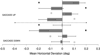
As found for more reflexive saccades, the presence of a distractor resulted in contralateral deviation of saccades for neurologically healthy participants, with a leftward deviation in the presence of right hemifield distractors and a rightward deviation in the presence of left hemifield distractors (see Figure 4a). However, as Figure 4b demonstrates, all upward saccades by HD participants deviated to the left, and all downward saccades deviated to the right, irrespective of distractor location.

Mean horizontal deviation by distractor condition for Control subjects—ENDOGENOUS saccades. Relative distractor position is indicated by black and white squares, with black representing the upper hemifield and white representing the lower hemifield. Negative values represent deviation to the left and positive values deviation to the right.

Mean horizontal deviation by distractor condition for HD patients—ENDOGENOUS saccades. Relative distractor position is indicated by black and white squares, with black representing the upper hemifield and white representing the lower hemifield. Negative values represent deviation to the left and positive values deviation to the right.
ANOVA revealed main effects of both Group [F(1,18) = 6.74, p < .025, η2 = 0.27] and Saccade Direction [F(1,18) = 4.91, p < .05, η2 = .21], a significant Group × Compatibility × Distractor Side interaction [F(1,18) = 4.47, p < .05, η2 = .20], and a significant Group × Distractor Side × Saccade Direction interaction [F(1,18) = 5.47, p < .05, η2 = .23].
Subsequent Group analyses for HD patients revealed a significant Distractor Side × Saccade Direction interaction [F(1,9) = 6.09, p < .05, η2 = .41]. For upward saccades, those with left side distractors deviated towards the distractor location (D = .06) whereas those with right side distractors deviated away from the distractor location (D = .13). For downward saccades, those with left side distractors deviated away from the distractor location (D = .08), and those with right side distractors deviated towards the distractor location (D = .17). This aberrant behavior was most evident for saccades in the presence of right side distractors (p = .01, η2 = .51).
For neurologically healthy participants, a significant Compatibility × Distractor Side interaction [F(1,9) = 5.56, p < .05, η2 = .38] was revealed. Although the Compatibility effect was normal in the presence of left side distractors (D = .09 > D = .04) and reversed in the presence of right side distractors (D = .05 < D = .16), no subsequent analyses reached significance.
Errors
Proportionately, HD participants (M = 6.38, SD = 3.84) responded erroneously to distractor stimuli no more often than neurologically healthy participants (M = 4.13, SD = 2.83).
DISCUSSION
In HD, a significant leftward horizontal deviation was evident for saccades directed upward, whereas a significant rightward horizontal deviation was evident for saccades directed downward, similarly suggesting dysfunctional supranuclear input. Also, it seems that irrespective of whether a saccade is stimulus-driven or intentionally generated, compatible target/distractor presentation confers no response time advantage for HD participants.
General Discussion
In two experiments presented here, the nature of distractor effects on the trajectory of vertical saccades was examined in adults with Huntington's disease (HD). Both visually-guided and endogenously driven saccades were characterized by anomalous deviation patterns, and unlike neurologically healthy participants, latencies revealed no response time advantage for saccades generated to compatible target/distractor locations for either saccade type.
In the absence of a distractor stimulus, saccades by HD participants displayed normal metrics and trajectory patterns, suggesting that the brainstem mechanisms responsible for generating the final directional commands to ocular motor neurons are relatively intact. However, the activity of these neurons is subject to regulation from numerous structures, and is dependent on the level of control necessary (i.e., exogenous vs. endogenous behavior and number of concurrent processes); these structures include the frontal and supplementary eye fields, as well as the SC, which receives frontal and parietal projections. All are regions that are intimately connected with the BG, the primary site of neuropathology in HD. Although the neural mechanisms responsible for the aberrant deviation patterns reported here are impossible to fathom based on behavioral analyses alone, the BG are inevitably implicated, whether directly, or indirectly via the numerous thalamic or collicular circuits, which traverse this region. Importantly, the nulcei that comprise the BG are primarily involved in the focusing of neural resources, synthesizing coarsely coded representations of sensory stimuli, and higher modulatory influences (i.e., motor intention) (McAuley, 2003), with the balance of activity over facilitatory and inhibitory pathways normally culminating in a behaviorally relevant response or movement. Alteration to this balance, as occurs in HD, results in inefficient response selection and poorly focused activity, particularly where behavior is internally rather than externally driven.
A strong leftward deviation for downwards exogenous saccades was revealed for HD participants, which correlated significantly with disease severity as well as number of CAG repeats, the length of which has been linked to the rate of disease progression (Aylward et al., 1997; Furtado et al., 1996). However, from the deviation evident in Figure 2a, it appears that these patients have actually preserved the general “pattern” of response in the presence of distractor stimuli, for upward and downward saccades but have incorporated an overall leftward shift unrelated to distractor presence.
This “shift” may well reflect an exaggerated compensatory strategy in HD, and is also evident in the second experiment, which required the generation of an endogenous saccade, the direction of which was determined by the interpretation of a central directional cue. Here the presence of a distractor, irrespective of location, similarly resulted in the deviation of saccades in the direction opposite to baseline trends. However, unlike upward saccades, horizontal deviations for downward saccades did not preserve the general pattern expected (that is if we presume an overall shift to the right and factor this into the response pattern). Responses appeared to be in the “same” direction as the distractor rather than the opposite direction. Arguably, shifting and patterning of trajectory may represent separable components of the movement deficits demonstrated by these HD patients, the former an exaggerated compensatory strategy, the latter indicating a true attentional or motor planning problem where actions are generated (or inhibited) internally rather than triggered by external stimuli. Although it may be argued that these behaviors cannot be solely a consequence of BG dysfunction in HD, given that neural degeneration may also be present in cortex (Gusella & MacDonald, 1997; Hersch & Ferrante, 1997), further investigation of this dissociation of motor deficits and compensatory strategies may well lead to a greater understanding of the impact of BG dysfunction on the range of behavioral anomalies seen in HD.
As anticipated, neurologically healthy participants generated shorter latency saccades in the presence of spatially compatible distractors (compared to spatially incompatible distractors) for both exogenous and endogenous saccades. Conversely, HD participants demonstrated no response time advantage for compatible target/distractor presentation for either saccade type. Typically, presentation of a distractor stimulus close to a subsequent target (e.g., the same hemifield) results in decreased reaction times to the target relative to presentation of distractor stimuli remote from the target. Threshold of activation appears to be reached more quickly with the combined facilitatory effects of overlapping distractor/target neural populations, prior to the onset of more volitionally generated inhibitory activity.
However, for HD participants, saccadic latencies were comparable across conditions, with relatively few erroneous responses to distractor stimuli. This suggests that these participants were either less influenced by distractor onset, affording no advantage of proximal target spatial overlap, or more strongly inhibited distractor-related activation. Not only is the former possibility inconsistent with a number of reports of increased distractibility in HD (Lasker et al., 1987; Leigh et al., 1983; Tian et al., 1991), but results from a concurrent study with this group demonstrating accelerated onset of inhibition of return (Fielding et al., 2006), suggest a relative precedence of inhibitory distractor-related activity where the spatial locations of distractor stimuli are clearly defined. This result may be a function of compensatory cortical rather than local BG inhibitory mechanisms, perhaps recruited where intruding motor programs interfere repeatedly over a range of essential behaviors. Notably, whereas distractor presentation was unpredictable, distractor locations were spatially defined at all times by the presence of the peripheral boxes that contained the distractors. As a final note, although it has been proposed that increased latencies might result in a reduction in deviation, with more time available to clearly define a target's location (McSorley et al., 2004), we found that differences in latency for both groups were not consistent with differences in deviation.
Further studies would ideally encompass a broader range of HD participants, verifying these results from a relatively small sample, also enabling comparison of medication effects, if any. However, it is clear that under specific circumstances people with HD are capable of suppressing inappropriate automatic responses, which may have implications for the development of adaptive strategies in the presence of seemingly irrepressible and disruptive behaviors. Whereas the absence of a response time advantage for compatible target/distractor presentation appears consistent with earlier work, irregularities in deviation require further investigation, potentially providing a clearer understanding of how the balance of activity over the BG is normally modulated to compensate for the presence of competing environmental activators.
ACKNOWLEDGMENTS
We gratefully acknowledge the help and cooperation of all participants who so willingly participated in this study, as well as Doctor Andrew Churchyard for his assistance with recruitment and assessment of HD participants. This research was supported, in part, by a Jean Gilmore bursary, awarded to J. Fielding by the AFWU-SA Inc. Trust Fund.