Introduction
The term inflammatory pseudotumour was first used in 1954 to describe a lesion that follows the clinical and radiological pattern of a neoplasm.Reference Umiker and Iverson1 It is not a single entity but rather a generic description applied to a non-specific, expanding, chronic inflammatory lesion. This broad definition has been used interchangeably with terms such as inflammatory myofibroblastic tumour, plasma cell granuloma, systemic fibrosis, xanthofibrogranulomatosis and multifocal fibrosclerosis.Reference Neild, Rodriguez-Justo, Wall and Connolly2
In 2001, Hamano et al. described an association between sclerosing pancreatitis and high levels of immunoglobulin (Ig) G4 complexes in the serum, compared with normal subjects and patients with chronic inflammatory conditions such as primary biliary cirrhosis.Reference Hamano, Kawa, Horiuchi, Unno, Furuya and Akamatsu3 This led to the proposal of a new entity characterised by extensive IgG4-positive plasma cell infiltration, together with cluster of differentiation 4 glycoprotein positive or cluster of differentiation 8 glycoprotein positive T lymphocytes, in the presence of sclerosing pancreatitis. Neild et al. conducted an extensive study on tissue from other organs with sclerosing disease, and found IgG4 present in serum and tissues.Reference Neild, Rodriguez-Justo, Wall and Connolly2 This relatively new condition has been named IgG4-related systemic sclerosing disease or hyper IgG4 disease.
Immunoglobulin G4 is the least abundant of the IgG subclasses to be expressed, accounting for only 3–6 per cent of total IgG in normal serum. It has a low affinity for target antigens and cannot activate the classic complement pathway, but it appears to be part of the desensitisation pathway. Pathologically, it produces fibrosis and obstructive phlebitis, leading to autoimmune manifestations mainly in the pancreas, biliary system, salivary glands, lungs, kidneys and prostate. Histologically, it is characterised by T-cell infiltration, fibrosis and numerous IgG4-positive plasma cells.Reference Kamisawa and Okamoto4 The quantity of IgG4-positive plasma cells appears to correlate with disease activity.Reference Neild, Rodriguez-Justo, Wall and Connolly2, Reference Hamano, Kawa, Ochi, Unno, Shiba and Wajiki5
We present a case of IgG4-related systemic sclerosing disease involving the temporal bone, pharynx, gall bladder, lungs, pelvis, omentum and eyes over the course of seven years. We also review the unusual clinical course and unique pathological and radiological features of this disease, together with suggestions from the literature for the management of both temporal bone and systemic disease.
Case report
A 58-year-old woman presented to the ENT clinic in 2008 with a five-month history of left-sided deafness and intermittent vertigo. Over the course of the previous seven years, she had undergone several operations for inflammatory masses thought to be due to inflammatory pseudotumour.
The initial presentation in 2001 had been to the ENT department, with a mass in the right parapharyngeal space centred on the tonsil. The mass had extended from the nasopharynx to the hypopharynx, causing airway obstruction, and had had an associated enlarged jugulo-digastric lymph node. Clinically, it had been thought to be due to carcinoma or lymphoma, but histological analysis had shown chronic inflammation. On further biopsy, the patient had developed airway obstruction requiring an emergency tracheostomy. Following this, the patient had been treated with oral steroids for six months. The inflammation had subsided and the patient had been decannulated. A diagnosis of inflammatory pseudotumour had been made, based on histological examination and the response to steroids.
In 2002, the patient had undergone cholecystectomy for symptoms of cholecystitis. A pre-operative chest X-ray had revealed a mass in the left upper lobe of the lung. A biopsy had demonstrated recurrence of the ‘pseudotumour’. Surgical management had been the preferred option. The operation had been complicated by excessive bleeding, leading to a left pneumonectomy (in 2003). Histological examination of resected tissue had revealed pseudotumour infiltrating blood vessels within the pleura, and satellite lesions within the lung.
Around the same time, an ophthalmological review for poor eyesight had revealed macular oedema. This had settled spontaneously over five months.
In 2004, the patient had developed a pelvic mass, treated surgically with a salpingo-oophorectomy and omentectomy. The histological report had described an inflammatory myofibroblastic tumour.
Over the course of the seven years, the patient had complained of intermittent hearing loss and tinnitus. She had had one set of grommets inserted for middle-ear effusion; this subsequently recurred and was treated conservatively. In 2008, she became acutely deaf and vertiginous. Examination revealed a mildly erythematous tympanic membrane and profound sensorineural hearing loss on the left side. She had a positive left fistula sign and rotated to the left with Unterberger's stepping test. A computed tomography scan showed a completely opacified left middle-ear cleft and mastoid. The tegmen and ossicular chain appeared intact; however, there was evidence of erosion of the mastoid air cells and a lateral semicircular canal fistula (Figure 1). A clinical diagnosis of cholesteatoma was made.
Axial computed tomography image showing left lateral semicircular canal dehiscence with total opacification of the left otomastoid cavity. L = left

A mastoid exploration revealed an extensive, firm, vascular mass filling the middle ear and mastoid. A large fistula was seen in the lateral semicircular canal, and the majority of the horizontal segment of the facial nerve canal was dehiscent. The stapes was absent over an open oval window. Remnants of the head of the malleus and the body of the incus were present. A modified radical mastoidectomy was performed to excise the lesion; however, complete excision was not possible. The initial histological examination suggested an inflammatory myofibroblastic tumour.
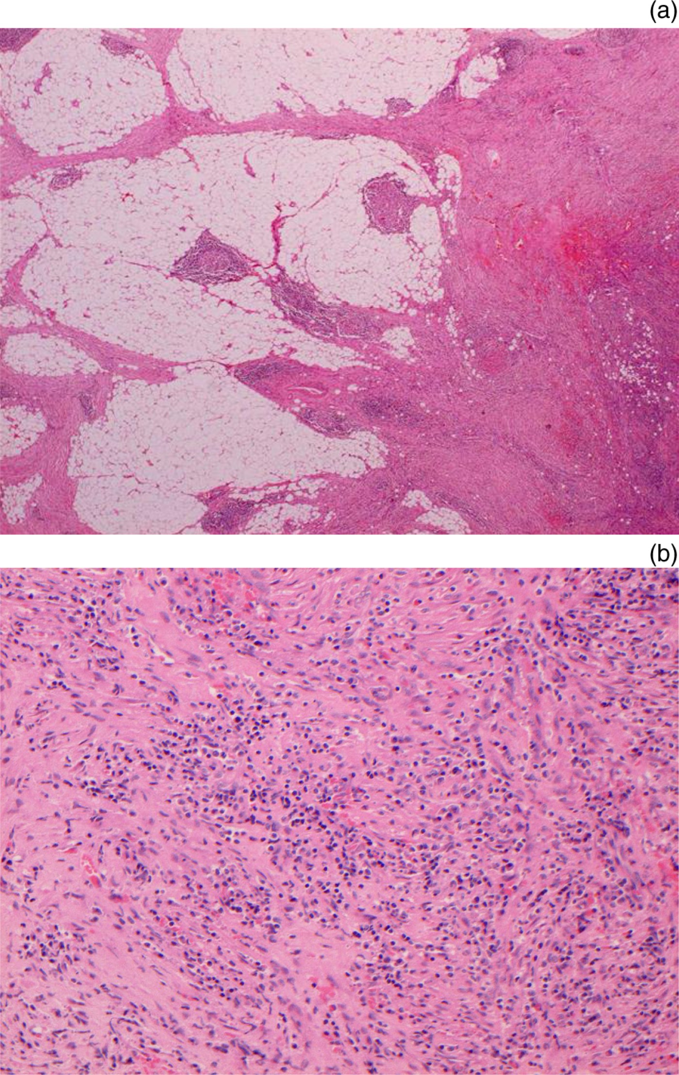
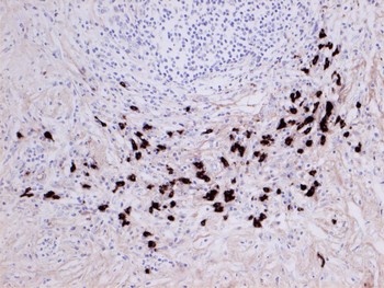
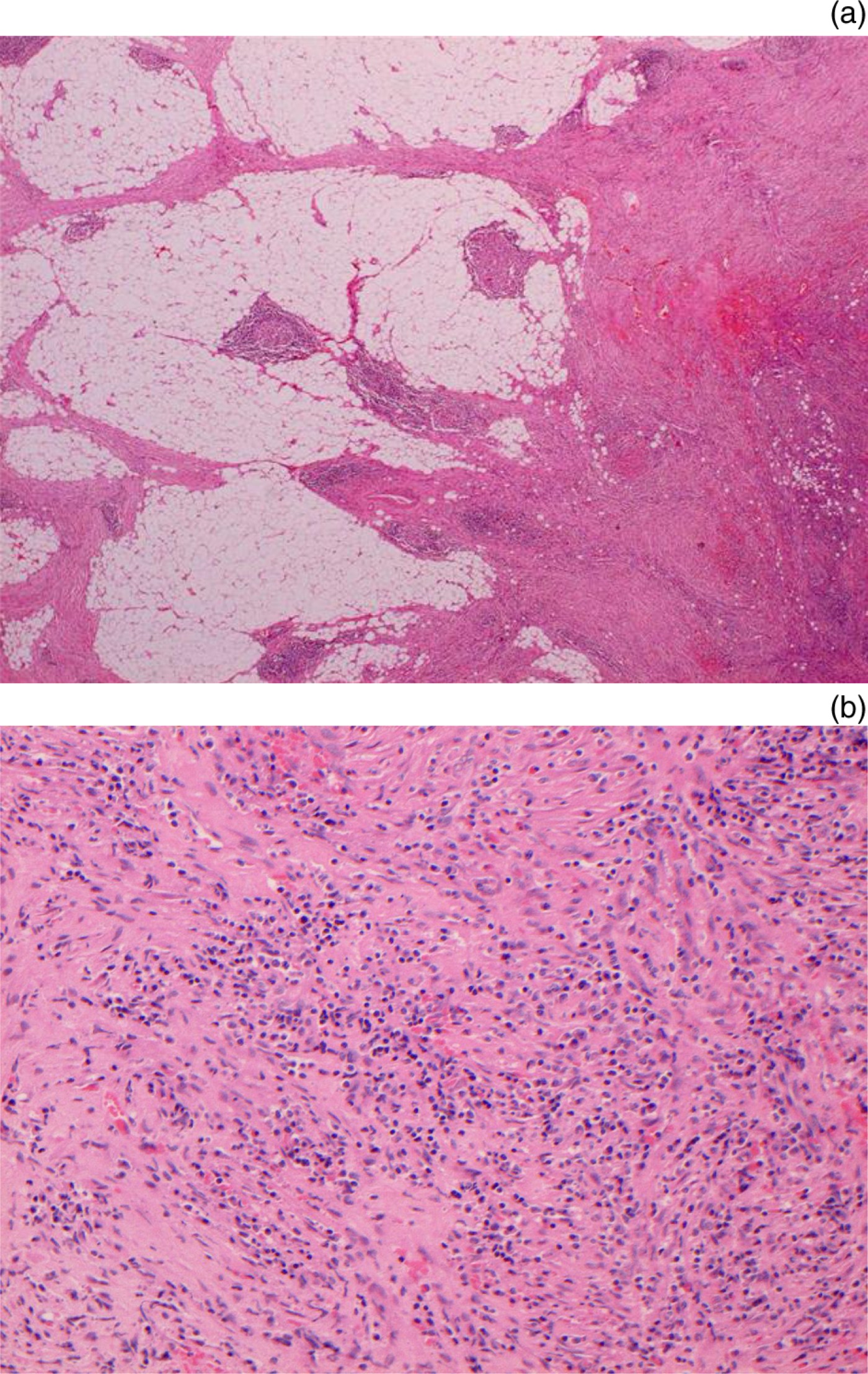
The patient was referred to the regional skull base multidisciplinary team. Histological review of the mastoid specimen revealed a diffuse infiltrate that did not correspond entirely to a diagnosis of inflammatory myofibroblastic tumour. The histology samples from previous resections were requested and analysed in detail by the histopathologist named in this paper (ER). This analysis revealed the same diffuse appearance in these specimens (Figure 2). Positive staining for IgG4 (Figure 3) led to the diagnosis of IgG4-related systemic sclerosing disease. This was confirmed by serum sample positivity for IgG4.
(a) Low power photomicrograph of omentum showing the poorly circumscribed, infiltrative nature of the lesion (H&E; ×10). (b) Medium power photomicrograph of temporal bone biopsy demonstrating fibrosis identical to that seen elsewhere, with an infiltrate of lymphocytes and plasma cells (H&E; ×20).
Photomicrograph of omentum showing immunohistochemical positivity for immunoglobulin (Ig) G4 in plasma cells, strong evidence for IgG4-related fibrosclerosing disease. A correlation with a raised serum IgG4 level during the active phase of disease would be diagnostic (×20).

This new diagnosis prompted a referral to the immunology team of the same institution. Systemic immunosuppressive treatment was initiated, in the form of mycophenolate mofetil and low dose prednisolone.
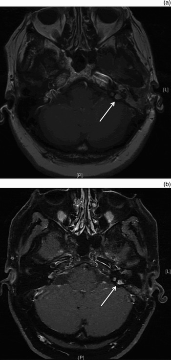
One year later, the patient remained well and in remission. The residual mass around the left jugular bulb was being reviewed at six-monthly intervals by magnetic resonance imaging (MRI) scanning. Serial imaging demonstrated a reduction in size and vascularity of the tumour, compared to its appearance prior to commencement of immunosuppressive treatment (Figure 4).
Interval, T1-weighted magnetic resonance imaging scans with gadolinium of temporal bones. (a) demonstrates disease (arrow) adjacent to left jugular bulb prior to treatment. (b) demonstrates decrease in the size of residual disease following six months of immunosuppressive treatment. L = left; P = posterior

Discussion
Immunoglobulin G4 related sclerosing disease is a new systemic entity and is commonly known to involve the pancreas and other retroperitoneal organs. Head and neck involvement has been described in the major salivary glands, the thyroid gland and ophthalmic region, but not, to date, in the temporal bone or larynx.Reference Neild, Rodriguez-Justo, Wall and Connolly2
Initially, this patient was thought to suffer from a systemic inflammatory pseudotumour, later relabelled as a multifocal inflammatory myofibroblastic tumour. The term inflammatory pseudotumour has been used to describe a pattern of bland fibroblastic and myofibroblastic spindle cell proliferation with prominent inflammation, particularly with plasma cells.Reference Schaeffer, Minai, Sharma, Kanne and Mohammed6 This probably represents a number of reactive entities. A proportion of these lesions have been shown to be true neoplasms – inflammatory myofibroblastic tumours.Reference Coffin, Watterson, Priest and Dehner7 Both diagnoses were used interchangeably in this case. However, these conditions usually present in the abdomen, retroperitoneum or lung, and tend toward local rather than distant or multifocal recurrence.Reference Gleason and Hornick8, Reference Meis and Enzinger9 Inflammatory myofibroblastic tumour typically occurs in children and young adults.
In the presented patient, the definitive diagnosis of IgG4-related systemic sclerosing disease was made after retrospective review of all histology specimens. The histological cellular constituents of this condition are identical to inflammatory pseudotumour and inflammatory myofibroblastic tumour, characteristically occurring together with obliterative phlebitis and, in the pancreas, periductal fibrosis plus a predominance of IgG4-positive plasma cells. However, when all our patient's biopsies were reviewed together, an infiltrative growth pattern with partial preservation of tissue architecture was observed, rather than the well defined mass seen in inflammatory myofibroblastic tumour. Additionally, there were more IgG4-positive plasma cells, eosinophils and lymphoid follicles than is typical for inflammatory myofibroblastic tumour. The spindle cells were anaplastic lymphoma kinase negative on immunohistochemical analysis, whereas up to 60 per cent of inflammatory myofibroblastic tumours are positive in this regard.Reference Cook, Dehner, Collins, Ma, Morris and Coffin10
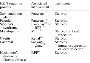
The recommended treatment of IgG4-related systemic sclerosing disease is corticosteroids.Reference Neild, Rodriguez-Justo, Wall and Connolly2, Reference Hamano, Kawa, Horiuchi, Unno, Furuya and Akamatsu3, Reference Tsuboi, Inokuma, Setoguchi, Shuji, Hagino and Tanaka11 This treatment has been advocated by Hamano and colleagues, who first described the disease, and by Neild et al., who found a significant reduction of plasma cells and absence of IgG4 expression in tissues after treatment of patients with corticosteroids.Reference Neild, Rodriguez-Justo, Wall and Connolly2, Reference Hamano, Kawa, Horiuchi, Unno, Furuya and Akamatsu3, Reference Hamano, Kawa, Ochi, Unno, Shiba and Wajiki5 Some authors have favoured the use of cytotoxic immunosuppression as an adjunct to steroid therapy when the head and neck region is affected (Table I). Surgical management is rarely indicated for extensive disease.
Reported head and neck manifestations of IgG4-related disease

Ig = immunoglobulin; H&N = head and neck; RPF = retroperitoneal fibrosis; MFF = multifocal fibrosis
Inflammatory pseudotumour affecting the temporal bone is rare, and as such the treatment strategy has not been established. Surgical resection is the treatment of choice in most reports due to the aggressive and unpredictable course.Reference Schonermark, Issing, Stover, Ruhand and Lenarz19–Reference Lee, Jung, Song, Yeo, Lee and Pang23 In one of the larger case series, Williamson et al. described complete surgical excision in eight cases and subtotal excision in one. Corticosteroids were administered to one patient with residual microscopic tumour, and external beam radiotherapy was used for residual or recurrent disease in another case.Reference Williamson, Paueksakon and Coker21 No long-term follow up exists for all treatment modalities. However, a review of two cases undergoing extensive surgical resection for erosive disease showed no recurrence at eight and 10 years, variously.Reference Strasnick and Vaughan20
Inflammatory pseudotumour can behave aggressively in the temporal bone, in contrast to the orbital region.Reference Williamson, Paueksakon and Coker21 Computed tomography and MRI are required to assess the extent of bone destruction and the inherent structure of the lesion. The recognised pattern is of a soft tissue mass characterised by low signal intensity on T2-weighted images and marked contrast enhancement. The tissue mass is often seen to spare the inner ear and the ossicles.Reference Gasparotti, Zanetti, Bolzoni, Gamba, Morassi and Ungari24 In the case described, the radiological and surgical findings demonstrated a more aggressive process.
Inflammatory pseudotumour of the temporal bone has been mostly treated surgically, followed by corticosteroids.Reference Strasnick and Vaughan20–Reference Lee, Jung, Song, Yeo, Lee and Pang23 However, there are cases of patients being treated with corticosteroids alone, with good results.Reference Schaeffer, Minai, Sharma, Kanne and Mohammed6, Reference Tsuboi, Inokuma, Setoguchi, Shuji, Hagino and Tanaka11, Reference Kasliwal, Suri, Gupta, Suri, Rishi and Sharma25, Reference Coulson, George, Biswas, Phelan and De26 Schaeffer et al. reported a case of recurrence following tapering reduction of corticosteroids, therefore supporting the need for surgery.Reference Schaeffer, Minai, Sharma, Kanne and Mohammed6 This case could also support an argument for the use of other immunosuppressive agents. None of the literature concerning pseudotumours in the temporal bone reported IgG4 staining. If performed, and in the absence of end-organ damage, this could have guided management strategies towards a more conservative approach. We would recommend a full review of all histological specimens in patients with a diagnosis of inflammatory pseudotumour or inflammatory myofibroblastic tumour. Consideration should be given to staining for IgG4, together with measurement of serum levels, especially in patients with unusual clinical or histological features such as multifocal distribution, poor circumscription with infiltrative margins, or non-typical constituent inflammatory cells.
• Immunoglobulin (Ig) G4 related systemic sclerosing disease is a multisystem, autoimmune inflammatory condition
• It closely resembles inflammatory pseudotumours and inflammatory myofibroblastic tumours
• The diagnosis is confirmed by immunohistochemical identification of IgG4-positive plasma cells, absence of anaplastic lymphoma kinase, and raised serum levels of IgG4
• Management is non-surgical, with corticosteroids and immunosuppressive agents
Our patient presented a diagnostic challenge, and underwent multiple operations under different specialties for various manifestations of the same disease. Her previous diagnosis of inflammatory pseudotumour, and latterly inflammatory myofibroblastic tumour, was replaced with a diagnosis of IgG4-related systemic sclerosing disease. The diagnosis of this autoimmune condition enabled the multidisciplinary team to avoid further surgery and to treat the patient with appropriate corticosteroids and immunosuppressive agents.