Introduction
Pigeon pea (Cajanus cajan (L.) Millsp.) seeds have a great potential as an important protein source (Cheva-Isarakul, Reference Cheva-Isarakul1992; Oshodi et al., Reference Oshodi, Olaofe and Hall1993) and every effort has been made to use pigeon pea seed meal (PM) for chickens (Mizubuti et al., Reference Mizubuti, Fonseca, Pinheiro, Motter, Van Dijk, Gois and Eleuterio1995), pigs (Batterham et al., Reference Batterham, Saini, Andersen and Baigent1993; Mekbungwan et al., Reference Mekbungwan, Yothinsirikul, Thongwittaya and Yamauchi1999) and lambs (Rao and Phillips, Reference Rao and Phillips2001). However, the efficacy of PM is limited by the presence of protease inhibitors such as trypsin and chymotrypsin inhibitors (Batterham et al., Reference Batterham, Saini, Andersen and Baigent1993). To improve the nutritional quality of PM, these antinutritional factors can be eliminated by heating (Singh, Reference Singh1988; Mekbungwan and Yamauchi, Reference Mekbungwan and Yamauchi2004), boiling (Rani et al., Reference Rani, Jood and Sehgal1996), roasting (Simoongwe, 1998), extraction (Benjakul et al., Reference Benjakul, Visessanguan and Thummaratwasik2000) and cooking (Aarti et al., Reference Aarti, Neelam and Saroj2001), resulting in improved protein and starch digestibility (Rani et al., Reference Rani, Jood and Sehgal1996). In our histological study (Mekbungwan and Yamauchi, Reference Mekbungwan and Yamauchi2004), the intestinal villi and epithelial cells of piglets fed raw PM showed an atrophic morphology owing to the presence of such antinutritional factors, resulting in decreased growth performance. Heating PM prevented the harmful effect of the antinutritional factors on villus histology, resulting in a growth performance comparable with the controls.
Additionally, charcoal powder has been used to reduce the adsorption of toxins from the gastrointestinal tract. Activated charcoal was reported to reduce the effects of dietary toxins by preventing intestinal absorption (Anjaneyulu et al., Reference Anjaneyulu, Rama Rao and Gopal Naidu1993). Also, a dietary 3 g/kg powder mixture of amorphous charcoal carbon and wood vinegar compound solution (CWVC) has been shown to produce more intestinal villus hypertrophy compared with the controls, resulting in improved feed efficiency in chickens (Samanya and Yamauchi, Reference Samanya and Yamauchi2001) and pigs (Mekbungwan et al., Reference Mekbungwan, Yamauchi and Sakaida2004b). Therefore, it was interesting to investigate the possibility that CWVC could reduce the protease inhibitory effect of dietary PM in piglet nutrition and encourage villus hypertrophy.
Material and methods
Animals and housing
Twenty-eight 30-day-old male castrated hybrid (0.25 Large White × 0.25 Landrace × 0.50 Duroc; average body weight = 14 kg) piglets were used in this study. Each piglet was placed in a pen with a 1.5 × 2.0 m floor area and given ad libitum access to feed and water. Weight gain and total feed intake were recorded. At the end of the experiment, three piglets per treatment were taken to a slaughterhouse; each piglet sedated with a 2-ml Nembutal (60 mg/ml pentobarbital sodium) intravenous injection and euthanased with a saturated injection of MgSO4 solution into the jugular vein. All the experiments were performed according to the humane care guideline for the care and use of laboratory animals established by the Faculty of Agriculture, Kagawa University (Miki, Japan).
Dietary treatment
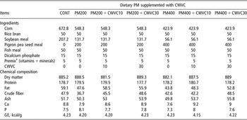
CWVC (Nekkarich®; 25 g/kg CP, 3 g/kg crude fibre, 134 g/kg ash, 39 g/kg calcium, 2 g/kg phosphorus and 286 g/kg water; pH 7.8; Miyazaki Midori Seiyaku Co., Ltd, Miyazaki, Japan) was produced as described below. Wood vinegar liquid obtained after cooling charcoal smoke from broadleaf trees by dry distillation at 300 to 450°C was kept for 2 to 3 years. Then the skimmed solution was distilled to remove harmful substances such as tar. This wood vinegar compound liquid was adsorbed onto amorphous charcoal carbon powder (1 : 4). The experimental design consisted of seven dietary treatment groups with four piglets in each group: unsupplemented diet (CONT), supplemented raw PM with 200 g/kg (PM200) and 400 g/kg (PM400) to basal diet, supplemented CWVC with 10 and 30 g/kg to PM200 and PM400 (PM200 + CWVC10, PM200 + CWVC30, PM400 + CWVC10 and PM400 + CWVC30) (Table 1).
The composition of experimental diets (g/kg) in 0, 200 and 400 g/kg pigeon pea seed meal (PM) (CONT, PM200 and PM400, respectively) diet, and 200 and 400 g/kg PM diets supplemented with 10 or 30 g/kg charcoal powder including wood vinegar compound liquid (CWVC) (PM200 + CWVC10, PM200 + CAVC30, PM400 + CWVC10 and PM400 + CWVC30, respectively)

†Premix supplies (per kg diet): vitamin A (3.333IU), vitamin D (667IU), vitamin E (0.33 mg), vitamin K3 (0.66 mg), vitamin B2 (1.67 mg), vitamin B12 (0.003 mg), calcium pantothenate (6.67 mg), cobalt (3.47 mg), copper (27.60 mg), iodine (0.77 mg), manganese (18.47 mg), zinc (50.00 mg) and Fe (60.00 mg).
Tissue sampling
After the piglets were euthanased, a mid-line incision was made to open the abdominal cavity and all the visceral organs were excised. Samples consisting of 5-cm sections of small intestine were taken at 15 cm from the stomach (regarded as the duodenum), in the middle of the small intestine (regarded as the middle part of the jejuno-ileum) and at 1 m from the ileo–caeco–colonic junction (regarded as the caudal part of the jejuno-ileum). Each section was ligated with a thread at both ends and a mixture of 30 ml/l glutaraldehyde and 40 ml/l paraformaldehyde in 0.1 mol/l cacodylate buffer (pH 7.4) was injected, taking care to avoid distension. Then, the sections were removed and kept in a bottle with the same fixative and prepared for light and scanning electron microscopy immediately.
Light microscopy
An 8 × 10-mm segment was cut from each 5-cm intestinal segment, fixed with Bouin’s fixative solution for one week at room temperature, embedded in paraplast and cut into 5-μm cross-sections. Every 10th section was collected and stained with haematoxylin-eosin. For villus height measurement, the villi (including the lamina propria) were chosen and the length from the villus tip to the bottom excluding the intestinal crypt was measured. Three villi were selected under 10 × 10 magnification for each section. Thirty values of villus height were counted from 10 sections per piglet, and the average of these values was expressed as the mean villus height for each piglet. To measure one cell area on the 5-μm cross-section, the area of the epithelial cell layer was randomly measured in the middle of the villi and the number of cell nuclei within this layer was counted. The area of the epithelial cell layer was then divided by this number. This measurement was employed in one to two fields per section. Fifteen samples were counted from 10 sections per piglet, and the average of these values was expressed as the mean cell area for each piglet. To measure the cell mitosis number per crypt, five crypts with almost the same size as one microscopic field (10 × 40 magnification) were randomly selected, the mitosis numbers were counted and then expressed as cell mitosis per one crypt. One to two fields per section were measured and 15 cell mitosis numbers were counted from 10 sections per piglet, and an average of these values was expressed as a mean cell mitosis number for each piglet. These measurements were recorded using an image analyser (Nikon Cosmozone lS, Nikon Co., Ltd, Tokyo, Japan). Finally, the mean of each intestinal parameter from the respective three piglets was expressed as the mean villus height, cell area and cell mitosis for one group.
Scanning electron microscopy
A 2 × 3 cm segment was cut from the 5-cm duodenal segment close to the light microscopic sample and slit longitudinally along the non-mesenteric side for its entire length. The intestinal contents were washed with 0.01 mol/l phosphate-buffered saline (pH 7.4). The tissue samples were pinned flat to prevent curling and fixed vertically with the mucosal surface facing downwards in the fixative mixture of 30 ml/l glutaraldehyde and 40 ml/l paraformaldehyde in 0.1 mol/l cacodylate buffer (pH 7.4) at room temperature for 1 h. The tissue block was further cut into a 3 × 10 mm rectangle and fixed for an additional 1 h. The pieces were rinsed with 0.1 mol/l sodium cacodylate buffer (pH 7.4) and post-fixed with 10 g/l osmium tetroxide in ice-cold buffer for 2 h. The specimens were dried in a critical-point drying apparatus. The dried specimens were coated with platinum and observed with a scanning electron microscope. After birth, as the exfoliative zone around the centre of the villus apical surface is the final stage of growth in epithelial cells of the intestinal crypt, we also observed the morphological alterations of epithelial cells around the central apical surface.
Gross anatomical protocol
After removing the tissue samples for microscopic examination, the remaining small intestine was divided into gross anatomical segments based on surface structural features: an intestinal part fixed to the abdominal cavity was prepared as the duodenum (showing thicker wall and slighter vessel networks on the serous surface than the jejuno-ileum; about 55 cm long), and a jejuno-ileal part of 2 m from the ileo–caeco–colonic junction to the rostral side was regarded as the caudal part of the jejuno-ileum (showing larger diameter but fewer aquare blood vessel networks than the rostral part of the jejuno-ileum). A remaining middle part was regarded as the middle part of the jejuno-ileum. Each part was cut, washed with 9 g/l NaCl solution to remove the intestinal contents and measured for length and weight. Visceral organs such as the liver, heart, spleen, kidney, stomach and lung were also weighed.
Statistical analysis
Data on growth performance, gross anatomy and light microscopic examination were statistically analysed by one-way analysis of variance (ANOVA), and significant differences among the treatments were determined with Duncan’s multiple range test using the Stat View® program (Abacus Concepts, Inc., Berkeley, CA, USA). Differences were considered significant at P < 0.05.
Results
Growth performance
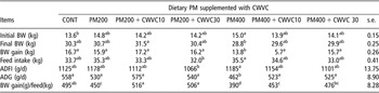
With increasing dietary PM levels, daily feed intake tended to increase. In contrast, daily body-weight gain tended to decrease, significantly in the PM400 group (P < 0.05), resulting in a significant decrease of feed efficiency in PM groups (P < 0.05) (Table 2). In general, body-weight gain and feed efficiency were higher (P < 0.05) in CWVC groups than in PM groups.
Growth performance of piglets fed dietary 0, 200 and 400 g/kg raw pigeon pea seed meal (PM) (CONT, PM200 and PM400, respectively) diets, and 200 and 400 g/kg PM diet supplemented with 10 or 30 g/kg charcoal powder including wood vinegar compound liquid (CWVC)(PM200 + CWVC10, PM200 + CAVC30, PM400 + CWVC10 and PM400 + CWVC30, respectively), (n = 4)

BW = body weight; ADFI = average daily feed intake; ADG = average daily gain; s.e. = standard error of mean.
a,b,c,dWithin a row, means without a common superscript letter differ (P < 0.05).
Gross anatomical observation
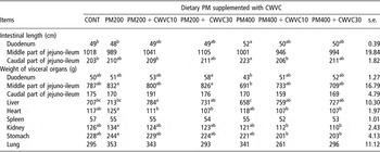
The duodenum and caudal part of the jejuno-ileum were longer (P < 0.05) in the PM400 group than in CONT (Table 3), but were similar to CONT in CWVC groups. Compared with CONT, PM200 and PM400 groups, the liver was heavier (P < 0.05), whereas the heart, kidney and stomach were lighter in CWVC groups; the weight of the heart in PM200 + CWVC10 and PM200 + CWVC30 groups was significantly decreased (P < 0.05).
Length of small intestine and weight of visceral organs of piglets fed dietary 0, 200 and 400 g/kg raw pigeon pea seed meal (PM) (CONT, PM200 and PM400, respectively) diets, and 200 and 400 g/kg PM diets supplemented with 10 or 30 g/kg charcoal powder including wood vinegar compound liquid (CWVC) (PM200 + CWVC10, PM200 + CAVC30, PM400 + CWVC10 and PM400 + CWVC30, respectively), (n = 3)

s.e. = standard error of mean.
a,b,cWithin a row, means without a common superscript letter differ (P < 0.05).
Light microscopic observations
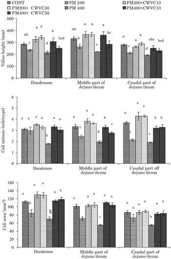
Villus height, cell area and cell mitosis number tended to decrease with increasing PM levels, and these values were lower (P < 0.05) in the PM400 group than in CONT for each intestinal segment (Figure 1). Most of these light microscopic parameters were higher in CWVC groups than in PM groups (P < 0.05).
Villus height, cell area and cell mitosis number in duodenum, middle part of the jejuno-ileum and caudal part of the jejuno-ileum in piglets fed dietary 0, 200 or 400 g/kg of raw pigeon pea seed meal (PM) (CONT, PM200 and PM400, respectively) diets, and 200 and 400 g/kg PM diets supplemented with 10 or 30 g/kg charcoal powder including wood vinegar compound liquid (CWVC) (PM200 + CWVC10, PM200 + CAVC30, PM400 + CWVC10 and PM400 + CWVC30, respectively), (n = 3). Values of light microscopic parameters are higher in CWVC groups than in PM groups. a,b,c,dMeans with different superscripts differ (P < 0.05) from each other.

Scanning electron microscopic observations
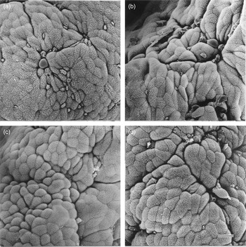
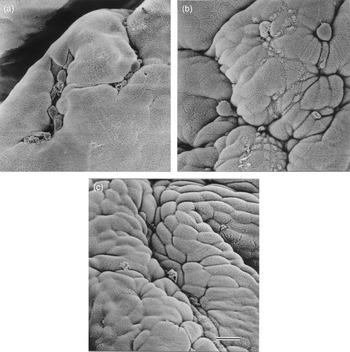
The duodenal villus apical surface in CONT (Figure 2a) was covered with comparatively protuberated epithelial cells, resulting in an inflated rough surface. On the villus tip of the PM200 group (Figure 2b), sunken parts (arrows) were frequently observed. However, in PM200 + CWVC10 (Figure 2c) and PM200 + CWVC30 groups (Figure 2d), clear cell protuberances developed, covering the villus apical surface and resulting in an inflated rough surface. In particular, the dome shaped epithelial cells of the PM200 + CWVC10 group protruded further into the lumen compared with CONT. In the case of the PM400 group (Figure 3a), epithelial cells changed to flat cells with unclear outlines, resulting in a smooth villus apical surface. These flat cells developed some protuberances in the PM400 + CWVC10 group (Figure 3b, arrows), which became clear on the whole villus apical surface in the PM400 + CWVC30 group (Figure 3c).
Duodenal villus apical surface of piglets fed dietary 0 (a) and 200 (b) g/kg raw pigeon pea seed meal (PM) diets, and fed dietary 200 g/kg PM diet supplemented with 10 (c) or 30 (d) g/kg charcoal-wood vinegar compounds (CWVC). Much more protuberated epithelial cells are seen in CWVC groups than in PM group. Scale bar = 32 μm, ×1058.

Duodenal villus apical surface of piglets fed dietary 400 g/kg raw pigeon pea seed meal (PM) diet (a), and fed dietary 400 g/kg PM diet supplemented with 10 (b) or 30 (c) g/kg charcoal-wood vinegar compounds (CWVC). One can see atrophied flat cells in PM group but hypertrophied protuberated cells in CWVC supplemented groups. Scale bar = 32 μm, ×1058.

Discussion
In this study, with increasing dietary PM levels, daily feed intake tended to increase. In contrast, daily body-weight gain tended to decrease, significantly in the PM400 group, resulting in a significant decrease of feed efficiency in PM groups. Nevertheless, the values of body-weight gain and feed efficiency were higher in CWVC groups compared with PM groups. Charcoal has been reported to reduce the effects of toxins in diets through adsorption (Anjaneyulu et al., Reference Anjaneyulu, Rama Rao and Gopal Naidu1993). When activated charcoal was added to diets containing aflatoxins or T-2 toxins, reductions in feed intake and body-weight gain of chickens were ameliorated (Dalvi and McGowan, Reference Dalvi and McGowan1984; Anjaneyulu et al., Reference Anjaneyulu, Rama Rao and Gopal Naidu1993; Edrington et al., Reference Edrington, Kubena, Harvey and Rottinghaus1997). The present CWVC would also eliminate the harmful effects of antinutritional factors in PM by adsorbing the toxins and thereby preventing their absorption by the intestine. These growth performances correspond with the histological intestinal alterations described later.
The PM400 group showed longer duodenum and caudal part of the jejuno-ileum, but decreased liver weight compared with CONT. Enlargement of the gastrointestinal tract was also observed in association with the various dietary arrangements (Iji et al., Reference Iji, Saki and Tivey2001; Brenes et al., Reference Brenes, Marquardt, Guenter and Viveros2002; Olkowski et al., Reference Olkowski, Classen, Wojnarowicz and Olkowski2005). PM includes protease inhibitors such as trypsin and chymotrypsin inhibitors, which decreased the activities of the pancreatic and small intestinal trypsin and chymotrypsin (Yen et al., Reference Yen, Jensen and Simon1977). The duodenum was reported as the site where the stimulation of pancreatic enzyme secretion is regulated (Corring, Reference Corring1974; Yen et al., Reference Yen, Jensen and Simon1977). The digestibility of crude protein and amino acids (Herkelman et al., Reference Herkelman, Cromwell, Stahly, Pferiffer and Knabe1992) was lowered by the trypsin inhibitors. A hyponutrient diet induced an increase in the length of all the intestinal sections not due to submucosal and serosal components, but the mucosa (Olkowski et al., Reference Olkowski, Classen, Wojnarowicz and Olkowski2005). Trypsin inhibitors induced pancreatic enlargement (Grant et al., Reference Grant, Dorward and Pusztai1993). From these cited references, the increased length of the intestine and decreased weight of the liver might be induced by the decreased use of nutrients in dietary PM diets due to the presence of trypsin and chymotrypsin inhibitors. Enlargement of the intestinal tract can be explained as an adaptive mechanism wherein by increasing the size of the intestine, the host increases its absorptive capacity in an attempt to extract maximum nutritional benefit from a poor-quality diet. However, these structural dissimilarities recovered to the CONT levels with inclusion of CWVC. From a nutritional and physiological standpoint, the mechanisms of lowered weight of heart, kidney and stomach associated with dietary CWVC are not completely understood in this study. However, it is speculated that these changes can also be explained as resulting from the adaptive mechanisms of the gastrointestinal tract in response to PM and CWVC diets.
Histology of the intestinal villi and epithelial cells on the villi apical surface in piglets (Mekbungwan et al., Reference Mekbungwan, Yamauchi and Thongwittaya2003) and chickens (Yamauchi et al., Reference Yamauchi, Buwjoom, Koge and Ebashi2006b) are well known to be affected by dietary feed components. Values of the present villus height, cell area and cell mitosis numbers were lower in the PM groups compared with CONT. Compared with a commercial diet, short villi were reported in pigs showing decreased body weight (Zijlstra et al., Reference Zijlstra, Whang, Easter and Odle1996) and low nutrient digestibility (Mekbungwan et al., Reference Mekbungwan, Thongwittaya and Yamauchi2004a), and in those fed a diet containing a higher percentage of soya beans (Nabuurs et al., Reference Nabuurs, Hoogendoorn, Van der Molen and Van Osta1993). These short villi correspond with reductions in activities of enzymes such as mucosal lactase and sucrase (Park et al., Reference Park, Monaco and Donovan1998), lactase and alkaline phosphatase (Zijlstra et al., Reference Zijlstra, Whang, Easter and Odle1996), alkaline phosphatase and disaccharidase (Lopez-Pedrosa et al., Reference Lopez-Pedrosa, Torres, Fernandez, Rios and Gil1998), and the total lactase phlorizin hydrolase and mucosal protein concentration (Dudley et al., Reference Dudley, Wykes, Dudley, Burrin, Nichols, Rosenberger, Jahoor, Heird and Reeds1998). The short villi were accompanied by reductions in the villus surface area (Park et al., Reference Park, Monaco and Donovan1998) resulting in a reduced absorptive function. Furthermore, with increasing dietary PM levels, the villus apical surface changed from being inflated and rough to smooth via the presence of sunken parts. As the cell area measured using light microscopy was reduced after feedings of PM200, these partially sunken parts are thought to be a consequence of reduced cell area. A further decrease in cell area in all epithelial cells might produce flat cells on the villus apical surface, resulting in a smooth surface seen in the PM400 group. Such flat cells were also reported in pigs undergoing a 3-day feed withdrawal (Mekbungwan et al., Reference Mekbungwan, Yamauchi and Thongwittaya2002). These studies suggest that the present decreased light microscopic parameters and the flat cells in PM groups might suggest histological atrophy. Raw PM has been reported to contain antinutritional factors such as trypsin and chymotrypsin inhibitors (Jambunathan and Singh, 1980; Godbole et al., Reference Godbole, Krishna and Bhatia1994; Rani et al., Reference Rani, Jood and Sehgal1996), resulting in decreased growth performance with increasing levels of raw PM (Mekbungwan and Yamauchi, Reference Mekbungwan and Yamauchi2004). The main reason for the present histologically atrophied features is thought to be the presence of antinutritional factors in PM. Conversely, the atrophied histological alterations seen in PM groups were found to be improved in CWVC groups. Long villi were reported in piglets that showed an increased body-weight gain (Zijlstra et al., Reference Zijlstra, Whang, Easter and Odle1996), in turkeys fed dietary amylase (Ritz et al., Reference Ritz, Hulet, Self and Tsao1995) and in chickens showing a high activity of amylase in the intestinal content (Samanya and Yamauchi, Reference Samanya and Yamauchi2002). It has been suggested that long villi result in increased surface area capable of greater absorption of available nutrients (Caspary, Reference Caspary1992), and that greater villus height and increased cell mitosis in the intestine are indicators of the activated function of the intestinal villi (Langhout et al., Reference Langhout, Schutte, Van Leevwen, Wiebenga and Tamminga1999; Yasar and Forbes, Reference Yasar and Forbes1999). In addition, on the villus apical surface of CWVC groups, protuberated cells were observed. Such cells were also reported in chickens showing a quicker recovery of body weight after refeeding sugar cane extract (Yamauchi et al., Reference Yamauchi, Buwjoom, Koge and Ebashi2006a) and in piglets showing high nutrient digestibility (Mekbungwan et al., Reference Mekbungwan, Thongwittaya and Yamauchi2004a). These studies suggest that the present high light microscopic parameters and the protuberated cells are much more hypertrophied in CWVC than in PM groups. In our previous studies, such histological intestinal recovery from the atrophied intestinal histology in raw PM groups was observed after feedings of heated PM diet in piglets (Mekbungwan and Yamauchi, Reference Mekbungwan and Yamauchi2004), and of the CWVC diet in piglets (Mekbungwan et al., Reference Mekbungwan, Yamauchi and Sakaida2004b) and chickens (Samanya and Yamauchi, Reference Samanya and Yamauchi2001). In the case of heated PM, the histological intestinal recovery was caused by a 99.15% decrease in the inhibition rate of trypsin in raw PM compared with 54.31% in heated PM (Mekbungwan and Yamauchi, Reference Mekbungwan and Yamauchi2004). The trypsin inhibitor in the raw PM has also been known to decrease by roasting (Simoongwe, 1998), cooking (Sharma and Sehgal, Reference Sharma and Sehgal1992) and heating (Mekbungwan and Yamauchi, Reference Mekbungwan and Yamauchi2004) PM. In the case of CWVC, the histological intestinal recovery was caused by the adsorbing effect of CWVC. Activated charcoal was reported to reduce the effects of dietary toxins by adsorbing them and thereby preventing their absorption from the intestine (Anjaneyulu et al., Reference Anjaneyulu, Rama Rao and Gopal Naidu1993). Amorphous charcoal carbon powder in CWVC adsorbed Salmonella enteritidis much more selectively than conventional Enterococcus faecium, and the wood vinegar compound liquid in CWVC inhibited the growth of S. enteritidis but accelerated the growth of E. faecium and Bifidobacterium thermophilum (Tana et al., Reference Tana, Watarai, Kodama and Iwakiri2003). Although we did not measure the inhibition rate of trypsin inhibitor in this study, trypsin activity in the jejunal content was significantly decreased after feedings of 300 g/kg dietary PM (P < 0.05) but tended to improve after supplementation of 10 g/kg CWVC to this 300 g/kg dietary PM in chickens (unpublished). These observations and the findings of similar studies in the literature lead to a general conclusion that the CWVC might eliminate the harmful effects of the antinutritional factors in PM on the villus and cellular functions, thereby activating intestinal function, resulting in a growth performance similar to CONT. The raw PM can be used by adding CWVC without heating.
In conclusion, histological intestinal villus alterations in piglets fed a PM or PM + CWVC diet demonstrate that the villi might be atrophied in the piglets fed PM due to the presence of antinutritional factors, resulting in decreased growth performance. CWVC might eliminate the harmful effects of the antinutritional factors on the villus function, resulting in normal growth performance.