Introduction
Opisthorchis viverrini, O. felineus and Clonorchis sinensis are liver flukes of man and piscivorous mammals that occur in many parts of East Asia and Eastern Europe. The number of people infected with liver flukes has been estimated at 45 million worldwide and the number of people at risk at over 600 million (Keiser & Utzinger, Reference Keiser and Utzinger2005; WHO, 2011). Liver flukes are foodborne trematodes that use Bithynia snails and cyprinid fish as first and second intermediate hosts, respectively. The human and animal final hosts become infected by eating raw or undercooked fish infected with metacercariae (Kaewkes, Reference Kaewkes2003; De & Le, Reference De and Le2011). In humans, the infection is associated with several hepatobiliary abnormalities. In addition, experimental and epidemiological evidence strongly implicate O. viverrini infection in the aetiology of cholangiocarcinoma (CCA) or bile duct cancer, which has one of the highest mortality rates of any cancer (Sripa et al., Reference Sripa, Brindley, Mulvenna, Laha, Smout, Mairiang, Bethony and Loukas2012).
Dogs and cats are important reservoir hosts of many foodborne trematodes, including Opisthorchis spp.; birds are reservoirs of some intestinal fluke species (Chai et al., Reference Chai, Murrell and Lymbery2005; Anh, Reference Anh2009; Anh et al., Reference Anh, Madsen, Dalsgaard, Phuong, Thanh and Murrell2010; Aunpromma et al., Reference Aunpromma, Tangkawattana, Papirom, Kanjampa, Tesana, Sripa and Tangkawattana2012). In 2003, Opisthorchis lobatus, a new species of Opisthorchis, was described in the intestines of ducks (Anas sp.) in Pakistan, and its metacercariae were found in red-tailed snakehead fish in Laos (Bilqees et al., Reference Bilqees, Shabbir and Parveen2003; Thaenkham et al., Reference Thaenkham, Nuamtanong, Vonghachack, Yoonuan, Sanguankiat, Dekumyoy, Prommasack, Kobayashi and Waikagul2011). Up to now, the epidemiology and zoonotic potential of this new species are not known.
In Vietnam, C. sinensis is prevalent in the north and O. viverrini in the south (De & Le, Reference De and Le2011). In November 2009, a small Opisthorchis-like fluke was found during routine necropsy in the bile ducts of domestic ducks in Binh Dinh province, in the south central coast region of Vietnam that is endemic for O. viverrini. Opisthorchis spp. had previously never been recorded in birds in Vietnam.
The objective of this study was to establish the taxonomic status of this fluke using morphological and molecular methods.
Materials and methods
Collection and examination of flukes
Twenty-five adult worms were collected in November 2011 from the bile ducts of two naturally infected domestic ducks (Anas platyrhynchos) during routine necropsy. The ducks originated from Phu My district, Binh Dinh province, Central Vietnam (14.005–14.024′N, 108.057–109.017′E).
Eleven worms were flattened between glass slides, fixed in polyvinyl alcohol–fixative–adhesive (AFA) solution and stained with Semichon's acetic carmine solution, before being cleared in xylene and mounted in DPX mountant on a glass slide. Stained worms were examined under a light microscopy: the shape and the sizes of the body, internal organs, suckers and eggs, and the ratios of body length to body width, and egg length to egg width, were taken into account for morphological examination. The identification key of the Opisthorchiidae family – Opisthorchis genus (Bray et al., Reference Bray, Gibson and Jones2008) and morphological descriptions from several authors (Bilqees et al., Reference Bilqees, Shabbir and Parveen2003; Kaewkes, Reference Kaewkes2003; Thaenkham et al., Reference Thaenkham, Nuamtanong, Vonghachack, Yoonuan, Sanguankiat, Dekumyoy, Prommasack, Kobayashi and Waikagul2011; Mordvinov et al., Reference Mordvinov, Yurlova, Ogorodova and Katokhin2012) were used to compare morphological characteristics of the duck worms with that of liver flukes from humans and other mammals.
Molecular analysis
Genetic markers used in this study include sequences of the second internal ribosomal spacer (ITS-2) and the cytochrome c oxidase subunit I (COI) of the mitochondrial DNA, obtained by polymerase chain reaction (PCR) and sequencing.
Total genomic DNA was extracted from a single piece of adult worms using the commercial Gentra Puregene Kit, according to the manufacturer's instructions (Qiagen GmbH, Hilden. Germany). Amplification was done from 5 μl of the extracted genomic DNA, using 0.4 μl of each primer: 3S: (5′-GGTACCGGTGGATCACTCGGCTCGTG-3′) and BD2 (5′-TATGCTTAAATTCAGCGGGT-3′) for the ITS2 marker (Bowles et al., 1993) and COI-OV-Hap-F (5′-GGGTTYGGTATRRTKAGWCAC-3′) and COI-OV-Hap-R (5′-AAACCAAGTRTCATGMAACAAAG-3′) for the COI marker (Thaenkham et al., Reference Thaenkham, Visetsuk, Dung and Waikagul2007), and 0.2 μl of GoTaq® DNA polymerase (Promega, Madison, Wisconsin, USA), plus 1.65 μl of MgCl2 25 mm and 0.2 μl deoxynucleoside triphosphates (dNTP) in a total volume of 25 μl. The PCR amplification was performed as follows: 40 cycles at 94°C for 15 s for denaturation; annealing 45 s at 50°C with primers BD2 and 3S and at 52°C with primers COI-Ov-Hap-F and -R; extension at 72°C for 45 s, final extension at 72°C for 10 min. Then, 5 μl of each PCR product was electrophoresed on agarose 2% in 0.5 × TBE buffer at 100 V for 20 min and visualized under ultraviolet light.
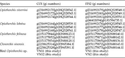
Next, PCR amplicons of both markers were purified on a column using QIAquick® PCR Purification Kit (Qiagen), cloned in JM109 Cells of the pGEM® T Vector Systems Kit (Promega) and sent to the VIB Genetic Service Facility (University of Antwerp, Belgium) for sequencing. The obtained DNA sequences were edited with BioEdit (Hall, Reference Hall1999), BLASTed on NCBI and aligned with sequences from O. viverrini, O. lobatus, O. felineus and C. sinensis (table 1).
DNA sequences of four Opisthorchis spp. and Clonorchis sinensis used for the alignment and construction of the phylogenetic trees.

DNA sequences were aligned with the MAFFT L-INS-I algorithm (v7.029b; Katoh & Standley, Reference Katoh and Standley2013). Phylogenetic trees were created with the MAFFT server (http://mafft.cbrc.jp/alignment/server/phylogeny.html) using neighbour joining (NJ; all gap-free sites) and bootstrap resampling set to 1000. The amino acid sequence was derived from the COI DNA sequences using transeq (Rice et al., Reference Rice, Longden and Bleasby2000) to verify whether changes in the DNA also resulted in amino acid changes.
Results
Morphological analysis
Based on Opisthorchis-specific morphological features, such as the presence of two extra-caecal chains of vitelline glands, running between the ventral sucker and the anterior or posterior testes, and the location of the ovary in the posterior third of the body, the duck fluke is to be classified as an Opisthorchis species. The duck fluke differs in a number of morphological characteristics from O. felineus and C. sinensis, such as the location of the uterus, which overlaps the ventral sucker entirely or partially, and the shape of the testes, which is lobate in Opisthorchis spp. and dendritic in C. sinensis.
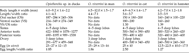
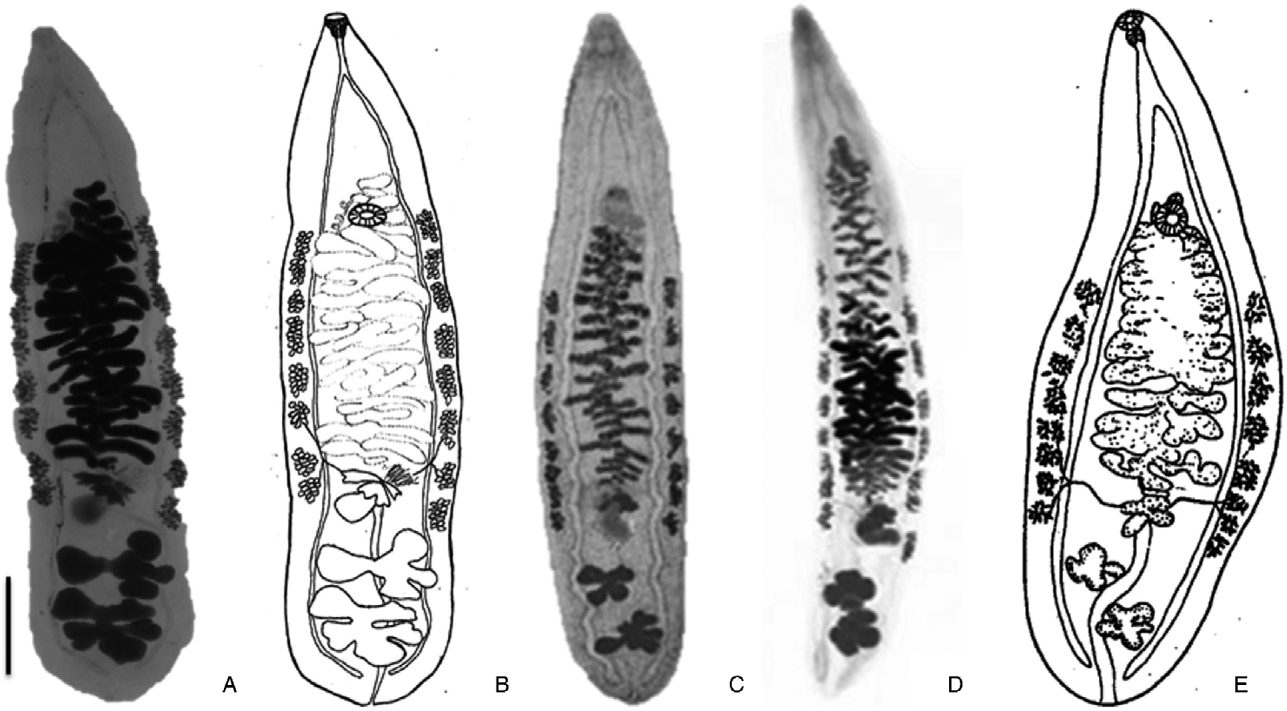
The morphological characteristics of the liver flukes of ducks are compatible with those of O. viverrini in human hosts, cats and an experimental hamster model (Mas-Coma & Bargues, Reference Mas-Coma and Bargues1997; Le, Reference Le2000; Kaewkes, Reference Kaewkes2003; Sohn et al., Reference Sohn, Shin, Yong, Eom, Jeong, Sinuon, Socheat and Chai2011) (table 2 and fig. 1). The body of the flukes is transparent and lanceolate; the ventral sucker is larger than the oral sucker, located in the anterior quarter of the body; the uterus, packed with oval-shaped eggs, loops irregularly between the ventral sucker and the ovary; the three-lobed ovary locates slightly submedian or median in the posterior third of the body; the seminal receptacle is voluminous, sited posteriorly to the ovary. The testes are large and located one after the other (tandem) with their extremities deeply lobed; they occupy almost the entire posterior body's width, overlapping both caeca laterally. The posterior testis is bigger than the anterior one. The excretory bladder lies posteriorly to the testes. The vitelline glands form two chains extra-caecally, and they extend between the ventral sucker and the testis. The caeca end blind in the posterior part of the body.
Morphological and morphometric comparison (in μm unless stated) of Opisthorchis sp. in ducks and O. viverrini in a man (Sohn et al., Reference Sohn, Shin, Yong, Eom, Jeong, Sinuon, Socheat and Chai2011), cat (Le, 2000) and hamster (Thaenkham et al., Reference Thaenkham, Nuamtanong, Vonghachack, Yoonuan, Sanguankiat, Dekumyoy, Prommasack, Kobayashi and Waikagul2011); all flukes are lanceolate in shape.
Intraspecific morphological variation of Opisthorchis viverrini in different hosts: (A) Opisthorchis sp. in birds from Vietnam; (B) schematic drawing of an Opisthorchis sp. in birds from Vietnam; (C) O. viverrini in a hamster (Sohn et al., Reference Sohn, Shin, Yong, Eom, Jeong, Sinuon, Socheat and Chai2011); (D) O. viverrini in a man from Cambodia (Sohn et al., Reference Sohn, Shin, Yong, Eom, Jeong, Sinuon, Socheat and Chai2011); (E) O. viverrini in a cat from Vietnam (Le, Reference Le2000). Scale bar = 1 mm.
However, morphometric data of the liver fluke of ducks are larger than those of O. viverrini in mammal hosts, except for the body length (table 2). The testes of the bird fluke are two to four times larger. Although the size of the oral and ventral suckers of the bird fluke is considerably larger, ratios of ventral to oral sucker are similar, with values of 1.36, 1.30 and 1.39 for bird flukes and O. viverrini in cat and hamster, respectively. The ratio of uterus egg length/width of the bird fluke is similar to that of O. viverrini in humans (ratio = 1.85 and 1.86, respectively). This ratio is higher in cat (ratio = 2.5) and lower in hamster (ratio = 1.27) flukes. These morphological variations may fall into the intraspecific variability of O. viverrini in different hosts.
Molecular analysis
BLASTN analysis and MAFFT alignments of VN1 and VN2 (ITS-2) showed a high degree of identity to the other O. viverrini sequences. A total of two mismatches (positions 145 and 329) and two gaps (six nucleotides) were observed. BLASTN reports 99.4% identity (no gaps, 355 aligned bases) and 97.8% identity (2 gaps, 361 aligned bases).
BLASTN analysis of VN11 and VN22 (COI) revealed a lower degree of identity to the other O. viverrini sequences, ranging from 89.3 to 90.6%. No gaps were observed, but BLASTN reported mutations all over the DNA sequence as well as 2–3 unaligned bases. The many mutations that were observed in the DNA sequences resulted in 3–5 amino acid changes.
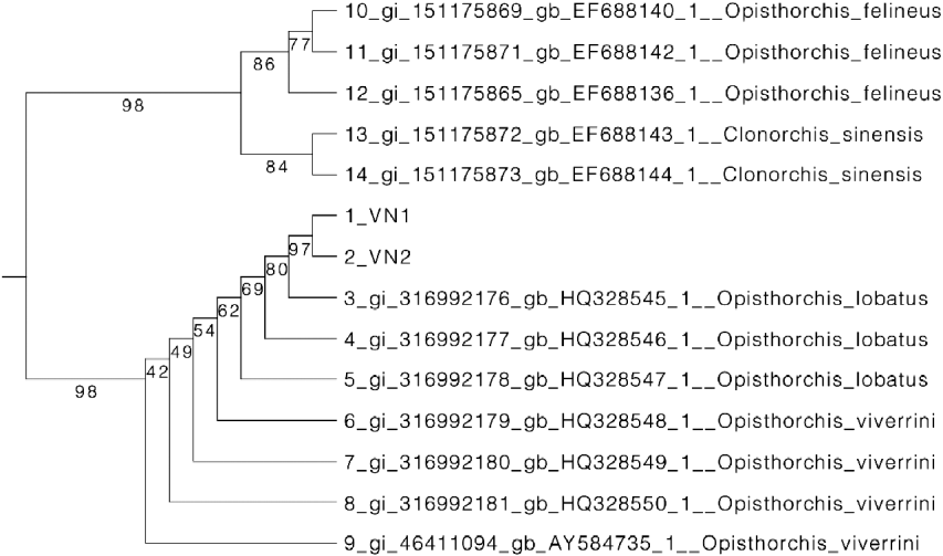
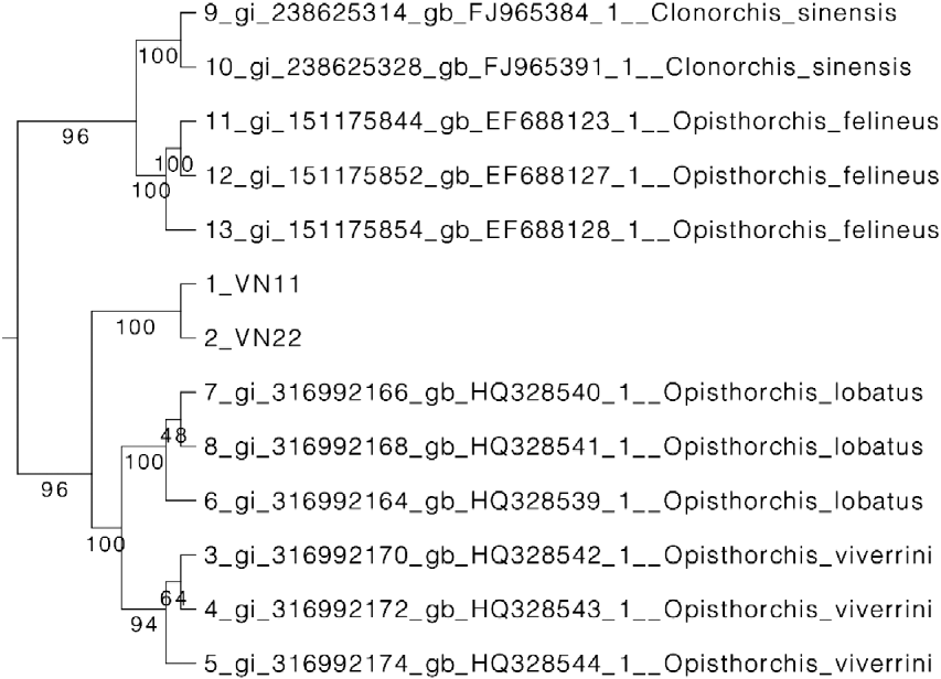
Analysis of the ITS-2 and COI phylogenetic trees reveals close resemblance between O. viverrini, O. lobatus and the unknown species; however, in both cases, the unknown species is always in a separate clade (figs 2 and 3).
Phylogenetic tree of the partial DNA sequences of ITS2 from the Opisthorchis sp. in ducks and O. viverrini, O. lobatus, O. felineus and Clonorchis sinensis sequences submitted to GenBank (table 1).
Phylogenetic tree of the partial DNA sequences of CO1 from the Opisthorchis sp. in ducks and O. viverrini, O. lobatus, O. felineus and Clonorchis sinensis sequences submitted to GenBank (table 1).
Discussion
Morphological examination and gene analysis of the liver flukes found in ducks in Central Vietnam show close similarity with O. viverrini and O. lobatus. Based on genetic analysis of isolates from different geographical areas, O. viverrini may be divided into at least two or six cryptic species; however, morphological variations within the species were not described until now (Sithithaworn et al., Reference Sithithaworn, Nuchjungreed, Srisawangwong, Ando, Petney, Chilton and Andrews2007; Laoprom et al., Reference Laoprom, Saijuntha, Sithithaworn, Wongkham, Laha, Ando, Andrews and Petney2009). In this study, we found some morphometric differences between the flukes of ducks and O. viverrini isolated from cat, human and hamster (fig. 1, table 1). In other trematode species, such as Fasciola hepatica, the final host species may influence both the morphology and the morphometric characteristics of adult worms and eggs, and the genetic characteristics (Valero et al., Reference Valero, Darce, Panova and Mas-Coma2001). In our study, analysis of the partial sequence of ITS2 of the rDNA, shows very close resemblance (97.4–99.4%) between the duck fluke and O. viverrini. On the mtDNA COI marker, a lower degree of identity between the duck fluke and published sequences of O. viverrini was found (~90%).
Although the observed differences in morphology and COI and ITS-2 DNA sequences may suggest that the liver flukes are a separate species in the Opisthorchis genus, they may also be due to adaptation of O. viverrini to a new/different host and the differences are then to be considered intra-specific variations.
Further studies should be done to demonstrate the life cycle of this O. viverrini-like species in birds and to investigate whether it could be transmitted to mammals. If that is the case, it would render this parasitic infection much more difficult to control. Indeed, birds could then act as a reservoir and migrate over long distances, easily bringing the infection to new areas. Hence, a large survey of O. viverrini in birds should be carried out.
Acknowledgements
We would like to thank the Japanese Government through the JJ/WBGSP (Joint Japan/World Bank Graduate Scholarship Program) and the Belgian Government through the DGD (Belgium Directorate-General for Development) scholarship for financial support. Mr Ko De Witte (Institute of Tropical Medicine, Antwerp, Belgium) and Ms Tran Thi Lam Ha (Sub-Animal Health Department of Binh Dinh province, Vietnam) are thanked for technical and logistic support.