Introduction
Hairy polyps, or dermoids, have classically been considered a form of primitive teratoma consisting of both mesoderm and ectoderm germ cells. They usually arise from the oropharynx or nasopharynx and, although rare, are the commonest congenital nasopharyngeal mass. The appearance is of a grey, pedunculated lesion, often presenting at birth, with symptoms of respiratory distress or feeding difficulties. The severity of symptoms is dependent on the size of the lesion, and occasionally these may go undetected until adult life.Reference Chaudhry, Lore, Fisher and Gambrino1
The aetiology of hairy polyps is unclear. They can be differentiated from teratomas by their lack of neoplastic potential. It is debatable whether these lesions represent true teratomas or are in fact developmental malformations of either the first or second branchial arch system.
We present what we believe to be the first reported association of a hairy polyp with a first branchial arch sinus, as evidence that hairy polyps are a malformation of the first branchial arch system.
Case report
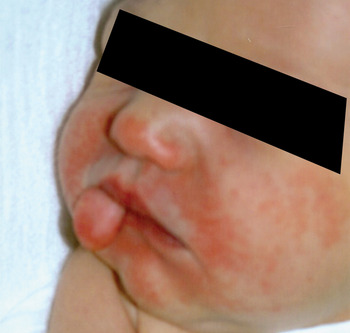
A 2-day-old, male infant was transferred from a district general hospital to the otolaryngology department at The General Infirmary at Leeds after his mother noticed a mass protruding from his mouth (Figure 1). He had had no health issues antenatally and was full-term.
The infant at first presentation, with the hairy polyp seen protruding from his mouth.

Apart from a soft tissue mass protruding from the left side of the mouth, examination was unremarkable.
The mass was excised from the left side of the posterior aspect of the soft palate on day four of life.
Histological examination revealed keratinising squamous epithelium surrounding a fibroconnective core, complete with clusters of hair follicles and sebaceous glands, features diagnostic of a hairy polyp.
The child made an uneventful recovery, and follow up at two and six months showed no signs of recurrence.
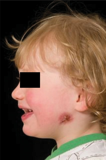
At the age of 16 months, the child was seen in our out-patient department having presented to his general practitioner with swelling and discharge from the angle of the left mandible. On questioning, the child's parents had noticed that he had had a small dimple in this region since birth, which had gradually increased in size and then started discharging fluid.
On examination, the overlying skin was discoloured and a tract could be palpated up to the external auditory canal (Figure 2).
The child at 16 months of age, with a discharging tract now evident.

A magnetic resonance imaging scan of the neck demonstrated a tract passing from the angle of the mandible into the superficial portion of the parotid gland and extending towards the external auditory meatus, in keeping with a type II first branchial cleft fistula.
The child subsequently underwent surgical excision of the sinus via a modified Blair's incision. This revealed a tract lying superficial to the facial nerve and extending towards the external ear canal. The tract was dissected clear of surrounding tissue and excised en bloc.
Discussion
Macroscopically, hairy polyps are sausage- or pear-shaped, grey, pedunculated lesions. Approximately 60 per cent originate from the nasopharynx, while the rest are found in the tonsillar region or the oropharynx. Microscopically, they are covered by squamous epithelium and associated skin appendages (i.e. sebaceous glands, sweat glands and hair follicles) and contain fibro-fatty tissue, cartilage and striated muscle.
The presenting symptoms are dependent on the size and location of the lesion, but patients are usually seen in the neonatal period with symptoms of respiratory distress or dysphagia.
Treatment usually involves prompt excision of the polyp, due to its potential to compromise the airway. Surgery is generally curative, although recurrence has been reported.Reference Haddad, Senders, Leach and Stool2
Arnold was the first to classify teratomas of the head and neck region, using a system based on the germ cell layers involved and the complexity of the lesion.Reference Arnold3 He proposed that these malformations could be classified into four main types, as follows. Dermoids (so-called hairy polyps) consist of two germ cell layers, mesoderm and ectoderm, and macroscopically appear as sausage- or pear-shaped, pedunculated growths. Teratomas are larger in size and originate from all three germ cell layers; they are commonly associated with other developmental skull deformities (e.g. anencephaly and palatal fissures). Teratoids are similar to teratomas but consist of much more poorly differentiated tissues. Finally, epignathi, also known as a parasitic fetus, comprise highly differentiated tissue usually attached to the sphenoid bone, and can contain organs and limbs.
This classification system, which is still widely used and accepted, takes the viewpoint that hairy polyps are a form of primitive teratoma. Teratomas are considered to be neoplastic as they display both progressive and disorganised growth. Hairy polyps, however, have limited growth potential, and to date there has not been a single reported case of a hairy polyp undergoing neoplastic change. They are six times more common in females, whereas teratomas display equal incidence in both sexes. Three cases of bilateral hairy polyps have been reported, suggesting that they may represent malformative lesions rather than true teratomas.Reference Chaudhry, Lore, Fisher and Gambrino1, Reference Franco, Florena, Lombardo and Restivo4, Reference Morgan5
Brown-Kelly is credited with reporting the first case of hairy polyp within the English language literature.Reference Brown-Kelly6 This author also reported 49 other cases from various world-wide sources, and noted a 10 per cent association with cleft palate, as well as associations with various branchial arch malformations including absence of the uvula and external ears, ankyloglossia, and facial hemihypotrophy. Brown-Kelly proposed that the site of the lesion was suggestive of a second branchial arch origin. Although we report the first case of a hairy polyp associated with a first branchial arch sinus, Burns et al. have reported a similar case of a hairy polyp associated with a second branchial arch sinus, supporting Brown-Kelly's theory.Reference Burns, Axon and Pahade7
• Hairy polyps are rare, congenital lesions
• They are unlikely to represent primitive teratomas
• The presented case supports the theory that they are branchial arch malformations
• Children with hairy polyps must be thoroughly examined to detect associated malformations
Schuring, however, considered the hairy polyp to be an accessory auricle originating from the first branchial arch system, due to its histological similarity.Reference Schuring8 Further histological evidence is provided by Heffner et al., who reviewed the histology of four pharyngeal teratomas and eight hairy polyps.Reference Heffner, Thompson, Schall and Anderso9 They noted that polyps containing cartilage resembled the fetal pinna both macroscopically and microscopically, in the arrangement of the cartilage as a curved plate of uniform thickness, an arrangement that was not seen in the teratomas. The theory of a first branchial arch origin was lent credence by a case report by Kanzaki et al. of a hairy polyp macroscopically resembling an auricle, and another case report by Boedts et al. of a hairy polyp located in the middle ear and mastoid cavity.Reference Kanzaki, Yamada, Fujimoto, Arata, Miyake and Ohbayashi10, Reference Boedts, Moerman and Marquet11
Taken together with previous reported cases, our case adds further weight to the argument that hairy polyps represent a developmental anomaly of the branchial arch system. Our case also highlights the importance of a thorough, systematic examination of such cases to ensure that associated branchial arch anomalies are not missed.
Conclusion
Hairy polyps are rare, congenital lesions that are unlikely to represent primitive teratomas. The presented case provides further evidence that they may be considered as malformations of the branchial arch system. When managing children with hairy polyps, a thorough examination is essential to ensure associated malformations are not missed.