Introduction
The increased spreading rate of epidemics as a result of a growing number of international and domestic flights as well as the population of countries, in particular cities, forced people to use both public and personal antimicrobial products [Reference Hollingsworth, Ferguson and Anderson1]. Since pathogenic microorganisms become more and more resistive against existing products, improvements to the antimicrobial agents are necessary [Reference Soares, Garcia, Roesch-Ely, da Costa, Giovanela and Aguzzoli2, Reference Ratha, Adarsh, Anand, Sinha, Diwan, Annapurna and Biswas3]. These antimicrobial agents are being used in diverse areas such as hygiene gels and surgical devices [Reference Barbut, Maury, Goldwirt, Boëlle, Neyme, Aman, Rossi and Offenstadt4, Reference Li, Leung, Yao, Song and Newton5]. Due to large surface area and high moisture sensitivity, microorganisms can easily multiply on fabrics. It is known that some bacteria can reach over 1 million cells from a single living cell within 7 h [Reference Gao and Cranston6]. These microorganisms not only cause hygiene problems and undesired odors, but also reduce the mechanical strength of the fabrics. Because of these reasons, antimicrobial textiles are extensively researched and also commercialized [Reference Zanoaga and Tanasa7, Reference Hong, Wen, Xiong and Hu8, 9].
Some materials are known for their natural antimicrobial activity. For example, silver (Ag) has been used for its superior antimicrobial efficacy since ancient times, even before the realization of microbes [Reference Alexander10]. Initially, bulk Ag and fibers coated with Ag thin films were used in the textile industry. However, the use of bulk Ag or fibers coated with Ag increased the cost of the final product. Moreover, antimicrobial effect is directly related to the Ag+ ion release rate, which can be increased with the surface area of Ag. Therefore, nanoparticles of silver (Ag NPs) have been investigated and used in antimicrobial textile applications by the early 2000s [Reference Dubas, Kumlangdudsana and Potiyaraj11, Reference Lee, Yeo and Jeong12, Reference Yeo and Jeong13, Reference Balantrapu and Goia14]. One of the first studies on the antibacterial activity of Ag NPs was conducted by Feng and coworkers. They investigated the antibacterial mechanisms of Ag NPs via scanning electron microscopy (SEM) and X-ray microanalysis and stated that deoxyribonucleic acid (DNA) loses its replication ability as a result of Ag+ treatment [Reference Feng, Wu, Chen, Cui, Kim and Kim15]. In another study, Morones et al. investigated the effect of Ag NP size on antibacterial properties. They showed that the decrease in Ag NP size increases the antibacterial efficacy since smaller Ag NPs can easily penetrate the bacteria cell membrane [Reference Morones, Elechiguerra, Camacho, Holt, Kouri, Ramírez and Yacaman16]. The discussion of whether the antibacterial effect is attributable to the nanostructure itself or Ag+ ions released from the nanostructure has been recently concluded. Xui and coworkers showed that the nanostructure effect can be neglected and that the antibacterial effect is mainly due to the released Ag+ ions [Reference Xiu, Zhang, Puppala, Colvin and Alvarez17]. However, the exact mechanism of the antibacterial effect of Ag+ ions is unclear. Two possible mechanisms are discussed in the literature [Reference Morones, Elechiguerra, Camacho, Holt, Kouri, Ramírez and Yacaman16]. The first suggested mechanism is the direct membrane damage as a result of the interaction between Ag+ ions and sulfur-containing proteins of the cell membrane [Reference Amro, Kotra, Wadu-Mesthrige, Bulychev, Mobashery and Liu18]. The second mechanism is the uptake of Ag+ ions through the cell. In this case, Ag+ ions interact with respiratory chain proteins and prevent DNA replication [Reference Rizzello and Pompa19]. Even though it is not clear which mechanism dominates in specific cases, these mechanisms are accepted as consecutively taking place.
With the synthesis of different Ag nanostructures, investigation of their antimicrobial activity received considerable attention. For example, silver nanowires (Ag NWs) can offer several advantages over Ag NPs when they are used in antimicrobial textiles. First of all, one-dimensional structure of NWs results in better interaction (or adhesion) between the coated surface (textile fibers) and the nanomaterials, which increases the durability of NWs against environmental effects, mechanical effects, and washing. If a nanomaterial is not durable enough on the host material (i.e., textile fiber), it may be easily detached during washing or laundering. This does not only limit the long-term use of that textile product, but also cause some environmental concerns [Reference Gorth, Rand and Webster20]. Secondly, Ag NWs show lower toxicity compared to Ag NPs, yet have an adequate antimicrobial effect [Reference George, Lin, Ji, Thomas, Li, Mecklenburg, Meng, Wang, Zhang, Xia, Hohman, Lin, Zinhk, Weiss and E Nel21, Reference Charehsaz, Coskun, Unalan, Reis, Helvacioglu, Giri and Aydin22]. Up to now, there are only a limited number of studies on the explanation of the antimicrobial effect of Ag NWs. For instance, Lei et al. used Ag NWs to fabricate antibacterial Konjac Galactomannan (KGM) matrix nanocomposite films. In this procedure, KGM granules and Ag NWs were solved/dispersed into water. Then, the mixture was poured into a Petri dish and vacuum-dried. It has been shown that even small amounts of Ag NW addition (1, 2.5, and 5 wt%) increases the thermal and mechanical performance of the KGM matrix. Moreover, antibacterial activity of the bare KGM matrix was negligible, whereas Ag NW/KGM nanocomposites showed promising antibacterial activity against Escherichia coli [Reference Lei, Tang, Luo, Duan and Zhu23]. More recently, Zhao and coworkers investigated antibacterial effects of graphene-wrapped Ag NWs deposited onto ethylene vinyl acetate/polyethylene terephthalate substrates. It has been shown that the fabricated antibacterial structures have death rates of 50, 45, and 40% against Gram-negative (Escherichia coli (E. coli)), Gram-positive bacteria (Staphylococcus aureus (S. aureus)), and yeast-like fungus (Candida albicans (C. albicans)), respectively [Reference Zhao, Deng, Chen, Lei, Hua, Peng and Yan24]. In another study, Jiang and Teng deposited Ag NWs onto polydimethylsiloxane (PDMS) substrates to examine their antibacterial activity against E. coli and S. aureus. Moreover, they also investigated the biocompatibility of these samples by the observation of the morphology of cytoskeletal structure of human dermal fibroblasts [Reference Jiang and Teng25]. Biocompatible Ag NW-deposited PDMS structures showed promising antibacterial activity against E. coli and S. aureus. In another study, 100% bacterial reduction against E. coli and S. aureus was shown by Satoungar and coworkers in 3 wt% Ag NW/polylactic acid (PLA) electrospun fibers. Moreover, a considerable decrease in the nanofiber surface resistance was observed, which shows the potential of Ag NWs/PLA nanocomposites also in the field of electronics [Reference Satoungar, Fattahi, Azizi and Mehrizi26]. Modification of conventional textiles using Ag NWs was previously studied in the literature for different purposes. For example, Cui et al. used Ag NWs to fabricate stretchable textile-based conductors. In this study, Ag NW-decorated conductive cupro fabrics showed good stretchability in the strain range of 0–190% [Reference Cui, Suganuma and Uchida27]. In another study, Hsu and coworkers evaluated the thermal properties of Ag NW-coated textiles. They showed that Ag NW-modified fabrics offered better thermal insulation than conventional textiles [Reference Hsu, Liu, Liu, Xie, Lee, Welch, Zhao and Cui28].
Almost all previous studies focused on the antibacterial activity of Ag NWs against S. aureus (ATCC#25923) and E. coli (ATCC#25922). However, to explore the potential of Ag NWs in antimicrobial applications, an extensive study is required against not only Gram-positive and Gram-negative bacteria, but also spore-forming bacterial and fungal agents. In this study, a facile dip and dry method was used to fabricate Ag NW-decorated cotton fabrics. As the most extensive study conducted so far, the antimicrobial activity of Ag NW-decorated cotton fabrics was investigated against a wide range of bacteria test strains, including a Gram-positive coccus (S. aureus), a Gram-negative bacillus (E. coli), a Gram-positive and spore-forming bacillus (B. cereus ATCC#14603), and a yeast-like fungus (C. albicans ATCC#90028). Effective antimicrobial duration of the fabrics was also investigated.
Results and discussion
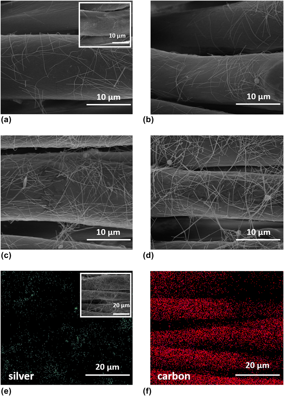
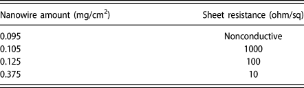
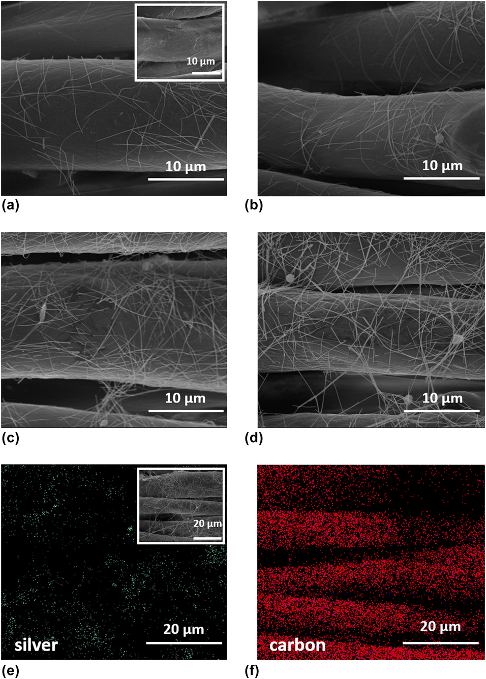
SEM images of Ag NW-modified cotton fabrics for each sample set with corresponding NW loadings of 0.095, 0.105, 0.125, and 0.375 mg/cm2 are shown in Fig. 1(a)–1(d), respectively. SEM image of bare cotton fabric is also shown as an inset in Fig. 1(a). It is clear that individual fibers can be easily decorated with Ag NWs. Moreover, Ag NW loading can be simply controlled through the number of dip and dry cycles. Changes in sheet resistance with respect to dip and dry cycles were already discussed in our previous work [Reference Doganay, Coskun, Genlik and Unalan29]. Changes in sheet resistance and the corresponding NW loading levels are given in Table I. Homogeneity of Ag NW-modified fabrics was observed via energy-dispersive X-ray spectroscopy (EDS) mapping technique, results of which are shown in Figs. 1(e) and 1(f). Dots in Fig. 1(e) represent Ag atoms, which show that NWs were homogenously decorated onto the fabrics (inset image in the same figure). Dots in Fig. 1(f) represent carbon atoms, which is the main element forming the molecular structure of cotton.
SEM images of Ag NW-decorated cotton fabrics with Ag NW loadings of (a) 0.095 (inset shows the SEM image for bare cotton fabric), (b) 0.105, (c) 0.125, and (d) 0.375 mg/cm2. EDS maps of Ag NW-decorated cotton fabrics at a NW loading of 0.375 mg/cm2 for (e) silver (inset shows the SEM image of EDS spot) and (f) carbon.
Ag NW loading and corresponding sheet resistance of 1 cm2 NW-decorated cotton fabrics.

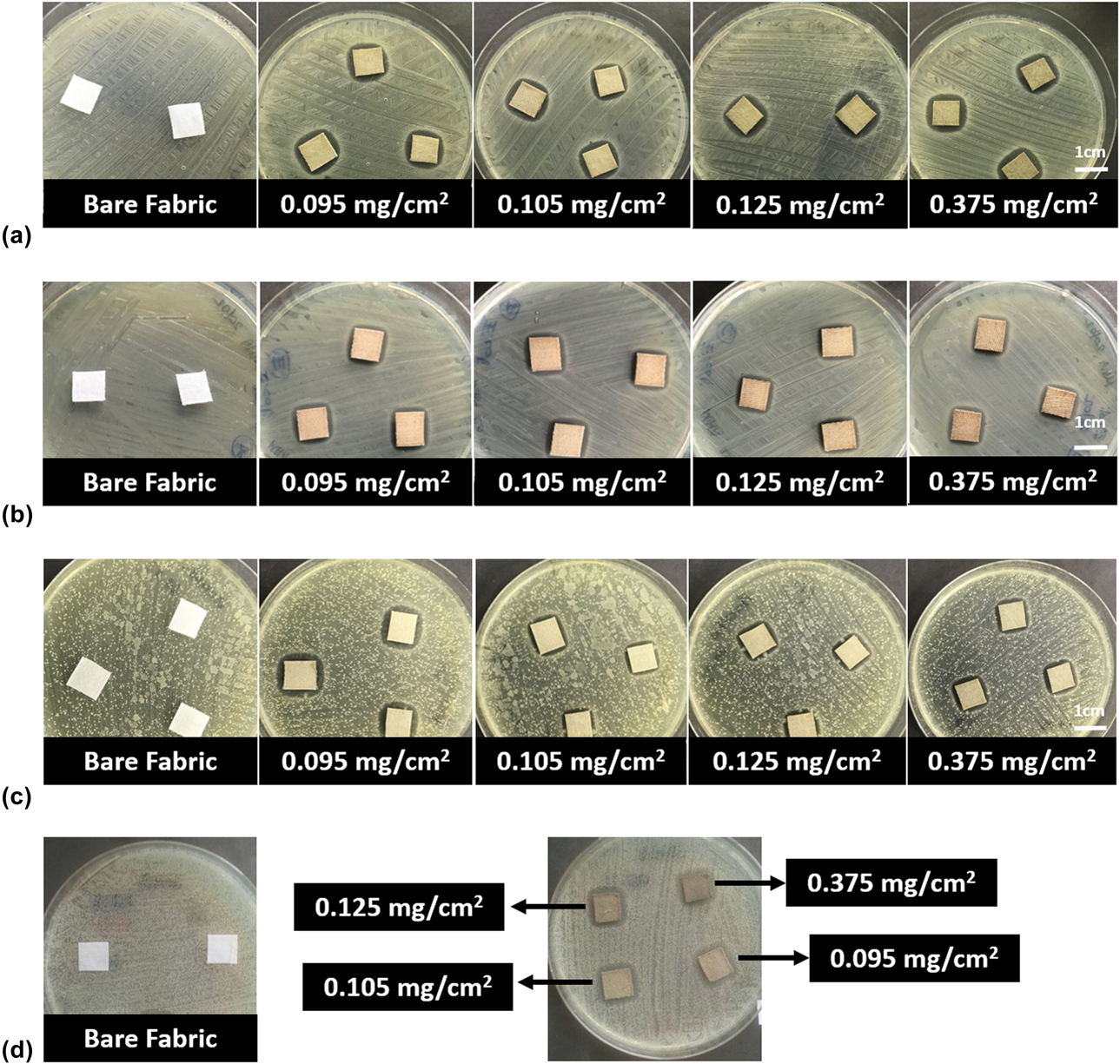
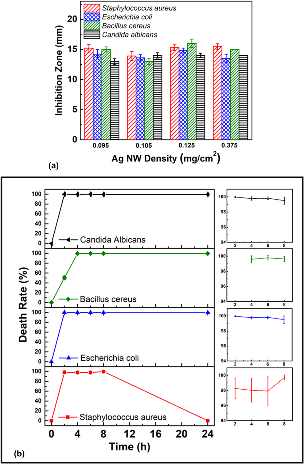
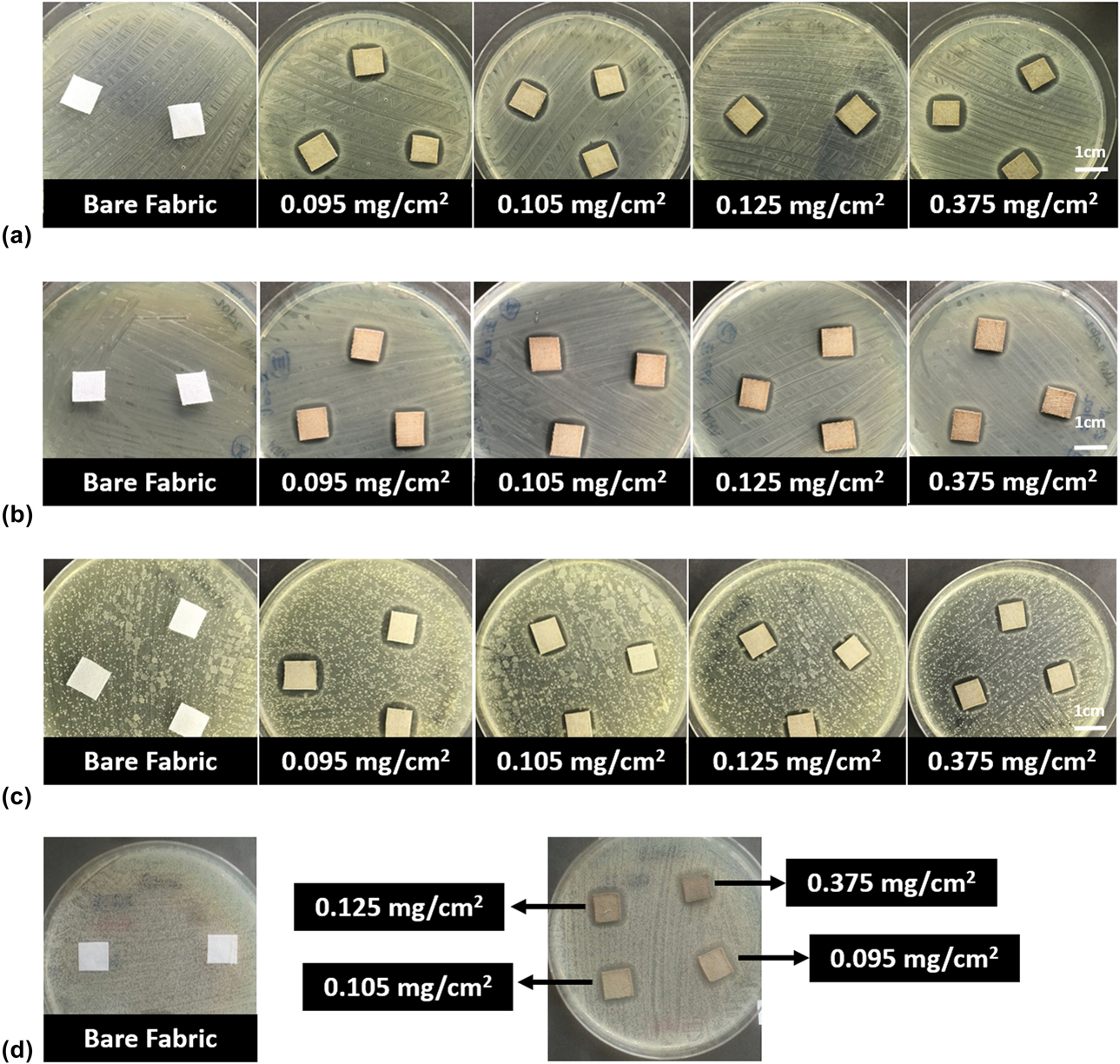
Photographs of bare cotton and Ag NW-decorated cotton fabrics following disk diffusion tests against Staphylococcus aureus (a Gram-positive coccus), Escherichia coli (a Gram-negative bacillus), Bacillus cereus (a Gram-positive and spore-forming bacillus), and Candida albicans (a yeast-like fungus) are provided in Figs. 2(a)–2(d), respectively. Ag NW-decorated fabrics showed encouraging antibacterial activity against all microorganisms used, whereas the bare cotton fabric had no effect against any of them. The size of the inhibition zones against all test strains is provided in Fig. 3(a). The inhibition zones can be described as the clear zones around the Ag NW-decorated fabrics on the Petri plates which contain bacterial suspensions cultured on suggested specific agar medium. Ag+ ions, while diffusing into the bacterial agar media, inhibit the bacteria and fungi grown on the agar at the meeting point, and there will be no bacterial growth until the concentration Ag+ ions is enough to kill the bacterial and fungal cells. The size of the inhibition zones was measured for the square samples, therefore; the inhibition zones are slightly lower at the edges of the samples. Size of the inhibition zones of Ag NW-modified fabrics against S. aureus with NW loadings of 0.095, 0.105, 0.125, and 0.375 mg/cm2 was 15, 14, 15, and 15 mm, respectively. This implied that NW loading has no significant influence on inhibition zones. A similar finding was reported by Hebeish et al. for Ag NP-decorated cotton textiles [Reference Hebeish, El-Naggar, Fouda, Ramadan, Al-Deyab and El-Rafie30]. Size of the inhibition zones of Ag NW-modified fabrics against E. coli with NW loadings of 0.095, 0.105, 0.125, and 0.375 mg/cm2 was 14, 14, 15, and 14 mm, respectively. Similarly, when tested against S. aureus strains, NW loading did not show any particular effect on the size of the inhibition zone, which was observed for Ag NP- and Ag NW-decorated textiles [Reference Satoungar, Fattahi, Azizi and Mehrizi26, Reference Messaoud, Chadeau, Brunon, Ballet, Rappenne, Roussel, Leonard, Oulahal and Langlet31]. It is expected that Ag NWs would show a wider inhibition zone against Gram-negative bacteria since Ag+ ions are positively charged. However, inhibition zones for S. aureus and E. coli were found to be more or less the same. Size of the inhibition zones of Ag NW-decorated fabrics against B. cereus with NW loading of 0.095, 0.105, 0.125, and 0.375 mg/cm2 was 15, 13, 16, and 15 mm, respectively. Size of the inhibition zones was found to be independent of the NW loading, similar to S. aureus and E. coli strains, especially when standard deviations were taken into consideration. Size of the inhibition zones of Ag NW-decorated fabrics against C. albicans with NW loading of 0.095, 0.105, 0.125, and 0.375 mg/cm2 was 13, 14, 14, and 14 mm, respectively. Similar to previous strains, NW loading had no influence on the size of the inhibition zones.
Photographs of bare cotton and Ag NW-modified fabrics following disk diffusion test against (a) Staphylococcus aureus, (b) Escherichia coli, (c) Bacillus cereus, and (d) Candida albicans.
(a) Size of inhibition zones for Ag NW-modified 1 cm2 cotton fabrics measured by disk diffusion method with respect to Ag NW loading. (b) Bacterial death rate with respect to contact time against S. aureus, E. coli, B. cereus, and C. albicans for 0.375 Ag NW-modified fabrics.
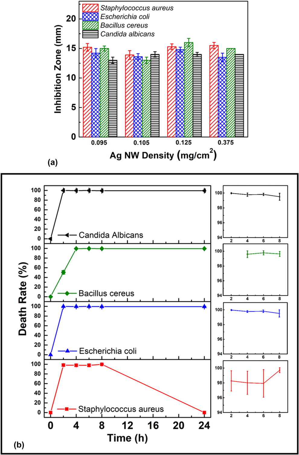
Effective antimicrobial duration is an important feature for antimicrobial fabrics. The fabrics should have sufficient efficacy for a long-term use. Therefore, antimicrobial effect of 0.375 mg/cm2 Ag NW-decorated fabric was tested with respect to time against all test strains and the results are compared in Fig. 3(b). Against S. aureus, Ag NW-modified cotton fabrics reached their maximum efficacy within 2 h, killed 99.9% of the bacteria, and maintained their activity for 8 h. Then, the antimicrobial activity started to decrease, and the bacterial concentration returned to the initial level after 24 h. The death rate of Ag NW-modified cotton fabrics against E. coli reached maximum (99.9%) within 2 h, and the fabrics maintained their activity for 24 h. Against B. cereus, the fabrics reached maximum efficiency within 4 h, and the maximum death rate reached 99.9%. Moreover, the fabrics maintained their activity for 24 h. The time required to reach the maximum efficiency was longer for B. cereus than for both S. aureus and E. coli. This can be attributed to the spore-forming characteristic of B. cereus. Bacterial colonies might protect themselves up to 4 h. On the other hand, death rate reached a maximum of 99.9% within 2 h and remained unchanged for 24 h against C. albicans.
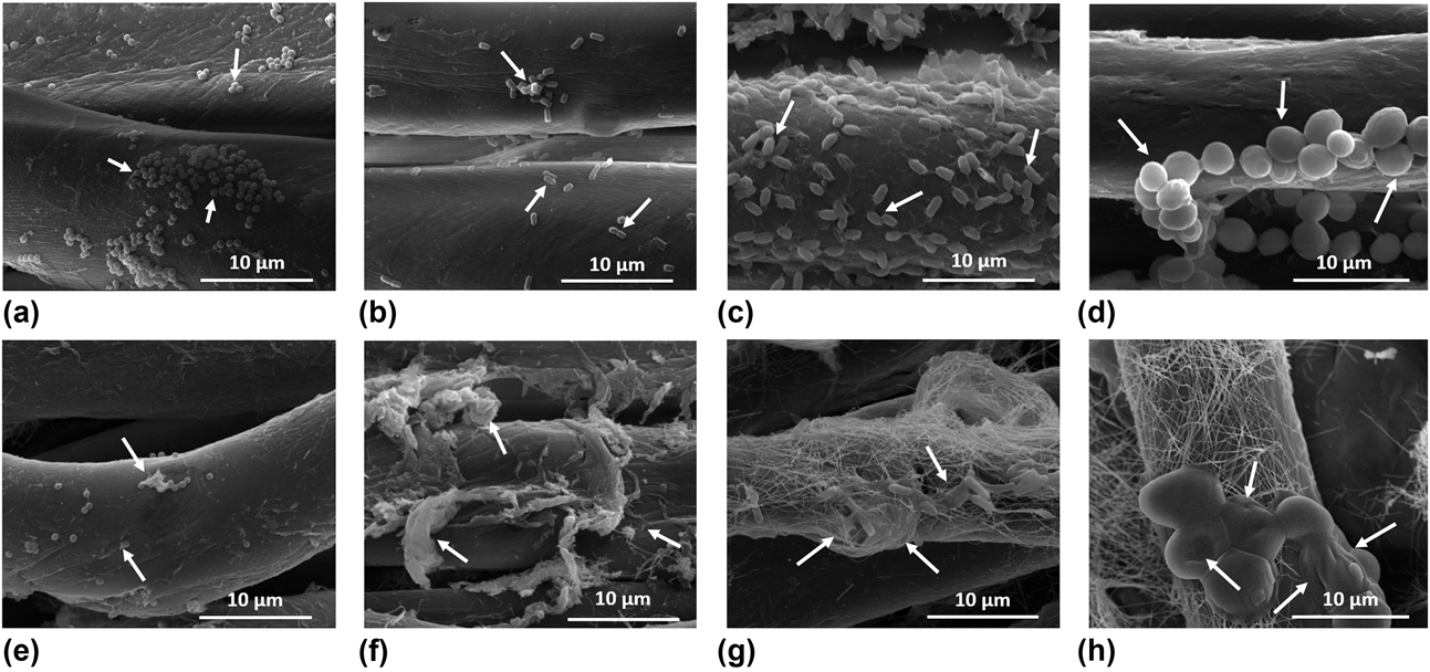
Morphological features of both living and dead bacteria were investigated via SEM to understand the antibacterial mechanism. SEM images of the bacteria on bare cotton and Ag NW-decorated cotton fabrics are shown in Figs. 4(a)–4(h). Perfect shapes of S. aureus, E. coli, B. cereus, and C. albicans [indicated by arrows in Figs. 4(a)–4(d), respectively] were observed on the bare cotton fabric, implying that all tested strains can easily survive and multiply on it. On the other hand, cellular malformation occurred when the strains encountered the NW-decorated fabrics. Final shapes of the dead S. aureus, E. coli, B. cereus, and C. albicans are indicated by arrows in Figs. 4(e)–4(h), respectively. In particular, due to the greater size of C. albicans, pit formations on the bacterial membrane are clearly observed [Fig. 4(h)]. These pit formations are consistent with the antibacterial mechanisms of the Ag NW as mentioned before.
SEM images of (a) and (e) S. aureus, (b) and (f) E. coli, (c) and (g) B. cereus, (d) and (h) C. albicans that are living (arrows show the actual shape of the strains) and dead (arrows show the cellular malformation of the strains). (a), (b), (c), and (d) are bare cotton fabrics, (e) and (f) are Ag NW fabrics with NW loadings of 0.095 mg/cm2 and (g) are (h) are Ag NW fabrics with NW loading of 0.375 mg/cm2.

Conducting textiles, as described in our previous work, can be used as mobile heaters with the help of Joule heating mechanism if an external potential is provided [Reference Doganay, Coskun, Genlik and Unalan29]. The results obtained in this work and our previous work clearly show that the deposition of Ag NWs makes fabrics multifunctional. Multifunctionality of the fabrics should not be limited only to these applications, but should be extended further with applications such as electromagnetic interference (EMI) shielding and wearable sensors.
Conclusions
Antimicrobial activity of Ag NW-modified cotton fabrics was investigated in this study. Dip and dry method was utilized as a facile, scalable, and reproducible way to modify cotton fabrics with Ag NWs. Moreover, Ag NW loading on the fabrics can be easily controlled with this method. Ag NW-decorated fabrics showed promising antibacterial activity against a wide range of strains. The inhibition zones of the 1 cm2 modified fabrics were found to be around 15 mm for each strain (S. aureus, E. coli, B. cereus, and C. albicans). Moreover, Ag NW loading was found to have no influence on the size of the inhibition zone for all strains. In addition, antibacterial efficacy was also investigated for all test strains. It was observed that the modified fabrics reach their maximum antimicrobial efficacy within the first 2 h against S. aureus, E. coli, and C. albicans. On the other hand, maximum efficacy was reached at 4 h for B. cereus, which was attributed to the spore-forming nature of B. cereus. Moreover, the modified fabrics maintained their activity up to 24 h against E. coli, C. albicans, and B. cereus and up to 8 h against S. aureus. To conclude, Ag NW-decorated cotton fabrics demonstrated herein showed promising antibacterial activity, which revealed the potential of Ag NWs as antibacterial modifiers in the textile industry due to their wide range, relatively long-term antimicrobial effect, and scalable production route. As shown, small amounts of Ag NW decoration enabled promising antimicrobial activity with a cost-effective way compared to conventional antimicrobial textiles. Moreover, the one-dimensional nature of Ag NWs ensured better interactions between the textile fibers that would increase washing durability (not reported here) as well as mechanical stability (not reported here) without a protection layer.
Experimental details
Synthesis and purification of Ag NWs
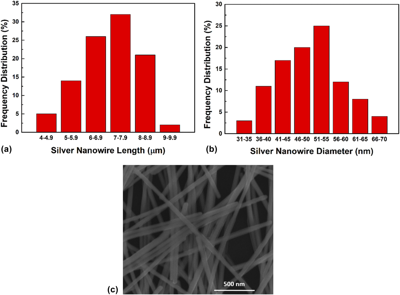
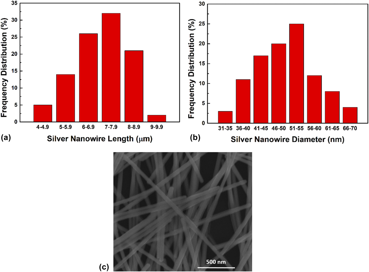
All chemicals were purchased from Sigma-Aldrich (St. Louis, MO) and used without further purification. Undyed 100% organic woven cotton fabrics and all glassware were cleaned using acetone (99.8%), isopropyl alcohol (99.8%), and finally deionized water (18.3 MΩ). Ag NWs were synthesized according to a previous study [Reference Coskun, Aksoy and Unalan32]. In a typical synthesis, a 10 mL and 0.45 M ethylene glycol (EG) solution of poly(vinylpyrrolidone) (PVP) (monomer-based calculation MW = 55000) was prepared, to which 7 mg of sodium chloride (NaCl) was added. Then, the solution was heated at 170 °C in a round flask within a silicon bath. Meanwhile, a 0.12 M silver nitrate (AgNO3) solution in 5 mL EG was prepared and added dropwise to the PVP solution by an injection pump (TOP-5300 Model Syringe Pump, TOP Corp., Tokyo, Japan) at a rate of 5 mL/h. The solution was stirred at a rate of 1000 rpm using a magnetic stirrer during the whole process. Upon the completion of dropwise addition, the NW solution was annealed for 30 min at 170 °C and finally air-cooled to room temperature. Then, to separate the polymer from the Ag NWs, the solution was diluted with acetone (at a ratio of 1:5) and centrifuged twice at 8000 rpm for 20 min. After that, the NWs were dispersed in ethanol and again centrifuged at 8000 rpm for 20 min. The final product was dispersed in ethanol for further characterization and processing. A final decantation process was performed to remove by-products, such as micro- and nano-sized Ag NPs. Frequency distributions of length and diameter and SEM image of Ag NWs are shown in Figs. 5(a)–5(c), respectively. The average length and diameter of the NWs utilized in this work were 8 µm and 50 nm, respectively.
Frequency distributions of (a) length and (b) diameter of Ag NWs used in this work. (c) SEM image of Ag NWs.
Ag NW modification onto fabrics
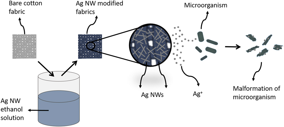
Dip and dry method is one of the most convenient ways to modify fabrics with nanomaterials, which is also scalable for large-scale production. Thus, this method was used to modify cotton fabrics with Ag NWs, details of which are described elsewhere [Reference Doganay, Coskun, Genlik and Unalan29]. Simply, 1 cm2 cleaned cotton fabrics were immersed into 20 mL of 0.2 mg/mL ethanolic Ag NW dispersions for 5 min and then dried at 80 °C for 10 min. This procedure was repeated until four different sets of samples in terms of Ag NW loading were obtained. Sheet resistance was measured with two probes along the side of the 1 cm2 square fabrics via Keithley 2400 Source Meter (Keithley Instruments, Inc., Solon, OH) A bare and nonconductive sample was used for reference. Resistance was measured at four different points, and an average value is reported. Resistance measurements allowed us to reproducibly deposit Ag NWs onto cotton fabrics as a control mechanism. Schematic representation of fabrication steps used in this work together with the antibacterial effect of Ag NW-decorated fabrics is presented in Fig. 6. Following decoration of Ag NWs onto fabrics, the exact Ag NW loading on cotton fabrics was determined via inductively coupled plasma mass spectrometry (ICP-MS). For this purpose, NW-coated fabrics were immersed into 20 mL of 10% nitric acid/deionized water mixture until a homogenous and clear solution was observed.
Schematic illustration of Ag NW decoration of fabrics and antibacterial mechanism of modified fabrics.

Antimicrobial analyses
Antimicrobial activities of 1 cm2 square fabrics against standard test strains of Staphylococcus aureus (S. aureus) ATCC#25923 as a Gram-positive coccus, Escherichia coli (E. coli) ATCC#25922 as a Gram-negative bacillus, Bacillus cereus (B. cereus) ATCC#14603 as a Gram-positive and spore-forming bacillus, and Candida albicans (C. albicans) ATCC#90028 as a yeast-like fungus were investigated via agar disk diffusion technique. After freshly culturing bacterial strains on Mueller–Hinton agar (MHA, Merck, Darmstadt, Germany) and C. albicans on Sabouraud Dextrose agar (SDA, Merck, Darmstadt, Germany) at 37 °C for 48–72 h, the grown colonies of the strains were harvested. The bacterial and fungal suspensions were prepared in sterilized test tubes containing 5 mL Mueller–Hinton broth (MHB, Merck, Darmstadt, Germany), and the final inoculums were adjusted to 1.5 × 10⁸ and 2 × 10⁸ CFU (colony-forming unit)/mL, respectively, according to 0.5 McFarland test standard, turbidometrically. Additionally, the inoculums were adjusted according to their optical density (OD450 nm: 0.600) spectrophotometrically. Then, 100 µL of each suspension was inoculated on the MHA for bacterial strains and on SDA for C. albicans. The fabrics were placed on these agar plates (at least ten samples each). They were incubated at 37 °C for 24–48 h, and after the incubation, the diameters (mm) of the inhibition zones of the samples were measured and calculated as CFU/mL. Fabrics were tested for their time-dependent killing capacity for 2, 4, 6, 8, and 24 h time intervals against the microorganisms. The same concentrations of the bacterial and fungal agents’ suspensions were placed in their specific broth media as mentioned before. Then, at least two samples of each fabric were put in the test tubes and incubated for the chosen time intervals at 37 °C. Afterward, the fabrics were taken out, washed twice with phosphate-buffered solution (PBS, pH: 7.0), transferred to another test tube containing sterilized 1 mL distilled water, and diluted 10−2 and 10−3 times. Then, 100 µL of samples from each test tube was cultured on specific agar plates as mentioned before. Following incubation, the grown colonies were counted and calculated as CFU/mL.
SEM analyses
The homogeneity of the NW modification was monitored via SEM analysis using FEI NOVA NANO SEM 430 microscope (FEI, Hillsboro, OR) with secondary image mode and EDX mapping techniques under an operating voltage of 5 kV. A thin gold layer (5–10 nm) was deposited onto the fabrics prior to SEM analysis. To understand the antibacterial mechanism, morphological features of both living and dead bacteria were investigated via SEM. Prior to SEM imaging, both living and dead bacteria were fixed onto fabrics to prevent deformation due to vacuum and voltage according to a procedure described in the literature [Reference Morones, Elechiguerra, Camacho, Holt, Kouri, Ramírez and Yacaman16]. Each sample was immersed into a 2.5% glutaraldehyde solution in phosphate-buffered saline (PBS) for 30 min, which is followed by 60, 70, 80, 90, and 100% ethanol/PBS solutions for 10 min immersion into each.
Acknowledgment
This work was financially supported by METU research funds through DKT-308-2018-3734 and BAP-07-02-2017-004-268.