Introduction
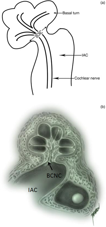
Inner-ear malformations affecting the bony labyrinth account for approximately 20 per cent of all cases of congenital sensorineural hearing loss (SNHL); the other 80 per cent are due to membranous labyrinth pathology.Reference Jackler, Luxford and House 1 Anomalies of the bony labyrinth can be diagnosed using computed tomography (CT) and magnetic resonance imaging (MRI), but cellular level pathologies of the membranous labyrinth cannot be diagnosed using conventional imaging methods. The bony cochlear nerve canal carries cochlear nerve fibres from the spiral ganglion to the internal auditory canal (Figure 1). It is considered narrow (stenotic) when the diameter at the mid-modiolus is less than 1.5 mm.Reference Miyasaka, Nosaka, Morimoto, Taiji and Masaki 2 A narrow bony cochlear nerve canal was recently defined as a bony labyrinth anomaly that causes SNHL, with or without accompanying inner-ear malformation.Reference Fatterpekar, Mukherji, Alley, Lin and Castillo 3
Schematic diagrams showing (a) the bony cochlear nerve canal (BCNC), internal auditory canal (IAC) and cochlear nerve, and (b) the normal cochlear structure and BCNC.

Owing to recent improvements in imaging technology, anomalies such as bony cochlear nerve canal atresia and stenosis can now be visualised. However, few studies have investigated bony cochlear nerve canal diameter. Nonetheless, rigorous assessment of this anatomical structure is important because stenosis of the bony cochlear nerve canal and internal auditory canal are commonly associated with cochlear nerve deficiency, which contraindicates cochlear implantation.
This study therefore aimed to (1) determine which inner-ear malformations commonly accompany bony cochlear nerve canal and internal auditory canal atresia and stenosis; and (2) investigate whether stenosis of the bony cochlear nerve canal and internal auditory canal correlate with cochlear nerve deficiency, as determined by CT and MRI findings.
Materials and methods
Patients
The study included 38 patients (59 ears) aged 1 to 19 (mean: 4.65 ± 4.26) years with SNHL due to inner-ear malformation who were followed up at Department of Otolaryngology – Head and Neck Surgery, Hacettepe University, and for whom temporal bone CT and MRI findings were stored in the hospital's Picture Archiving System (‘PACS’; General Electrics, Chicago, IL, USA) database. Those who underwent CT and/or MRI at a different institution were excluded. All patients diagnosed via temporal bone CT with bony cochlear nerve canal stenosis or atresia were included. A cut-off value of 1.5 mm for the bony cochlear nerve canal diameter was based on earlier studies.Reference Miyasaka, Nosaka, Morimoto, Taiji and Masaki 2 , Reference Stjernholm and Muren 4 Internal auditory canals with mid-point diameters of less than 2 mm were considered stenotic.Reference Valvassori and Palacios 5 – Reference Masuda, Usui and Matsunaga 7 Patients lacking a bony cochlear nerve canal due to ear anomalies such as Michel's deformity, cochlear aplasia, common cavity and rudimentary otocyst were excluded.
The control group comprised 36 patients (72 ears) aged 0–15 years with normal labyrinth findings upon CT investigation for trauma, tympanic membrane perforation or chronic otitis media. These patients did not have SNHL (based on audiological examination) or any inner-ear pathology. Subsequent management and treatment modalities, including hearing aids, medical treatment, cochlear implantation and auditory brainstem implantation, were noted.
Imaging
All SNHL and control patients underwent CT and MRI in the Radiology Department, Hacettepe University. High-resolution CT was performed in the axial plane using a four-channel multidetector CT scanner (Somatom Plus 4 Volume Zoom, Siemens, Erlangen, Germany); all images had 0.5-mm collimation and were 0.5 mm thick. Reformatted axial images parallel to the lateral semi-circular canal and coronal images were recorded. MR examinations were performed with either a 3T (Allegra, Siemens, Erlangen, Germany) or a 1.5 T scanner (Symphony, Siemens, Erlangen, Germany), using a standard head coil. The standard temporal bone protocol included axial and sagittal-oblique three-dimensional (3D) constructive interference in steady state (‘CISS’; Siemens) or DRIVE (Philips) imaging.
Image evaluation
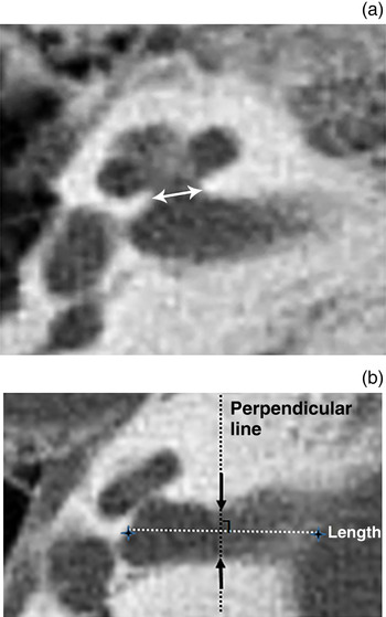
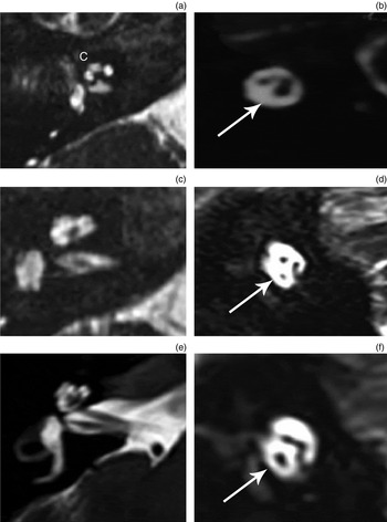
All CT and MRI scans were retrospectively reviewed by a neuroradiologist and two otolaryngologists experienced in temporal bone imaging. The bony cochlear nerve canal at the mid-modiolus was measured using calipers on axial CT images (shown in Figure 2a). All bony cochlear nerve canals were categorised as normal, stenotic or atretic. The internal auditory canal diameter was evaluated in axial CT sections by measuring the width at the centre of the canal (as shown in Figure 2b). Cochlear nerves were evaluated on axial and sagittal-oblique T2-weighted MRI images, and classified as aplastic, hypoplastic or normal, according to their size relative to the facial nerve (Figure 3). They were defined as aplastic if they could not be seen in the internal auditory canal and hypoplastic if smaller than the facial nerve inside the internal auditory canal. Each cochlea was examined separately and cochlear anomalies were defined according to Sennaroğlu's classification system.Reference Sennaroğlu 6
Axial high-resolution computed tomography sections showing (a) the bony cochlear nerve canal diameter at the mid-modiolar level (white arrow) and (b) the internal auditory canal diameter at the centre of the canal (indicated by the perpendicular line).

Sagittal (left) and oblique (right) 3 Tesla magnetic resonance imaging sections showing (a,b) an aplastic cochlear nerve, (c,d) a hypoplastic cochlear nerve and (e,f) a normal cochlear nerve. White arrows indicate the cochlear nerve; c = cochlear nerve

Audiological and statistical evaluation
Hearing status determined by otoacoustic emission (OAE), auditory brainstem response (ABR; at a click stimulus intensity of 99 dB nHL), audiography and subjective audiological test was obtained from patient records. Statistical evaluation was performed using IBM SPSS Statistics for Windows version 21.0 (IBM, Armonk, NY, USA). The study protocol was approved by the Hacettepe University School of Medicine Ethics Committee (GO 14/462).
Results
Inner-ear measures
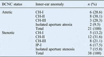
Of the 59 ears in the SNHL group, the bony cochlear nerve canal was atretic in 21 and stenotic in 38. Of the 21 ears with an atretic bony cochlear nerve canal, 19 had cochlear hypoplasia: 6 had type I, 8 had type II and 5 had type III. The other two cochleas had a normal appearance and were classified as having isolated bony cochlear nerve canal atresia. Of the 38 ears with stenotic bony cochlear nerve canal, 25 had cochlear hypoplasia: 5 had type I, 12 had type II, and 8 had type III. A further seven had incomplete partition: six had type I and one had type II. The remaining six cochleas had a normal appearance and were classified as having isolated bony cochlear nerve canal stenosis. The distribution and frequency of anomalies are shown in Table I.
Distribution of inner-ear anomalies accompanying bony cochlear nerve canal stenosis or atresia

χ2 test, p = 0.287. BCNC = bony cochlear nerve canal; CH-I = cochlear hypoplasia type 1; CH-II = cochlear hypoplasia type 2; CH-III = cochlear hypoplasia type 3; IP-I = incomplete partition type 1
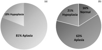
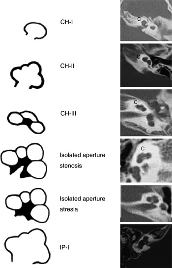
All 21 ears with bony cochlear nerve canal atresia had cochlear nerve deficiency: the cochlear nerve was aplastic in 18 (81 per cent) and hypoplastic in 4 (19 per cent). Of the 38 ears with bony cochlear nerve canal stenosis, the cochlear nerve was aplastic in 24 (63.1 per cent), hypoplastic in 8 (21 per cent) and normal in the remaining 6 (15.8 per cent). Cochlear nerve status in ears with atretic and stenotic bony cochlear nerve canals is shown in Figure 4. The commonest anomaly accompanying cochlear nerve aplasia in 41 ears was cochlear hypoplasia type II (n = 15 ears, 37 per cent), followed by cochlear hypoplasia type III (n = 9 ears, 22 per cent) and cochlear hypoplasia type I (n = 8 ears, 20 per cent). Overall, three ears (7 per cent) had incomplete partition type I. Schematic diagrams of these inner-ear malformations are shown in Figure 5.
Pie charts showing the proportions of cochlear nerves according to status in ears with atretic (a) and stenotic (b) bony cochlear nerve canals.

Schematic representations of inner-ear malformations, including isolated aperture atresia and stenosis (left), and axial high-resolution computed tomography images (right). c = cochlear nerve; CH-I = cochlear hypoplasia type 1; CH-II = cochlear hypoplasia type 2; CH-III = cochlear hypoplasia type 3; IP-I = incomplete partition type 1

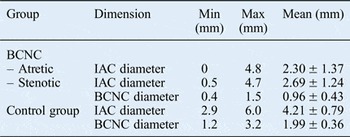
The mean internal auditory canal diameter of the 72 ears in the control group was 4.21 ± 0.79 mm (range 2.9–6.0 mm). In the study group, the mean internal auditory canal diameter was 2.32 ± 1.40 mm (range 0–4.8 mm) in ears with bony cochlear nerve canal atresia and 2.45 ± 1.24 mm (range 0.2–4.7 mm) in ears with bony cochlear nerve canal stenosis. Therefore, this measure was significantly higher in the control group than in the SNHL group (p < 0.05). Internal auditory canal and bony cochlear nerve canal measurements for both groups are shown in Table II. The cochlear nerve was deficient in all 16 ears with stenosis of both the bony cochlear nerve canal and internal auditory canal. In the six ears with bony cochlear nerve canal stenosis and a normal cochlear nerve, none of the internal auditory canals was stenotic.
Internal auditory canal and bony cochlear nerve canal measures

Kruskal–Wallis variance analysis, p = 0.00. Min = minimum; Max = maximum; IAC = internal auditory canal; BCNC = bony cochlear nerve canal
Hearing and other abnormalities
In all, 31 of the 39 SNHL patients underwent OAE testing. Of these, 9 had positive OAE findings bilaterally, with no ABR response, and another 29 patients had no ABR response bilaterally. Associated systemic syndromes in the SNHL group included Goldenhar syndrome (three patients) and coloboma, heart defects, atresia choanae, growth retardation, genital abnormalities and ear abnormalities (‘CHARGE’) syndrome (two patients). Two patients in each study group had a cleft palate/lip, motor mental retardation or a central nervous system anomaly (such as microcephaly).
Treatment modalities
A total of 11 patients in the SNHL group underwent cochlear implantation and 10 underwent auditory brainstem implantation. One patient initially underwent cochlear implantation in the right ear, but subsequently underwent auditory brainstem implantation on the contralateral side due to insufficient auditory–verbal development. In this patient, the diameters of the right and left bony cochlear nerve canals were 1.1 mm and 1.2 mm, respectively, that is, stenotic. Eight patients did not undergo cochlear or auditory brainstem implantation: in five, hearing loss detection was delayed and three had unilateral hearing loss.
Discussion
A narrow internal auditory canal is commonly thought to indicate a hypoplastic or aplastic cochlear nerve. However, the internal auditory canal diameter alone may be insufficient to indicate cochlear nerve deficiency, and a second measurable parameter is also required to predict cochlear nerve status, especially in cochlear implant candidates. The normal internal auditory canal diameter is 2–8 mmReference Valvassori and Palacios 5 – Reference Masuda, Usui and Matsunaga 7 ; a recent report indicates that an internal auditory canal is stenotic when its diameter is 2 mm or less.Reference Sennaroğlu 6 Computed tomography is the method recommended for assessing the internal auditory canal diameter, whereas MRI is recommended for determining whether the internal auditory canal contains cochlear nerve fibres.Reference Valvassori and Palacios 8 , Reference Sakina, Goh, Abdullah, Zulfiqar and Saim 9
In the present study, the minimum internal auditory canal diameter in the SNHL group was 0 mm (i.e. atretic), the maximum diameter was 4.8 mm and the mean diameter was 2.37 ± 1.40 mm. The cochlear nerve was aplastic or hypoplastic in all patients in the SNHL group with an internal auditory canal diameter of less than 2 mm. These findings suggest that, in addition to internal auditory canal diameter, the bony cochlear nerve canal diameter might be a useful marker for evaluating the cochlear nerve status. A correlation between bony cochlear nerve canal stenosis and internal auditory canal stenosis was previously reported.Reference Fatterpekar, Mukherji, Alley, Lin and Castillo 3 , Reference Adunka, Roush, Teagle, Brown, Zdanski and Jewells 10 The bony cochlear nerve canal diameter appears to be a reliable marker of the presence and status of the cochlear nerve. However, a normal cochlea and internal auditory canal diameter do not indicate that the cochlear nerve is normal. In the SNHL group, 8 out of 59 ears (14 per cent) had a normal cochlear shape, even though the cochlear nerve was deficient. Adunka et al. reported a normal internal auditory canal diameter in 56 per cent of cochlear nerve aplasia patients and a normal cochleovestibular anatomy in 42 per cent.Reference Adunka, Roush, Teagle, Brown, Zdanski and Jewells 10 In the present study, eight patients with cochlear nerve deficiency had a normal cochlear structure.
In a histopathological study, Henderson et al. reported a mean bony cochlear nerve canal diameter of 2.26 ± 0.25 mm at the mid-modiolar level in normal temporal bones.Reference Henderson, Wilkins, Huang, Kenna and Gopen 11 Cochlear nerve hypoplasia or aplasia should be considered when radio-anatomical findings indicate a bony cochlear nerve canal diameter of less than 1.4 mm (according to Stjernholm and MurenReference Stjernholm and Muren 4 ) or 1.5 mm (according to Miyasaka et al.Reference Miyasaka, Nosaka, Morimoto, Taiji and Masaki 2 ). In the present study, the cochlear nerve was of normal size in only 6 out of 59 ears (10 per cent) with a bony cochlear nerve canal diameter of 1.5 mm or less. Therefore, both the present study and current literature indicate that the bony cochlear nerve canal and internal auditory canal are complementary structures that should be examined via CT in all cochlear implant candidates.
Masuda et al. reported in 69 SNHL patients that bony cochlear nerve canal stenosis was accompanied by internal auditory canal stenosis, suggesting a strong correlation between these two entities.Reference Masuda, Usui and Matsunaga 7 The present findings support Masuda and colleagues’ study: cochlear nerve deficiency was observed in 16 ears with bony cochlear nerve canal stenosis or atresia accompanied by internal auditory canal stenosis. The most common anomaly accompanying cochlear nerve aplasia in the present study was cochlear hypoplasia (n = 40 ears). Yi et al. reported that patients with bony cochlear nerve canal and internal auditory canal stenosis had smaller than normal cochlear dimensions.Reference Yi, Lim, Kang, Park, Park and Lee 12 However, further research is necessary to clarify the relationship between cochlear hypoplasia and cochlear nerve deficiency. Normal hair cell development can be completed in the absence of neural innervationReference Malgrange, Belachew, Thiry, Nguyen, Rogister and Alvarez 13 ; however, spiral ganglion and inner-ear innervation depend on neurotrophic factors released by hair cells. Similarly, cochlea development may be normal in the absence of a cochlear nerve. This hypothesis is supported by the finding that 9 out of 41 ears with cochlear nerve aplasia had a normal cochlear structure. Moreover, cochlear hypoplasia was detected in 44 out of 59 ears (75 per cent) with a bony cochlear nerve canal diameter of 1.5 mm or less; cochlear hypoplasia type II was most common. Insufficient growth factor production by a hypoplastic cochlea may adversely affect neural development, resulting in cochlear nerve deficiency.
In the present study, 17 patients had unilateral bony cochlear nerve canal atresia or stenosis without contralateral ear anomalies (vs 21 bilaterally affected patients). Thus, unilateral bony cochlear nerve canal atresia or stenosis might cause single-sided deafness. This possibility should always be considered when planning cochlear implant surgery for patients with single-sided deafness to avoid cochlear implantation in patients with a deficient cochlear nerve.
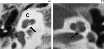
The cochlear nerve can be aplastic or hypoplastic even when temporal bone CT shows a normal cochlea. Thus, a normal cochlear shape does not always indicate normal cochlear nerve structure and function. In the present study, 9 out of 41 cochlear nerve aplasia patients (22 per cent) and 1 out of 12 (8 per cent) cochlear nerve hypoplasia patients had a normal cochlear structure. In these patients, the anomaly was defined as isolated bony cochlear nerve canal atresia and isolated bony cochlear nerve canal stenosis, respectively (Figure 6). Although cochlear aperture anomalies were described in the Sennaroğlu classification system (2013 revision), these new terms may need to be added to the classification system in the future.Reference Sennaroğlu, Ozkan and Aslan 14
Axial high-resolution computed tomography images showing normal cochlear anatomy with (a) isolated aperture atresia and (b) isolated aperture stenosis (indicated by arrows).

Recommended imaging methods for cochlear implant candidates are CT alone, MRI alone, or a combination of both.Reference Vaid and Vaid 15 If only CT is to be used, the diameters of the bony cochlear nerve canal and internal auditory canal can be used to estimate cochlear nerve status. In patients scheduled for cochlear implantation, MRI should also be performed if possible, even if no cochlear anomaly is evident by temporal bone CT. As the cochlear nerve is less likely to be normal in the presence of isolated bony cochlear nerve canal atresia or stenosis, auditory rehabilitation and language development after cochlear implantation is likely to fail in patients with undetermined cochlear nerve status. In addition, patients with cochlear implantation failure should not undergo MRI because of artefacts resulting from the implant.
In 2006, Buchman et al. screened 65 auditory neuropathy patients and noted an aplastic cochlear nerve in 18 per cent.Reference Buchman, Roush, Teagle, Brown and Zdanski 16 They also observed cochlear microphonics in 70 per cent of ears with cochlear nerve aplasia, despite a negative ABR, thus proving that hair cells can function in the absence of a cochlear nerve. In the present study, an ABR could not be identified in nine patients with positive OAE test results. Physiologically, transiently evoked otoacoustic emissions (TEOAEs) represent the response of the cochlea's external hair cells and can therefore be identified in individuals with a normal cochlea but an abnormal cochlear nerve. In itself, a normal TEOAE does not guarantee normal hearing and a normal cochlear nerve so, even in healthy neonates, hearing screening should involve both TEOAE and automated ABR tests.
Bilateral labyrinth aplasia, cochlear aplasia, a narrow or aplastic internal auditory canal, absence of the cochlear nerve, bony cochlear nerve canal aplasia, or any combination of these malformations causes prelingual total hearing loss that is impossible to correct by cochlear implantation.Reference Kaga 17 , Reference Papsin 18 As hearing loss is prelingual in such patients, age at cochlear implantation and the decision-making process for implantation surgery are critically important for a successful outcome. Auditory brainstem implantation positively affects the development of auditory–verbal abilities in paediatric patients.Reference Sennaroğlu, Ziyal and Atas 19 – Reference Colletti, Shannon and Colletti 21 When a cochlear anomaly, or a stenotic bony cochlear nerve canal and internal auditory canal, are detected via pre-operative temporal bone CT, cochlear implantation should not be performed before establishing that the cochlear nerve is normal. Unfortunately, in the presence of a hypoplastic cochlear nerve, it is difficult to predict whether auditory–verbal abilities will develop sufficiently after cochlear implant surgery. If a patient does not benefit from a cochlear implant, then an auditory brainstem implant should be considered a treatment option; however, during this critical period, the patient will develop auditory–verbal abilities in the absence of auditory stimuli. In such cases, auditory brainstem implantation into an ear with a severe anomaly and hypoplasia and cochlear implantation into an ear with a milder anomaly would provide binaural hearing. Simultaneous cochlear and auditory brainstem implantation should therefore be considered in cases of bilateral cochlea and cochlear nerve anomalies.
-
• A normal cochlea does not guarantee a normal cochlear nerve
-
• The bony cochlear nerve canal diameter is a marker of cochlear nerve status
-
• A narrow or atretic bony cochlear nerve canal with otherwise normal cochlear anatomy may be termed isolated bony cochlear nerve canal atresia or stenosis
-
• All patients with cochlear nerve deficiency had bony cochlear nerve canal stenosis or atresia and internal auditory canal stenosis
-
• As otoacoustic emission testing does not guarantee normal hearing and a normal cochlear nerve, neonates should also undergo automated auditory brainstem response testing
The present study has a few limitations. One limitation is the small cohort size due to excluding some patients who underwent CT and/or MRI at another institution. Since there is no generally accepted cochlear nerve measurement method, another limitation is that the cochlear nerve was measured manually (diameter and density) and assessed by comparison with the facial nerve. As imaging technologies continue to develop, it may soon be possible to assess the cochlear nerve in a more objective and definitive way. In addition, not all MRI examinations used a 3 Tesla scanner, although this had no effect on visualisation of the cochlear nerve within the internal auditory canal.
Conclusion
The present study found that the cochlear nerve was hypoplastic or aplastic when the bony cochlear nerve canal diameter was less than 1.5 mm and the internal auditory canal diameter was less than 2 mm. The bony cochlear nerve canal and internal auditory canal are complementary structures, and both should be assessed by temporal bone CT to predict cochlear nerve status and plan treatment.
Acknowledgements
The authors would like to thank Dr S Jafarov for his excellent work in preparing the schematic drawings.